Avoiding False-Positive Glaucoma Diagnosis in Myopic Eyes: Clinical Importance of OCT Scan Diameter
Saadet Gültekin Irgat, Ramazan Demirel, Ecem Ulutürk, Alpaslan Koç, Fatih Özcura, Özlem Arık

TL;DR
This study shows that using a 4.1 mm OCT scan diameter helps avoid false glaucoma diagnoses in myopic eyes by better distinguishing natural myopic thinning from actual glaucomatous damage.
Contribution
The study identifies the optimal OCT scan diameter (4.1 mm) for minimizing false-positive glaucoma diagnoses in myopic eyes.
Findings
RNFLT decreases significantly with larger scan diameters, especially in nasal quadrants.
The 4.1 mm scan (C2) showed the best discrimination between myopic and control eyes in nasal regions.
A shift in OCT color codes from green to yellow/red was most noticeable at the outer 4.7 mm scan (C3).
Abstract
Background/Objectives: Diagnosing glaucoma in myopic eyes remains challenging because myopia-related structural changes can mimic glaucomatous damage on optical coherence tomography (OCT). This study aimed to identify the optimal circular scan diameter for differentiating physiological myopic thinning from glaucomatous loss by analyzing retinal nerve fibre layer thickness (RNFLT) and colour-code distribution across three scan diameters. Methods: In this prospective cross-sectional study, 204 eyes (41 controls, 44 mild myopia, 66 moderate myopia, and 53 high myopia) were examined using spectral-domain OCT (Spectralis, Heidelberg). Three concentric circumpapillary scans centred on the Bruch’s membrane opening were obtained: C1 = 3.5 mm, C2 = 4.1 mm, and C3 = 4.7 mm. Global and sectoral RNFLT were evaluated in seven anatomical regions (TS, NS, N, NI, TI, T, and G). Statistical analyses…
Genes, proteins, chemicals, diseases, species, mutations and cell lines named across the full text — each resolved to its canonical identifier and authoritative record.
Click any figure to enlarge with its caption.
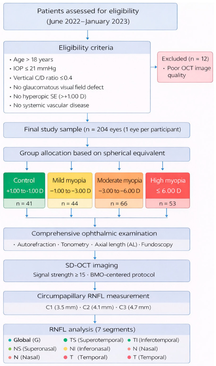 Figure 1
 Figure 2
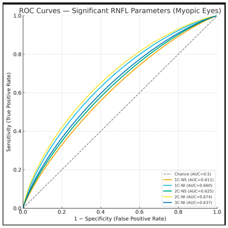 Figure 3
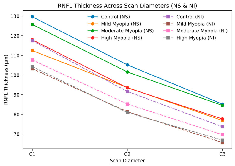 Figure 4
Peer Reviews
No public reviews on file for this paper yet. If you reviewed it on a platform where reviews are public (OpenReview, ICLR, NeurIPS, ICML), you can paste yours below so the community can read it here.
Videos
No videos yet. Explain this paper in a talk, walkthrough, or lecture? Add one.
Taxonomy
TopicsGlaucoma and retinal disorders · Ophthalmology and Visual Impairment Studies · Retinal Diseases and Treatments