Predictive Value of a Radiomics-Derived Risk Score for Local Progression in T3 Laryngeal Cancer: A 10-Year Single-Center Retrospective Cohort Study
Caglar Eker, Muhammed Dagkiran, Emin Demirel, Burak Mete, Hasan Suat Arslantas, Omer Kaya, Bedir Kaya, Elvan Onan, Naqibullah Mohammadi, Mustafa Mert Gedik, Ilda Tanrisever Pehlivan, Merve Gizem Gonullu, Ozgur Surmelioglu

TL;DR
This study developed a radiomics-based risk score from CT scans to predict local progression in T3 laryngeal cancer after treatment, showing strong performance in identifying high-risk patients.
Contribution
A novel radiomics-derived risk score was developed and validated for predicting local progression in T3 laryngeal cancer after chemoradiotherapy.
Findings
The radiomics score was an independent predictor of local progression with a hazard ratio of 2.38 per 1-point increase.
Higher radiomics scores were associated with significantly shorter local progression-free survival at 1, 3, and 5 years.
The model demonstrated high discrimination with a C-index of 0.855.
Abstract
Background/Objective: Local progression after concurrent chemoradiotherapy in T3 laryngeal carcinoma (LC) remains difficult to predict using conventional clinical assessment alone. This study aimed to develop a radiomics-derived risk score from routine post-treatment contrast-enhanced CT and evaluate its prognostic value—together with clinical variables—for predicting local progression-free survival (LPFS). Methods: In this single-center retrospective cohort, 67 patients with pathologically confirmed T3-stage LC treated with chemoradiotherapy were included. All patients underwent contrast-enhanced CT at baseline and 3 months after treatment completion; radiomics analysis was performed using post-treatment CT with 3D manual segmentation of the primary tumor. A total of 111 radiomic features were extracted (shape, first-order, and texture). Features with AUC > 0.60 were screened, and six…
Genes, proteins, chemicals, diseases, species, mutations and cell lines named across the full text — each resolved to its canonical identifier and authoritative record.
Click any figure to enlarge with its caption.
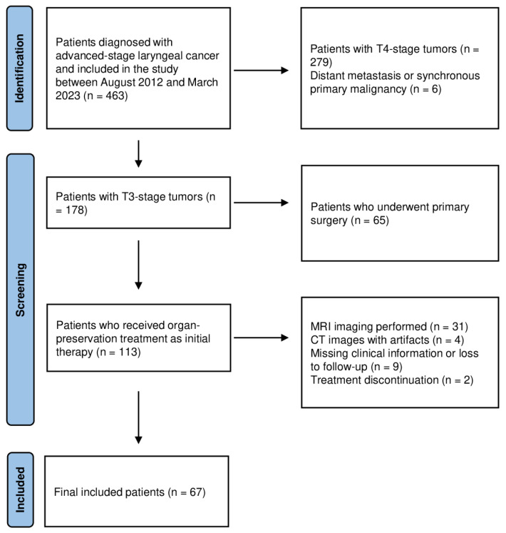 Figure 1
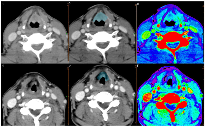 Figure 2
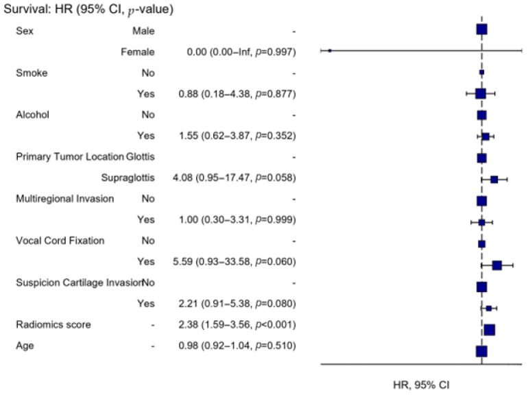 Figure 3
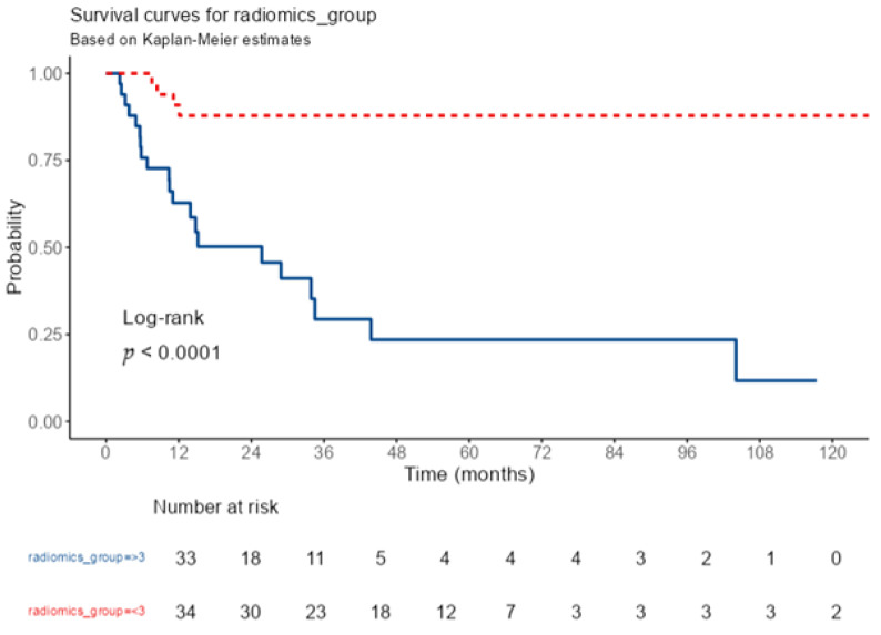 Figure 4
Peer Reviews
No public reviews on file for this paper yet. If you reviewed it on a platform where reviews are public (OpenReview, ICLR, NeurIPS, ICML), you can paste yours below so the community can read it here.
Videos
No videos yet. Explain this paper in a talk, walkthrough, or lecture? Add one.
Taxonomy
TopicsRadiomics and Machine Learning in Medical Imaging · Head and Neck Cancer Studies · Esophageal Cancer Research and Treatment