Assessing Impact of Data Quality in Early Post-Operative Glioblastoma Segmentation
Ragnhild Holden Helland, David Bouget, Asgeir Store Jakola, Sébastien Muller, Ole Solheim, Ingerid Reinertsen

TL;DR
This study examines how image and annotation quality affect deep learning models for early post-operative glioblastoma segmentation, finding that high-quality data improves performance but limits generalization to lower-quality data.
Contribution
The study quantifies the impact of data quality on model performance in early post-operative glioblastoma segmentation using a curated dataset with expert evaluations.
Findings
Models trained on high-quality images did not generalize well to low-quality images.
High-quality annotations achieved similar performance as the full dataset using only two-thirds of the data.
Both image and annotation quality significantly affect model performance in early post-operative segmentation.
Abstract
Quantification of the residual tumor from early post-operative magnetic resonance imaging (MRI) is essential in follow-up and treatment planning for glioblastoma patients. Residual tumor segmentation from early post-operative MRI is particularly challenging compared to the closely related task of pre-operative segmentation, as the tumor lesions are small, fragmented, and easily confounded with noise in the resection cavity. Recently, several studies successfully trained deep learning models for early post-operative segmentation, yet with subpar performances compared to the analogous task pre-operatively. In this study, the impact of image and annotation quality on model training and performance in early post-operative glioblastoma segmentation was assessed. A dataset consisting of early post-operative MRI scans from 423 patients and two hospitals in Norway and Sweden was assembled, for…
Genes, proteins, chemicals, diseases, species, mutations and cell lines named across the full text — each resolved to its canonical identifier and authoritative record.
Click any figure to enlarge with its caption.
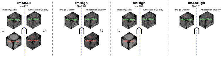 Figure 1
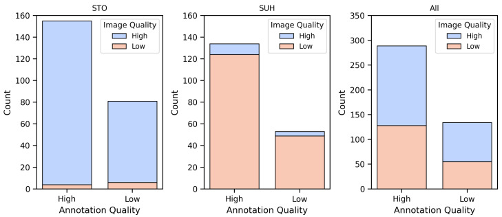 Figure 2
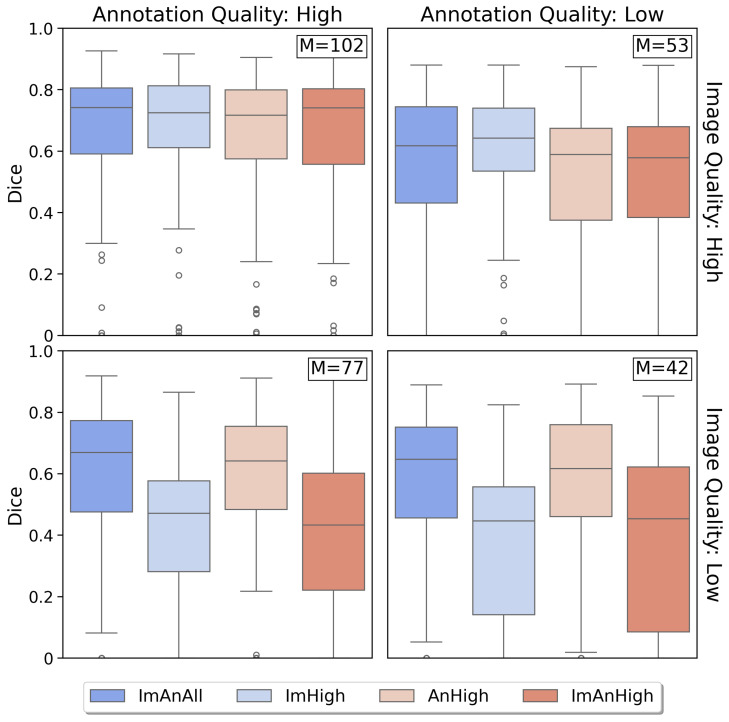 Figure 3
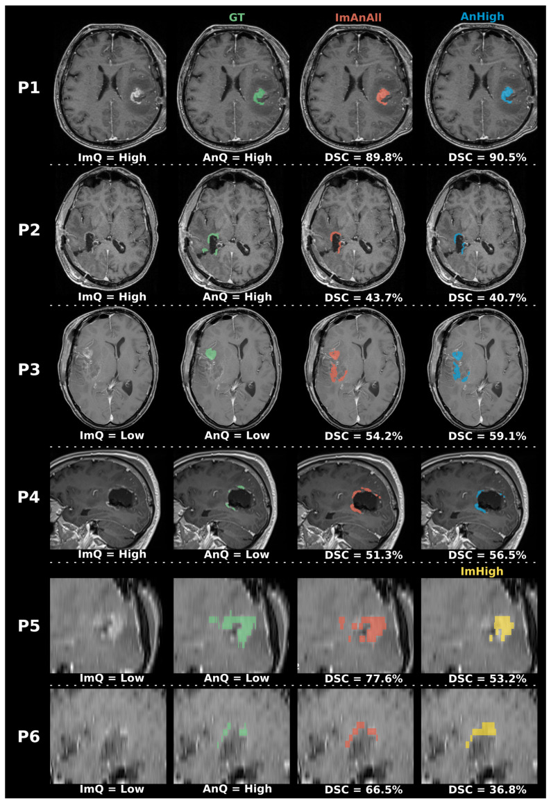 Figure 4
Peer Reviews
No public reviews on file for this paper yet. If you reviewed it on a platform where reviews are public (OpenReview, ICLR, NeurIPS, ICML), you can paste yours below so the community can read it here.
Videos
No videos yet. Explain this paper in a talk, walkthrough, or lecture? Add one.
Taxonomy
TopicsGlioma Diagnosis and Treatment · Radiomics and Machine Learning in Medical Imaging · Medical Imaging and Analysis