Development of an Artificial Intelligence-Based Chromosome Interpretation System for Amniotic Fluid Karyotyping
Kuan-Han Wu, Hsuan-Wei Huang, Chia Yun Lin, Hsu-Tung Huang, Tzuo-Yau Fan, Yueh-Peng Chen, Yung-Chiao Chang, Te-Yao Hsu, Kuo-Chung Lan

TL;DR
This paper introduces an AI system that automates chromosome analysis in prenatal diagnosis, reducing manual labor and increasing efficiency.
Contribution
A modular AI workflow is developed for automated chromosome interpretation in amniotic fluid karyotyping, achieving high accuracy.
Findings
The AI system achieved high classification accuracy across training, validation, and testing cohorts.
The overlap-recognition module effectively reduced errors in composite chromosome regions.
The workflow successfully generated draft karyotypes from unsorted images with expert-level concordance.
Abstract
Conventional G-banded karyotyping remains indispensable in prenatal diagnosis but continues to rely on labor-intensive, expertise-dependent visual examination. To address these challenges, we developed a modular artificial intelligence (AI) workflow that automates chromosome interpretation from amniotic fluid metaphase images. The system integrates image denoising, chromosome segmentation, overlap screening, and morphology-based classification, and was trained using 13,223 clinical cases comprising more than 50,000 manually annotated chromosomes. Across training, temporal validation, and independent testing cohorts, classification accuracy remained consistently high (97.45%, 96.95%, and 95.72%, respectively). The overlap-recognition module further reduced downstream errors by reliably identifying composite chromosome regions. When applied to unsorted metaphase images from a later…
Genes, proteins, chemicals, diseases, species, mutations and cell lines named across the full text — each resolved to its canonical identifier and authoritative record.
Click any figure to enlarge with its caption.
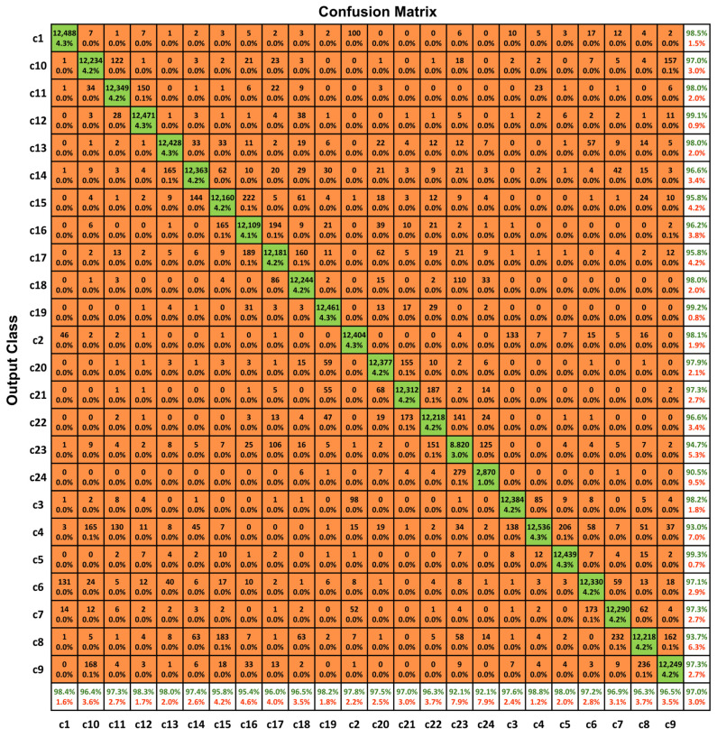 Figure 1
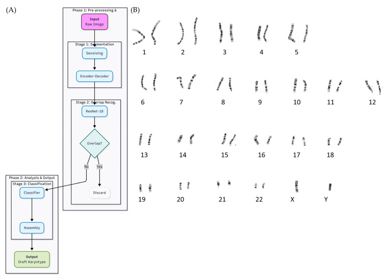 Figure 2
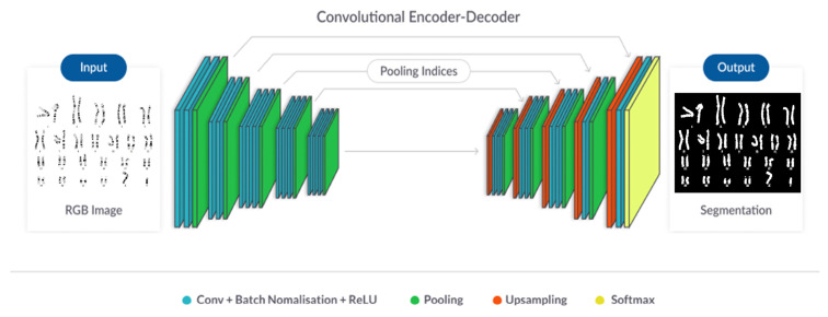 Figure 3
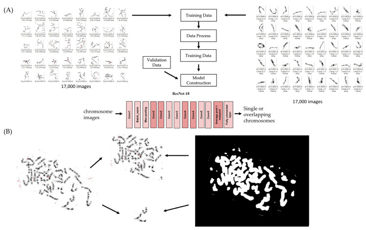 Figure 4
- —Chang Gung Memorial Hospital
Peer Reviews
No public reviews on file for this paper yet. If you reviewed it on a platform where reviews are public (OpenReview, ICLR, NeurIPS, ICML), you can paste yours below so the community can read it here.
Videos
No videos yet. Explain this paper in a talk, walkthrough, or lecture? Add one.
Taxonomy
TopicsPrenatal Screening and Diagnostics · Genomic variations and chromosomal abnormalities · Fetal and Pediatric Neurological Disorders
1. Introduction
Amniocentesis remains a cornerstone of prenatal diagnosis by enabling direct cytogenetic analysis of fetal chromosomes through conventional G-banded karyotyping [1,2]. Despite advances in molecular techniques and non-invasive prenatal testing (NIPT), karyotyping continues to play an indispensable role in detecting numerical abnormalities, balanced structural rearrangements, and large-scale chromosomal alterations that are not reliably identified by sequencing-based approaches [3,4]. However, manual karyotype interpretation is labor-intensive, time-consuming, and highly dependent on the expertise of trained cytogeneticists, creating increasing challenges in routine clinical practice [5].
Early attempts to automate chromosome analysis focused primarily on idealized metaphase spreads, often assuming complete sets of well-separated chromosomes [6]. These systems typically relied on handcrafted features or early artificial neural network classifiers and demonstrated limited robustness when confronted with overlapping chromosomes, variable staining quality, or structural abnormalities commonly encountered in prenatal specimens [7]. More recent artificial intelligence-based pipelines, including CNN-based detection frameworks such as Mask-R-CNN-derived models used for chromosome segmentation and deep learning enumeration frameworks such as DeepACEv2 [8], have shown promising performance under controlled experimental conditions. Nevertheless, many of these approaches depend on pre-segmented chromosomes or assume ideal metaphase preparations, limiting their applicability to routine prenatal datasets characterized by heterogeneous image quality and frequent chromosome overlap [9].
Given these limitations, artificial intelligence offers an opportunity to streamline chromosome interpretation while reducing manual workload under real-world clinical conditions [10]. In this study, we present an end-to-end AI-assisted workflow designed for chromosome interpretation from amniotic fluid metaphase images obtained during routine clinical practice. Rather than relying on a single artificial neural network for generic image recognition, the proposed system adopts a modular design that integrates chromosome segmentation, overlap recognition, morphology-based classification, and draft karyotype assembly. This design enables transparent processing at each stage and facilitates expert review within routine prenatal cytogenetic workflows.
2. Results
2.1. Classification Accuracy Across Datasets
The chromosome-classification model was trained and evaluated using temporally separated datasets collected over multiple years. Classification accuracy remained consistently high across the training, validation, and independent testing cohorts, reaching 97.45%, 96.95%, and 95.72%, respectively (Table 1). These results indicate stable classification performance across datasets acquired under different time periods and routine laboratory conditions.
2.2. Performance Metrics for Individual Chromosome Classes
Detailed performance metrics for individual chromosome classes are summarized in Table 2, allowing per-class evaluation of the model’s behavior beyond overall accuracy. Across most chromosome classes, both PPV and NPV remained high, indicating reliable identification of target chromosomes and effective exclusion of non-target classes. Performance variations were primarily observed among chromosomes with similar morphology and G-banding patterns.
2.3. Error Distribution and Confusion Matrix Analysis
Analysis of the confusion matrix (Figure 1) revealed that classification errors were not randomly distributed but were predominantly confined to morphologically similar chromosome classes. Most misclassifications occurred between neighboring autosomes with comparable size and G-banding patterns, such as chromosomes 14 and 15 or chromosomes 21 and 22. In contrast, large or structurally distinct chromosomes demonstrated consistently high classification accuracy.
Importantly, no systematic bias toward over- or under-prediction of specific chromosome classes was observed, as reflected by the dominance of diagonal elements across the matrix. These error patterns closely mirror the challenges encountered during manual karyotype interpretation and suggest that the observed misclassifications reflect biologically plausible ambiguities rather than systematic classification failure.
2.4. Overlap Recognition Performance
The overlap-recognition module was evaluated to determine its ability to distinguish between isolated chromosomes and overlapping chromosome regions. The model accurately identified overlapping regions across a range of metaphase images, enabling effective filtering of ambiguous chromosome segments prior to downstream classification. This performance supports reliable handling of complex metaphase images containing overlapping chromosomes.
2.5. Application to Unsorted Metaphase Images
To evaluate real-world applicability, the complete AI workflow was applied to an independent set of unsorted G-banded metaphase images. As shown in Figure 2, the system processes raw metaphase inputs without manual preselection and executes the same three-stage pipeline used during model development.
In Stage 1, raw metaphase images undergo denoising followed by automated extraction of chromosome-containing regions using a convolutional encoder–decoder segmentation network. In Stage 2, each segmented region is evaluated by a ResNet-18-based overlap-recognition module, which removes overlapping or composite chromosome structures to prevent downstream misclassification. Representative examples of segmentation refinement and overlap detection are shown in Figure 2B.
In Stage 3, isolated chromosomes are classified into 24 morphological categories and automatically assembled into a draft karyotype. When applied to unsorted clinical metaphase images, the workflow reliably extracted individual chromosomes, filtered ambiguous regions, and generated draft karyotypes consistent with expert interpretations. These results demonstrate that the proposed system can operate effectively on uncurated metaphase images and is suitable for routine prenatal cytogenetic practice.
3. Discussion
This study presents an AI-assisted workflow designed to support the interpretation of G-banded chromosomes from amniotic fluid samples in routine prenatal cytogenetics. By integrating chromosome segmentation, overlap recognition, morphology-based classification, and draft karyotype assembly, the proposed system aims to reduce the manual effort required for chromosome sorting while preserving expert oversight throughout the diagnostic process.
Across temporally separated cohorts collected over multiple years, the chromosome-classification module demonstrated consistently high performance under routine laboratory conditions. Although classification accuracy decreased modestly from the training to the independent testing cohort, this trend likely reflects increased heterogeneity in staining quality, chromosome spreading, and overlap patterns commonly encountered in real-world prenatal datasets. Importantly, most misclassifications occurred among chromosomes with similar size and banding characteristics, mirroring challenges faced during manual karyotype interpretation and suggesting that the observed errors were morphologically plausible rather than systematic.
In the context of prenatal cytogenetics, the system is explicitly intended to function as a clinical decision-support tool rather than an autonomous diagnostic platform. From this perspective, performance metrics such as positive predictive value (PPV) and negative predictive value (NPV) provide particularly relevant information. High PPV indicates that chromosomes assigned to a given class are likely to be correct, thereby reducing the burden of manual verification, whereas high NPV helps ensure that true homologs are not inadvertently excluded during karyotype assembly. Together, these metrics align closely with routine laboratory workflows and expert review practices.
The incorporation of an overlap-recognition module represents a key design feature of the proposed workflow. Overlapping chromosomes are a frequent source of ambiguity in automated cytogenetic analysis, as segmentation errors can propagate into downstream classification. By identifying and filtering overlapping regions prior to classification, the workflow limits such error propagation and supports more reliable draft karyotype assembly. This modular strategy differs from earlier automated karyotyping systems that assumed idealized metaphase spreads and lacked mechanisms to address structural complexity in prenatal samples.
Compared with previously reported AI-based chromosome-analysis pipelines, which often rely on pre-segmented inputs or controlled image conditions [11], the present study emphasizes applicability to unsorted metaphase images obtained during routine clinical practice. The ability to generate draft karyotypes directly from these images represents an important step toward practical integration into prenatal cytogenetic workflows, where initial image quality and chromosome arrangement are highly variable.
Several limitations of this study should be acknowledged. First, all data were derived from a single institution, and performance may vary under different staining protocols, imaging systems, or laboratory practices. Second, although the dataset was large, rare structural abnormalities were underrepresented, and additional data will be required to fully assess system performance in these cases. Finally, the workflow does not independently diagnose chromosomal abnormalities but instead provides draft karyotypes that require expert verification.
Overall, this study demonstrates that an AI-assisted workflow can meaningfully enhance the efficiency of prenatal cytogenetic analysis without replacing expert interpretation. By streamlining the most labor-intensive steps of karyotyping and supporting expert review, such systems may help address increasing clinical workloads while maintaining diagnostic reliability.
4. Materials and Methods
4.1. Data Sources
Metaphase images were collected from 13,223 amniotic fluid samples processed at the Cytogenetics Laboratory of Kaohsiung Chang Gung Memorial Hospital (CGMH) between 2014 and 2020. The dataset included both normal and abnormal karyotypes encountered in routine prenatal diagnosis. For model development, the images were divided into separate temporal cohorts for training (2014–2015), validation (2016), and independent testing (2017–2018). Additional cases collected in 2019 were used for workflow evaluation with unsorted metaphase images. The number of cases and images included in each cohort is summarized in Table 3. All samples were anonymized before analysis.
4.2. Chromosome Annotation
Chromosomes were manually annotated by trained cytogeneticists to generate ground-truth labels for supervised learning. For each metaphase image, individual chromosomes were delineated using polygonal masks or bounding boxes. Regions containing overlapping or entangled chromosome structures were additionally annotated and labeled as overlapping regions.
In total, more than 50,000 chromosome instances across 24 biologically defined classes (22 autosomes and the X and Y chromosomes) were curated. These annotations were used for training and validating the segmentation, overlap-recognition, and chromosome-classification modules.
4.3. Model Components
4.3.1. Denoising Module
Raw G-banded metaphase images were first subjected to image denoising to reduce background noise and enhance chromosome contours. This preprocessing step aimed to improve image quality prior to segmentation by generating clearer chromosome boundaries and suppressing staining artifacts.
4.3.2. Segmentation Module
Chromosome segmentation was performed to extract candidate chromosome regions from denoised metaphase images. A convolutional neural network-based segmentation model was trained to distinguish chromosome foreground from background regions. Morphological postprocessing was subsequently applied to refine chromosome edges and remove residual artifacts.
Representative examples of the chromosome segmentation process, including raw inputs, segmentation masks, and refined outputs, are shown in Figure 3.
4.3.3. Overlap Recognition Module
To resolve ambiguities caused by overlapping or closely apposed chromosomes, an overlap-recognition module was developed to distinguish isolated chromosomes from overlapping chromosome structures. Separate annotated datasets were curated specifically for this task: 17,000 regions containing true overlapping chromosomes and 17,000 regions containing single, non-overlapping chromosomes. These regions were generated from the segmentation output and manually reviewed by trained cytogenetic technologists to ensure high-quality labeling.
A ResNet-18-based binary classifier was trained on this dataset to output the probability that a segmented region represents either a single chromosome or an overlapping structure. This module operates as a mandatory quality-control gate within the overall workflow, filtering out ambiguous regions before they proceed to downstream analysis.
The training structure of this module, its integration within the multi-stage pipeline, and representative examples of its output categories are shown in Figure 4, which illustrates both the dataset construction workflow (Panel A) and the ResNet-18 architectural schematic with output behavior (Panel B).
4.3.4. Classification Module
Segmented chromosome regions identified as isolated were subsequently processed by the chromosome-classification module. Each chromosome region was classified into one of 24 biologically defined categories corresponding to the 22 autosomes and the X and Y chromosomes.
The classification model output a predicted chromosome class together with an associated confidence score. These outputs were later used during draft karyotype assembly for expert review rather than for autonomous diagnosis.
4.4. Performance Evaluation
Model performance was evaluated at the individual-chromosome-instance level. Each segmented chromosome was treated as an independent instance, and classification accuracy was defined as the proportion of correctly classified chromosome instances among all evaluated instances within each dataset.
Evaluation metrics included sensitivity, specificity, precision, recall, F1 score, positive predictive value (PPV), and negative predictive value (NPV). These metrics were calculated as follows: sensitivity = TP(TP + FN); specificity = TN/(TN + FP); precision (PPV) = TP/(TP + FP); recall = TP/(TP + FN); NPV = TN/(TN + FN); and F1 score = 2 × (precision × recall)/(precision + recall). TP, FP, TN, and FN denote true positives, false positives, true negatives, and false negatives, respectively.
Overall classification accuracy across datasets is summarized in Table 1, and detailed per-class performance metrics are provided in Table 2.
4.5. System Overview
The proposed system follows a modular end-to-end workflow for chromosome interpretation from amniotic fluid metaphase images. The workflow consists of chromosome segmentation, overlap recognition, morphology-based chromosome classification, and draft karyotype assembly. Each module was trained independently using dedicated annotated datasets, enabling transparent processing and evaluation at each stage.
5. Conclusions
This study presents an artificial intelligence-assisted workflow that supports conventional G-banded karyotyping in prenatal cytogenetics. By integrating chromosome segmentation, overlap recognition, and morphology-based classification, the system reduces the need for manual chromosome sorting and addresses common sources of error in routine analysis.
The workflow achieved consistently high classification accuracy across multiple real-world clinical cohorts and successfully generated draft karyotypes from unsorted metaphase images that closely matched expert interpretation. Rather than operating as an autonomous diagnostic tool, the system functions as a clinical decision-support aid, improving efficiency while preserving expert oversight.
The reference list from the paper itself. Each links out to its DOI / PubMed record.
- 1Gregg A.R. Skotko B.G. Benkendorf J.L. Monaghan K.G. Bajaj K. Best R.G. Klugman S. Watson M.S. Noninvasive prenatal screening for fetal aneuploidy, 2016 update: A position statement of the American College of Medical Genetics and Genomics Genet. Med.2016181056106510.1038/gim.2016.9727467454 · doi ↗ · pubmed ↗
- 2Underwood M.A. Gilbert W.M. Sherman M.P. Amniotic fluid: Not just fetal urine anymore J. Perinatol.20052534134810.1038/sj.jp.721129015861199 · doi ↗ · pubmed ↗
- 3Wapner R.J. Martin C.L. Levy B. Ballif B.C. Eng C.M. Zachary J.M. Savage M. Platt L.D. Saltzman D. Grobman W.A. Chromosomal microarray versus karyotyping for prenatal diagnosis N. Engl. J. Med.20123672175218410.1056/NEJ Moa 120338223215555 PMC 3549418 · doi ↗ · pubmed ↗
- 4Liu X. Liu S. Wang H. Hu T. Potentials and challenges of chromosomal microarray analysis in prenatal diagnosis Front. Genet.20221393818310.3389/fgene.2022.93818335957681 PMC 9360565 · doi ↗ · pubmed ↗
- 5Kumar S. Kiso A. Kithan N.A. Chromosome banding and mechanism of chromosome aberrations Cytogenetics-Classical and Molecular Strategies for Analysing Heredity Material Intech Open London, UK 2021
- 6Beksac M.S. Eskiizmirliler S. Cakar A.N. Erkmen A.M. Dagdeviren A. Lundsteen C. An expert diagnostic system based on neural networks and image analysis techniques in the field of automated cytogenetics Technol. Health Care 1996321722910.3233/THC-1996-34038705397 · doi ↗ · pubmed ↗
- 7Stanley R.J. Keller J.M. Gader P. Caldwell C.W. Data-driven homologue matching for chromosome identification IEEE Trans. Med. Imaging 19981745146210.1109/42.7121349735908 · doi ↗ · pubmed ↗
- 8Xiao L. Luo C. Yu T. Luo Y. Wang M. Yu F. Li Y. Tian C. Qiao J. Deep AC Ev 2: Automated Chromosome Enumeration in Metaphase Cell Images Using Deep Convolutional Neural Networks IEEE Trans. Med. Imaging 2020393920393210.1109/TMI.2020.300764232746135 · doi ↗ · pubmed ↗