Antibiotic-Driven Evolutionary Engineering in Salmonella Heidelberg Reveals Genomic Signatures of Attenuation
Ruy D. Chacón, Manuel Ramírez, Claudete S. Astolfi-Ferreira, Antonio J. Piantino Ferreira

TL;DR
This study shows how exposing Salmonella Heidelberg to antibiotics leads to genetic changes that may reduce its virulence, offering insights into vaccine development.
Contribution
The study identifies specific genomic mutations and functional networks linked to attenuation in Salmonella Heidelberg through antibiotic-driven evolutionary engineering.
Findings
Mutant strains showed resistance to streptomycin and rifampicin with distinct genetic mutations.
Genes like rpoS, ascD, and rpoB were linked to antibiotic resistance and virulence attenuation.
Network analysis revealed functional connections in regulatory and metabolic pathways affected by mutations.
Abstract
Background/Objectives: Salmonella Heidelberg (SH) is a globally distributed pathogen associated with gastrointestinal disease in humans and animals and frequently affects poultry. Among the classic strategies used in vaccine development, evolutionary engineering enables the generation of attenuated bacterial strains through exposure to selective pressures such as antibiotics. In this study, spontaneous antibiotic-resistant mutant strains of SH were generated by exposure to high concentrations of streptomycin and rifampicin, after which their phenotypic and genotypic characteristics were evaluated. Methods: The wild-type strain SA628 wt was subjected to continuous and discontinuous selection under antibiotic pressure. Phenotypic characterization included biochemical profiling and antibiotic susceptibility testing. Whole-genome sequencing was performed to identify genetic changes…
Genes, proteins, chemicals, diseases, species, mutations and cell lines named across the full text — each resolved to its canonical identifier and authoritative record.
Click any figure to enlarge with its caption.
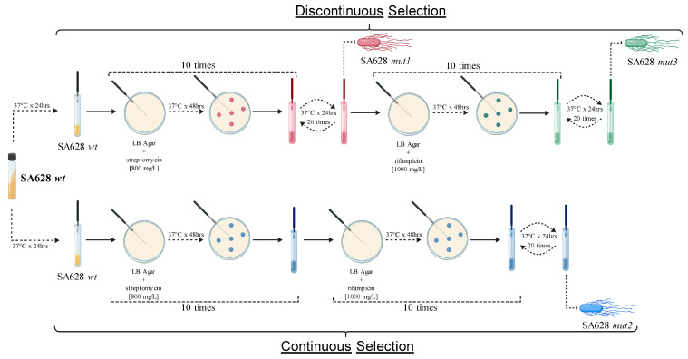 Figure 1
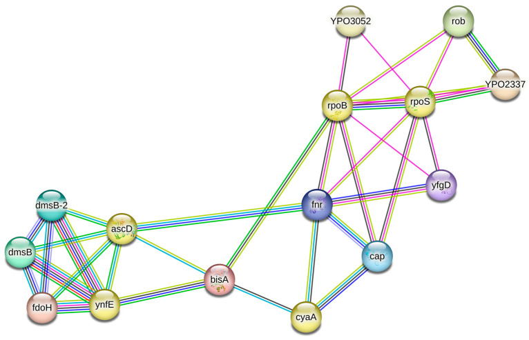 Figure 2
 Figure 3
- —CAPES (Coordenação de Aperfeiçoamento de Pessoal de Nível Superior–Brasil)
- —Conselho Nacional de Pesquisa e Desenvolvimento Tecnológico (CNPq)
Peer Reviews
No public reviews on file for this paper yet. If you reviewed it on a platform where reviews are public (OpenReview, ICLR, NeurIPS, ICML), you can paste yours below so the community can read it here.
Videos
No videos yet. Explain this paper in a talk, walkthrough, or lecture? Add one.
Taxonomy
TopicsSalmonella and Campylobacter epidemiology · Bacterial Genetics and Biotechnology · Burkholderia infections and melioidosis
1. Introduction
Salmonella Heidelberg (SH) is a member of Salmonella enterica subsp. enterica and is included in serogroup B on the basis of its antigenic profile (1,4,5,12:r:1,2) according to the Kauffmann–White classification system [1]. SH is a globally distributed serovar and is frequently associated with clinical cases in humans, primarily because of the consumption of contaminated poultry-derived food products. It is capable of causing severe gastroenteritis [2]. Moreover, compared with other serovars, SH is known for having variable virulence profiles and may exhibit greater invasiveness in human infections [3,4].
For several decades, SH has been frequently reported in clinical and epidemiological studies involving isolates from commercial poultry worldwide [5,6,7,8]. In Brazil, the presence of SH is no less concerning, as it remains among the most prevalent serovars in poultry farms across the country [4,9,10,11]. Studies have demonstrated genomic similarities and relationships with strains from other countries, suggesting the potential introduction and dissemination of strains of diverse origins into Brazilian poultry farms [12,13].
Moreover, SH frequently exhibits antibiotic resistance, and this issue is exacerbated by the emergence of multidrug-resistant strains, which pose an imminent threat to food safety and cause significant economic losses in the poultry industry [10,14,15,16,17]. These resistance traits can be transferred and disseminated via mobile genetic elements, particularly plasmids [11,12,13].
Several preventive measures against salmonella infections include competitive exclusion using nonpathogenic organisms and the development of vaccines [17,18]. Currently, prophylactic protection against SH is achieved through vaccination with attenuated strains of Salmonella Typhimurium (ST) [19]. Although live vaccines are commercially available for poultry use, the mechanisms underlying attenuation in these strains remain poorly understood. Moreover, numerous outbreaks in vaccinated flocks have been reported in recent years [20,21].
Certain antibiotic resistance mechanisms have been investigated as potential means of attenuating Salmonella for use as live vaccines. Some studies have reported reduced virulence in bacterial strains resistant to antibiotics such as rifampicin, streptomycin, and nalidixic acid [22,23,24]. Commercially, two classic vaccines have employed evolutionary engineering to attenuate Salmonella strains as antibiotic mutants. The vaccine AviPro SALMONELLA VAC E (Lohmann Animal Health GmbH & Co. KG, Cuhaven, Germany) is based on an S. Enteritidis (SE) strain attenuated through evolutionary selection, characterized by a prolonged generation time, increased quinolone sensitivity, and enhanced bacterial membrane permeability. This strain survives long enough within the host to stimulate immunity but cannot persist in the environment. However, it is resistant to rifampicin and streptomycin [25]. Similarly, AviPro SALMONELLA VAC T (Lohmann Animal Health GmbH & Co. KG, Cuhaven, Germany) is based on an ST strain attenuated by evolutionary selection, exhibiting resistance to nalidixic acid and rifampicin [26].
In a previous study, Revolledo and Ferreira [27] applied evolutionary engineering to Brazilian strains of ST, SE, and SH, exposing them to high concentrations of nalidixic acid, streptomycin, and rifampicin, successfully selecting spontaneous mutant strains resistant to these antibiotics. These strains exhibited reduced fecal shedding, lower invasiveness and persistence in organs, and no clinical signs of disease in infected chicks.
This study produced and selected spontaneous antibiotic-resistant mutant strains of SH through exposure to high concentrations of streptomycin and rifampicin. It then evaluated the phenotypic and genotypic characteristics of these strains by employing biochemical characterization, antimicrobial susceptibility profiling, genomic analysis, and variant calling.
2. Materials and Methods
2.1. Salmonella Heidelberg Isolate
The isolate SA628, identified as SH in a previous study [11], was selected because it is naturally susceptible to streptomycin and rifampicin. This isolate originated from a fecal sample collected from breeder hens in São Paulo, Brazil, in 2006. It is part of the microbiological collection of the Laboratory of Avian Diseases (FMVZ-USP, São Paulo, Brazil).
2.2. Generation of Antibiotic-Resistant Mutant Strains
The isolate SA628, defined as the wild-type strain (SA628 wt), was treated with high concentrations of streptomycin and rifampicin following the methodology described by Revolledo and Ferreira [27]. In the aforementioned study, the first mutant strain (SH/LABOR/USP/08) was generated and renamed SA628 mut1. Compared with the parental wild-type strain, this mutant strain exhibited significantly reduced fecal shedding and organ invasion and showed no clinical signs in inoculated chicks [27]. Collectively, these observations were consistent with a biologically attenuated phenotype under in vivo conditions. In this study, we aimed to elucidate the genomic features of this strain, and we continued to apply additional selection processes to potentially increase its attenuation.
Briefly, an inoculating loopful of the parental strain SA628 wt was plated on LB agar (Difco™, BD, Franklin Lakes, NJ, USA) and incubated at 37 °C for 48 h in the presence of streptomycin [800 mg/L]. Each selection cycle was initiated with an estimated bacterial load of approximately 10^8^ CFU. After exposure, the colonies were transferred to LB broth and incubated at 37 °C for 24 h. This process involved ten consecutive cycles of antibiotic exposure. To assess mutation stability, the colonies were subcultured 20 times in LB broth at 37 °C for 24 h each. After the 20 antibiotic-free passages were completed, the persistence of resistance was evaluated phenotypically using the minimum inhibitory concentration (MIC) method described in Section 2.4. Additionally, to introduce a second resistance marker and increase the selection pressure with the goal of increasing the degree of attenuation, all previous procedures were repeated using rifampicin [1000 mg/L]. The antibiotic concentrations used were deliberately selected to be well above the minimum inhibitory concentration of the parental strain to impose stringent selective pressure and favor the emergence of high-level resistant mutants, as previously described [27]. A 48 h incubation period on LB agar was chosen to allow the recovery and detection of resistant colonies with potentially reduced growth rates under strong antibiotic stress. The selection experiments were conducted as a single lineage-based evolutionary process, without parallel independent replicates, in which mutant strains were derived sequentially under defined selective regimes for downstream phenotypic and genomic characterization. On the basis of this experimental framework, two distinct evolutionary pathways were implemented. The first, referred to as discontinuous selection, involved antibiotic exposure interspersed with antibiotic-free stabilization passages, resulting in a stepwise evolutionary trajectory (SA628 mut1 → SA628 mut3). In contrast, continuous selection consisted of sustained antibiotic pressure without intermediate stabilization passages, leading to the independent selection of resistant mutants (SA628 mut2) (Figure 1).
2.3. Biochemical and Molecular Characterization and Monitoring
Each antibiotic-resistant mutant generated was monitored for biochemical and molecular stability. For biochemical characterization, the isolates were analyzed using EPM, MILi, and Simmons’ citrate tests from the Enterokit B system (Probac, São Paulo, Brazil), as well as on MacConkey agar plates (Difco™, BD) incubated aerobically at 37 °C for 24 h.
For molecular characterization, DNA was extracted using the DNeasy Blood and Tissue Kit (Qiagen, Hilden, Germany). The concentration and quality of the extracted DNA were assessed using a NanoDrop™ spectrophotometer (Thermo Scientific™, Waltham, MA, USA).
PCR was performed using the primers and conditions described by Park and Ricke [29] to identify and confirm the isolate as SH at each passage. All PCR products were visualized by loading 9 µL of the product and 1 µL of loading dye onto a 1.5% agarose gel, using a 100 bp DNA ladder (Invitrogen, Carlsbad, CA, USA) as the molecular weight marker and applying 10 V per cm of gel. Final visualization and image acquisition were performed using a UV gel documentation system. While PCR was used to confirm the isolates as SH at each passage, the stability of the antibiotic resistance markers was evaluated phenotypically only after completion of the 20 antibiotic-free passages by reassessing antimicrobial susceptibility using the MIC procedure described in Section 2.4.
2.4. Antimicrobial Susceptibility Profile
The antimicrobial susceptibility profiles of the parental strain and the mutant strains were evaluated using the minimum inhibitory concentration (MIC) method with precoated AviPro^®^ PLATE (Lohmann Animal Health, Cuxhaven, Germany) according to the manufacturer’s recommendations [30]. AviPro^®^ PLATE is designed to determine the activity of antimicrobial agents against Gram-positive and Gram-negative bacteria in poultry flocks and includes markers used to discriminate between field Salmonella strains and Salmonella metabolic drift mutants contained in AviPro^®^ Salmonella live vaccines (erythromycin, rifampicin, and streptomycin). For the purposes of this study, only the results of ten antimicrobials relevant to Salmonella (amoxicillin, enrofloxacin, ceftiofur, cefpodoxime, cefotaxime, tetracycline, neomycin, trimethoprim/sulfamethoxazole, colistin, and doxycycline) and two markers of Salmonella metabolic drift mutants (streptomycin and rifampicin) were evaluated. The MIC ranges tested, and their interpretive scales are detailed in Table S1.
A total of 50 µL of bacterial suspension from a single clonal isolate was diluted in 10 mL of Mueller–Hinton broth (Difco™, BD), and 100 µL was distributed into each well of the AviPro^®^ PLATE. The plate was then incubated at 37 °C for 18–24 h, and growth was visually assessed by the presence or absence of bacteria in each well.
In addition, antimicrobial susceptibility was evaluated using the disk diffusion (DD) method in accordance with the Clinical and Laboratory Standards Institute (CLSI) M100 guidelines [31]. This approach was applied to assess phenotypic resistance patterns in relation to specific genetic mutations identified in this study, as described in Section 3.2. For this purpose, 100 µL of the same diluted bacterial suspension used for MIC testing was spread onto Mueller–Hinton agar plates (Difco™, BD). Antimicrobial disks (Sensifar, Cefar Diagnóstica Ltda., São Paulo, Brazil) were then placed equidistantly on the agar surface. The plates were incubated at 37 °C for 24 h, after which the inhibition zones were measured and interpreted. The antimicrobials tested included ciprofloxacin (CIP, 5 µg), enrofloxacin (ENR, 5 µg), fosfomycin (FOS, 200 µg), and nalidixic acid (NAL, 30 µg). The diameter breakpoint interpretive criteria are provided in Table S2.
2.5. Whole-Genome Sequencing and Bioinformatic Analysis
Whole-genome sequencing of the bacterial isolates was carried out using the Illumina™ MiSeq platform (San Diego, CA, USA), starting from 500 ng of genomic DNA with an OD260/280 ratio between 1.8 and 2.0 and an OD260/230 ratio between 1.2 and 2.0. Libraries were prepared using the Nextera XT DNA Library Preparation Kit (Illumina™) for paired-end sequencing (2 × 150 bp), targeting ~100× coverage for the SH genomes, which are approximately 4.8 megabases in size.
Read quality was assessed using FastQC v0.11.9 [32], and low-quality reads were trimmed using Trimmomatic v0.36 [33]. De novo genome assembly was performed with SPAdes v3.15 [34]. Assembly quality statistics—including the genome size, number of contigs, N50 value, and G+C content—were evaluated using QUAST v4.1 [35]. Genome annotation was performed using PGAP v6.3 [36].
The presence of antimicrobial resistance genes and virulence factors was assessed using ABRicate v1.0.1 (https://github.com/tseemann/abricate; accessed on 2 February 2022) with reference to the Comprehensive Antibiotic Resistance Database (CARD) [37] and the Virulence Factor Database (VFDB) [38]. Salmonella pathogenicity islands (SPIs) were identified using SPIFinder v2.0 [39].
All the genomes were mapped against the reference genome UFPRLABMOR1 (GenBank: NZ_CP020101). Comparative variant-calling analysis was performed to identify differences between the genomes of the wild-type strain and the mutant strains using SNIPPY v4.4.5 [40], with the annotated genome of the wild-type strain set as the reference. Genes containing mutations were analyzed using the STRING v12.0 platform [41]. A full STRING network was constructed, in which edges represent both functional and physical protein associations. Network edges were defined on the basis of evidence, with line colors indicating the type of supporting interaction. Active interaction sources included text mining, experimental data, curated databases, co-expression, genomic neighborhood, gene fusion, and co-occurrence. A medium confidence interaction score (≥0.400) was applied as the minimum threshold for inclusion in the network. To facilitate network interpretation, k-means clustering was applied to identify a defined number of clusters on the basis of centroid similarity. STRING analyses were used for functional contextualization and hypothesis generation.
3. Results
3.1. Generation of Antibiotic-Resistant Mutant Strains, Biochemical Characterization and Antimicrobial Susceptibility Profiles
After the application of selection pressure and the assessment of the phenotypic stability of the antibiotic resistance markers (streptomycin and rifampicin), three spontaneous mutant strains, designated SA628 mut1, SA628 mut2, and SA628 mut3, were obtained (Figure 1). These strains were isolated from two independent selective pressure protocols that differed in the temporal structure of antibiotic exposure: a discontinuous selection process, characterized by repeated cycles of antibiotic exposure followed by stabilization passages, and a continuous selection process, in which antibiotic pressure was maintained without stabilization phases (Figure 1).
Phenotypic analysis revealed similar biochemical profiles among the wild-type and mutant strains. All the strains tested positive for acid production, gas production, H_2_S production, motility, lysine decarboxylase activity, and Simmons’ citrate utilization, whereas all the strains tested negative for lactose metabolism, L-tryptophan deaminase (LTDa), urease, and indole production (Table 1). The only observed difference was in SA628 mut2, which was isolated after continuous selection and exhibited a reduced capacity for gas production compared with that of SA628 wt.
With respect to antimicrobial susceptibility evaluated by the MIC plate, the parental strain SA628 wt exhibited intermediate susceptibility to enrofloxacin. The mutant strains retained this intermediate phenotype while they acquired additional resistance traits. Specifically, strain SA628 mut1 additionally showed resistance to streptomycin (as expected), whereas strains SA628 mut2 and SA628 mut3 exhibited resistance to both rifampicin and streptomycin (as expected) (Table 1). In contrast, antimicrobial susceptibility assessed using the disk diffusion (DD) method was consistent across all the strains tested: susceptibility to fosfomycin, intermediate susceptibility to ciprofloxacin and enrofloxacin, and complete resistance to nalidixic acid (Table 1).
3.2. Genomic Characterization
Whole-genome sequencing of the parental strain SA628 wt and its derived mutants revealed that general genomic features, including multilocus sequence typing (MLST) and core-genome MLST (cgMLST) profiles, were preserved. These findings were supported by high-quality reads (Phred scores ranging from 36.4 to 37.0) and genome coverage depths between 132× and 209× (Table 2).
The composition of virulence genes, pathogenicity islands (SPIs), plasmids, resistance genes (ARGs), and associated point mutations in known genes was also analyzed. As shown in Table 3, the presence of these genetic elements and the evaluated point mutations was consistently maintained.
3.3. Comparative Genomics and Variant Calling
Comparative genomic analysis and variant calling enabled the identification of genetic mutations arising from both discontinuous and continuous selection of the analyzed strain. Under discontinuous selection, the SA628 mut1 strain exhibited spontaneous mutations in the rpoS gene, characterized by an insertion with a frameshift variant, as well as in the ynfE and cyaA genes, both of which presented single-nucleotide polymorphisms with missense variants (Table 4). The SA628 mut3 strain, derived from SA628 mut1, presented additional spontaneous mutations in the ascD gene, consisting of a single-nucleotide polymorphism with a stop-gain variant, and in the rpoB gene, which presented a single-nucleotide polymorphism with a missense variant, in addition to the previously observed alterations in rpoS, ynfE, and cyaA (Table 5). Notably, the mutation identified in cyaA in the SA628 mut3 strain differed from that observed in SA628 mut1, consisting of a single-nucleotide polymorphism that resulted in a stop-gain variant.
Under continuous selection, the SA628 mut2 strain presented spontaneous missense variants in the ybiO and rpoB genes. Additional stop-gain mutations were identified in the iscU and rsmG genes (Table 6).
3.4. Genetic Network Analysis
To explore the potential impact of the detected mutations and affected genes, we performed a protein–protein interaction network analysis under both discontinuous and continuous selection conditions. In discontinuous selection, the mutated genes rpoS, rpoB, ascD, ynfE, and cyaA were directly or indirectly interconnected within a single genetic network comprising three major functional subclusters (Figure 2). The first subcluster corresponds to global transcriptional regulation, with the genes rpoS (a global regulator) and rpoB positioned as central regulatory hubs connected to other key transcription factors and stress-response elements (rob, fnr, yfgD). The second subcluster includes ascD and ynfE, which are part of the anaerobic metabolism module and interact with components of the DMSO reductase and formate dehydrogenase systems (dmsB, dmsB-2, and fdoH). The third subcluster is centered on cyaA, which is linked to cap and fnr, consistent with its role in cAMP production and its integration with global metabolic and oxygen-responsive regulators.
In the case of continuous selection, the protein–protein interaction network constructed from the mutated genes (rpoB, rsmG, iscU, and ybiO) shows that these loci are distributed across two main interaction modules, with limited but defined connectivity between them (Figure 3). One module is associated with DNA topology and transcriptional processes. Within this module, the mutated gene rpoB appears as a central node that interacts with gyrA and gyrB, which encode the two subunits of DNA gyrase. The network also includes the mutated gene rsmG, which is directly connected to gyrA, gyrB, and rpoB. This placement reflects the established role of rsmG as a 16S rRNA methyltransferase linked to ribosome function and translational accuracy and its proximity to genes involved in transcription and DNA topology. A second module corresponds to iron–sulfur (Fe–S) cluster biogenesis. The mutated gene iscU is positioned at the center of this subcluster and strongly interacts with known Fe–S assembly components, including hscB, yfhF, and yfhJ. These interactions are consistent with the canonical Isc system involved in the assembly and delivery of Fe–S clusters to target proteins. The mutated gene ybiO appears as a peripheral node with a single direct interaction, forming a minimal subnetwork separated from the two main modules. Its isolated position in the network is consistent with its annotation as a poorly characterized protein and indicates limited integration within the core interaction clusters detected under continuous selection.
4. Discussion
SH is among the serovars most frequently associated with human salmonellosis worldwide [42,43]. In recent years, concern over SH has increased because of the emergence of strains with multidrug resistance and enhanced virulence [44,45]. Poultry vaccines include mainly antigens from the serovars S. Gallinarum, SE, and ST [46,47], providing only partial cross-protection against SH [19]. Given the risk that SH poses to both human and animal health, as well as the absence of commercially available vaccines specifically targeting this serovar, research aimed at understanding its virulence and antibiotic resistance mechanisms, along with the development of attenuated strains, is highly relevant.
The effects of certain classes of antibiotics, such as ansamycins, aminoglycosides, and quinolones, on bacterial virulence have been explored in several studies. For example, Staphylococcus aureus and Francisella tularensis strains resistant to rifampicin exhibit reduced virulence in mice [22,23]. This knowledge has been applied in the production of attenuated antibiotic-resistant strains of ST, SH, and SE, which exhibit reduced invasion, colonization, and fecal shedding in challenged birds, including the SA628 mut1 strain studied in this report [47,48].
In the present study, we observed a few biochemical changes that may indicate phenotypic and metabolic alterations in the mutant SH strains. Moreover, we confirmed the emergence and preservation of resistance to the antimicrobial selection markers streptomycin and rifampicin. Intermediate susceptibility to ciprofloxacin and enrofloxacin, together with resistance to nalidixic acid, was already present in the wild-type SH strain and remained unchanged in all the mutants, indicating that these phenotypes were not driven by the selection process. These profiles are consistent with preexisting mutations in gyrA (S83F) and parC (T57S), which were previously detected in Brazilian Salmonella isolated from poultry [49,50]. Similarly, although fosA7 was detected in the SH genome, it did not result in a measurable fosfomycin resistance phenotype, in agreement with previous reports showing that chromosomally encoded fosA7 in Salmonella spp. may be phenotypically reduced because of limited expression or the absence of complementary resistance mechanisms [11,51,52]. Overall, these resistance traits represent intrinsic features of the parental strain rather than adaptive responses to antimicrobial pressure.
Furthermore, the molecular and genomic characteristics appear to be stable with respect to the composition of SPIs, antibiotic resistance genes (ARGs), and plasmids. In Salmonella, loss of virulence has been reported in strains resistant to streptomycin, rifampicin, and nalidixic acid, followed by the accumulation of compensatory mutations that promote survival and adaptation rather than restore virulence [24]. Although high antibiotic concentrations and prolonged exposure may impose additional selective forces, the experimental design employed in the present study focused on generating stable resistant lineages rather than characterizing stress-related kinetics or transient adaptive responses. Because the method used here, selection under spontaneous pressure, is less drastic than that of mutagenic agents, which are more aggressive, we expected to obtain more compensatory mutations than deleterious mutations.
Whole-genome sequencing of the studied strains, using a variant-calling approach, allowed the identification of mutations in the ascD, cyaA, iscU, rpoB, rpoS, rsmG, ybiO, and ynfE genes. The ynfE gene is part of the ynfEFGH-dmsD operon [53] and encodes the catalytic subunit of an enzyme with selenate reductase activity, which is activated under anaerobic conditions and may influence infection and pathogenesis processes [54].
The cyaA gene encodes an adenylate cyclase associated with the type III secretion system and plays important roles in virulence, host colonization, environmental persistence, energy production, and metabolism [55,56,57]. Knockout deletions of cyaA have been linked to reduced virulence in ST [58]. SE and SH are capable of generating polymorphisms in cyaA, which has been suggested as a dynamic evolutionary mechanism to adapt to environmental changes without the loss of critical functions [57,59]. Interestingly, cyaA mutations have also been reported in E. coli strains resistant to fosfomycin and fosfomycin derivatives [60,61].
The rpoS gene encodes the sigma factor of RNA polymerase and is well known for its role in adaptation to stress conditions [62,63], including antibiotics and biocides [64], by influencing the regulation of other genes [65,66]. It also contributes to virulence and biofilm formation in ST [67,68]. One way it controls population balance is through the generation of mutants under oxidative stress and DNA damage [69]. Because it is considered a master regulator of the stress response, knockout studies of this gene have been conducted for vaccine development [70,71].
The ascD gene encodes a catalytic protein involved in the biosynthesis of cytidine diphosphate (CDP) sugars, including 3,6-dideoxyhexoses, through clusters of enzymes, including ascD (ddhD, rfbI) [72,73]. In addition to sugar biosynthesis, it is involved in the biosynthetic pathway of O-antigens in some enterobacteria, such as Yersinia and Salmonella [74,75]. The mutation detected in strain SA628 mut3 is a stop codon, which could have significant effects on antigenic conformation and bacterial metabolism.
The rpoB gene, which encodes the beta subunit of RNA polymerase, is well known for its association with rifampicin resistance, particularly within a 69-base pair hotspot described mainly in M. tuberculosis and E. coli [76,77]. It has also been linked to the selection of natural mutants under exposure to novobiocin and nalidixic acid [78]. As part of the bacterial response to antibiotics and stress, rpoB mutations may contribute to the overregulation of efflux pumps, compensatory effects to offset fitness costs, and increased survival during prolonged starvation [79]. The SA628 mut3 strain harbors the missense mutation His526Leu, previously reported in rifampicin-resistant M. tuberculosis strains from Brazil, France, and China [80,81]. This alteration provides direct evidence that selection pressure favors spontaneous mutations in rifampicin-exposed populations.
Notably, the cyaA mutation observed in SA628 mut3 affects the same amino acid position as the mutation in SA628 mut1 but leads to different outcomes: although both mutations are transversions, SA628 mut1 carries a missense substitution, whereas SA628 mut3 contains a stop codon, likely causing a more severe effect on gene expression. The presence of two distinct mutations at the same site suggests that it is a mutation hotspot susceptible to selective pressure.
The ybiO gene encodes a mechanosensitive ion channel transmembrane protein involved in protecting bacteria from hypoosmotic shock, and its expression is regulated by NaCl levels and by rpoS [82]. The SA628 mut2 strain carried a missense mutation in ybiO as well as in rpoB. The Gln513Leu (Q513L) mutation in rpoB has previously been reported in rifampicin-resistant M. tuberculosis strains [83,84].
Two of the identified mutations in SA628 mut2 were stop codons. The rsmG gene encodes the 16S ribosomal subunit methyltransferase G, which modulates growth and morphology under stress and has been associated with loss of fitness and virulence in ST [85], as well as increased resistance to streptomycin and neomycin [86].
The iscU gene is part of the iron–sulfur cluster assembly system, which contributes to redox reactions, gene regulation, and several metabolic pathways [87,88]. In bacteria, these proteins are essential for maintaining virulence by altering metabolism during iron starvation and host-induced oxidative stress, thereby enabling infection, invasion, and intracellular survival within macrophages [89]. Notably, iscU mutants exhibit reduced expression of SPI-1 and the type III secretion system (T3SS) [90]. In the strain SA628 mut2, this gene has a premature stop codon (Cys106*). Its deleterious effect on SPI-1 activity directly impacts key processes related to Salmonella metabolism and virulence, making this strain a candidate for further in vivo evaluation.
Although the mutations identified by variant calling arose independently under discontinuous and continuous selection regimes, network analysis revealed that the affected genes were not randomly distributed across the genome but instead clustered within defined regulatory and metabolic interaction modules. This pattern suggests that adaptive mutations preferentially target central nodes within the cellular network rather than being uniformly dispersed across the genome, a phenomenon consistently reported in experimental evolution studies [91,92,93].
From an evolutionary perspective, these observations support the concept that mutations arise stochastically. However, their persistence under selection appears to be constrained by the underlying regulatory and metabolic architecture of the cell. Global regulators such as rpoB and rpoS control large regulons and have well-documented pleiotropic effects on stress responses, metabolism, and antibiotic susceptibility, making them recurrent targets of selection in diverse bacterial systems [92,93]. Similarly, mutations affecting iron–sulfur cluster assembly (iscU) or ribosomal function (rsmG) can influence multiple downstream processes, including redox balance, respiration, and translational fidelity, thereby providing broad adaptive leverage under sustained stress conditions [94,95]. Together, the network-level coherence observed in this study suggests that adaptation proceeds along predictable functional trajectories shaped by pleiotropy and network connectivity rather than through the random accumulation of unrelated mutations, a pattern increasingly recognized as a hallmark of bacterial adaptive evolution [96,97].
On the basis of these findings, we propose that Salmonella activates a stress-responsive gene regulatory network that promotes the expression of alternative metabolic pathways and adaptive mechanisms. In parallel, disruptions in core metabolic functions and in the expression of essential genes—such as those encoding RNA polymerase subunits—may influence downstream regulatory cascades involved in pathogenesis. These effects may contribute to reduced infectivity and virulence, which is consistent with an attenuated phenotype.
This study has several limitations. Functional validation of individual mutations was not performed, and causal links between specific genetic changes and attenuation could not be conclusively established. The network analyses were predictive in nature and used for contextual interpretation. Fitness and compensatory evolution were not quantitatively evaluated under antibiotic-free or host-relevant conditions. Finally, although attenuation has been demonstrated in vivo in a previous study, biosafety and regulatory considerations preclude any immediate application of antibiotic-resistant strains without further investigation.
5. Conclusions
In this study, spontaneous mutant strains were successfully selected from an SH isolate through exposure to high concentrations of streptomycin and rifampicin. Mutations detected in the genes ascD, cyaA, iscU, rpoB, rpoS, rsmG, ybiO, and ynfE likely represent compensatory bacterial responses to antibiotic-induced stress, resulting in genetic changes with potential phenotypic implications. Many of these mutations are commonly associated with metabolic processes, bacterial survival under stress conditions, and the modulation of infection and pathogenesis-related functions, including potential reductions in intracellular survival and anaerobic metabolism.
Given their genomic characteristics, the mutant strains described here represent promising candidates for further in vivo evaluation of pathogenesis and potential vaccine development. Moreover, the identification and characterization of genes altered through spontaneous mutation selection provide valuable targets for future biotechnological applications and for the rational generation of targeted mutants.
The reference list from the paper itself. Each links out to its DOI / PubMed record.
- 1Grimont P.A. Weill F.-X. Antigenic Formulae of the Salmonella Serovars WHO Collaborating Centre for Reference and Research on Salmonella 9th ed.WHO Geneva, Switzerland 20071166
- 2Antony L. Behr M. Sockett D. Miskimins D. Aulik N. Christopher-Hennings J. Nelson E. Allard M.W. Scaria J. Genome Divergence and Increased Virulence of Outbreak Associated Salmonella enterica Subspecies Enterica Serovar Heidelberg Gut Pathog.2018105310.1186/s 13099-018-0279-030603048 PMC 6304783 · doi ↗ · pubmed ↗
- 3Foley S.L. Johnson T.J. Ricke S.C. Nayak R. Danzeisen J. Salmonella Pathogenicity and Host Adaptation in Chicken-Associated Serovars Microbiol. Mol. Biol. Rev.20137758260710.1128/MMBR.00015-1324296573 PMC 3973385 · doi ↗ · pubmed ↗
- 4Webber B. Borges K.A. Furian T.Q. Rizzo N.N. Tondo E.C. Santos L.R.D. Rodrigues L.B. Nascimento V.P.D. Detection of Virulence Genes in Salmonella Heidelberg Isolated from Chicken Carcasses Rev. Inst. Med. Trop. Sao Paulo 201961 e 3610.1590/s 1678-994620196103631340248 PMC 6648003 · doi ↗ · pubmed ↗
- 5Lin D. Yan M. Lin S. Chen S. Increasing Prevalence of Hydrogen Sulfide Negative Salmonella in Retail Meats Food Microbiol.2014431410.1016/j.fm.2014.04.01024929875 · doi ↗ · pubmed ↗
- 6Jarquin C. Alvarez D. Morales O. Morales A.J. López B. Donado P. Valencia M.F. Arévalo A. Muñoz F. Walls I. Salmonella on Raw Poultry in Retail Markets in Guatemala: Levels, Antibiotic Susceptibility, and Serovar Distribution J. Food Prot.2015781642165010.4315/0362-028X.JFP-15-11726319717 · doi ↗ · pubmed ↗
- 7Wang Y. Liu C. Zhang Z. Hu Y. Cao C. Wang X. Xi M. Xia X. Yang B. Meng J. Distribution and Molecular Characterization of Salmonella enterica Hypermutators in Retail Food in China J. Food Prot.2015781481148710.4315/0362-028X.JFP-14-46226219361 · doi ↗ · pubmed ↗
- 8Shimojima Y. Nishino Y. Fukui R. Kuroda S. Suzuki J. Sadamasu K. Salmonella Serovars Isolated from Retail Meats in Tokyo, Japan and Their Antimicrobial Susceptibility J. Food Hyg. Soc. Jpn. (Shokuhin Eiseigaku Zasshi)20206121121710.3358/shokueishi.61.21133390528 · doi ↗ · pubmed ↗