Infrarenal Abdominal Aortic Cystic Adventitial Disease Detected on Ultrasound and Magnetic Resonance Imaging
Corrado Tagliati, Alessia Quaranta, Fiammetta Ventura, Fabiola Principi, Enrico Paci

TL;DR
This paper reports the first case of abdominal aortic cystic adventitial disease detected using ultrasound and MRI in an asymptomatic 55-year-old man.
Contribution
This is the first documented case of abdominal aortic cystic adventitial disease identified using both ultrasound and MRI.
Findings
An abdominal ultrasound revealed a para-aortic anechoic lesion without color Doppler signals.
MRI showed a cystic lesion consistent with cystic adventitial disease.
Abstract
Here, we describe a case of an asymptomatic 55-year-old male patient who underwent an abdominal ultrasound examination which showed a para-aortic anechoic lesion without color Doppler signals. A previously performed magnetic resonance imaging examination showed a cystic lesion with features consistent with cystic adventitial disease. To the best of our knowledge, this is the first case of abdominal aortic cystic adventitial disease detected with ultrasound and magnetic resonance images.
Genes, proteins, chemicals, diseases, species, mutations and cell lines named across the full text — each resolved to its canonical identifier and authoritative record.
Click any figure to enlarge with its caption.
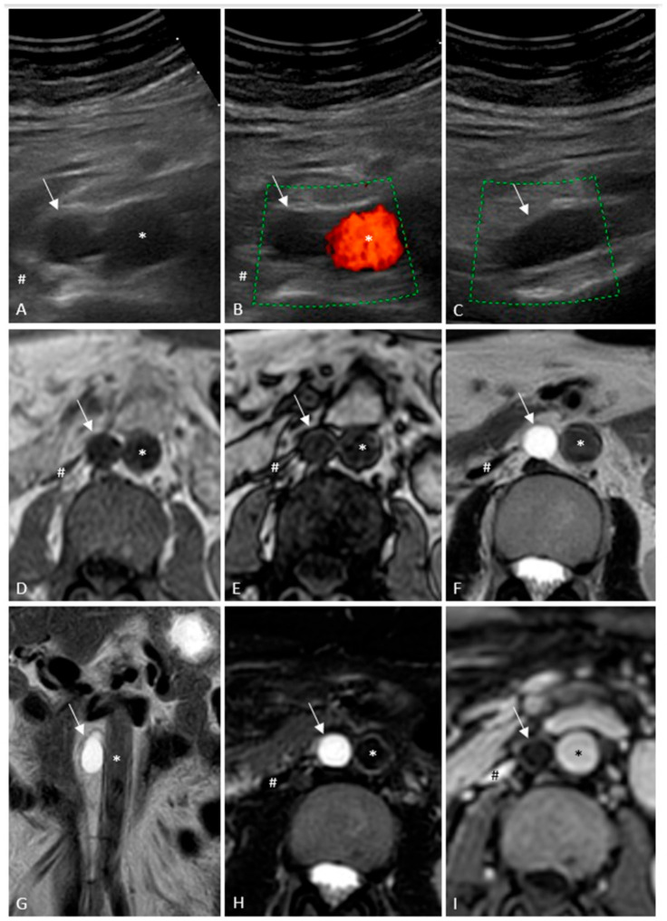 Figure 1
Peer Reviews
No public reviews on file for this paper yet. If you reviewed it on a platform where reviews are public (OpenReview, ICLR, NeurIPS, ICML), you can paste yours below so the community can read it here.
Videos
No videos yet. Explain this paper in a talk, walkthrough, or lecture? Add one.
Taxonomy
TopicsMuscle and Compartmental Disorders · Abdominal Surgery and Complications · Appendicitis Diagnosis and Management