CBCT in Evaluation of Root Canal Preparation—A Scoping Review
Andreia Vidal, Ana Moura Teles, Miguel Cardoso, Maria Bartolomeu, Rita Noites

TL;DR
This scoping review evaluates how CBCT is used to assess root canal preparation techniques, highlighting variability in study designs and the need for standardized methods.
Contribution
The paper provides a comprehensive overview of CBCT's role in comparing root canal instrumentation techniques and identifies gaps in current research methodologies.
Findings
Rotary instruments like ProTaper Next® and XP-Endo Shaper® showed favorable shaping trends in studies.
There is significant heterogeneity in study designs and outcome measurements across the reviewed literature.
Standardized methodologies and further research, especially on manual techniques, are needed.
Abstract
Cone-beam computed tomography (CBCT) is widely utilized in endodontics for evaluating root canal shaping outcomes, offering critical three-dimensional imaging capabilities. This study aims to assess the differences in apical and root canal preparation across various instrumentation techniques using CBCT. A systematic search of the Medline database (via PubMed) and Web of Science was performed up to 12 April 2025, yielding a total of 70 studies, with 45 full-text articles assessed for eligibility; 28 were included in the review. Studies showed great heterogeneity in experimental design, anatomical variables, and outcome measurements. The results indicate that rotary instruments, such as ProTaper Next® and XP-Endo Shaper®, were reported more frequently or showed favorable shaping trends in individual studies. Although rotary systems often appeared advantageous, conclusions were limited by…
Genes, proteins, chemicals, diseases, species, mutations and cell lines named across the full text — each resolved to its canonical identifier and authoritative record.
Click any figure to enlarge with its caption.
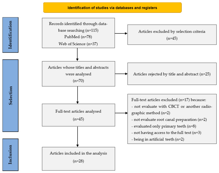 Figure 1
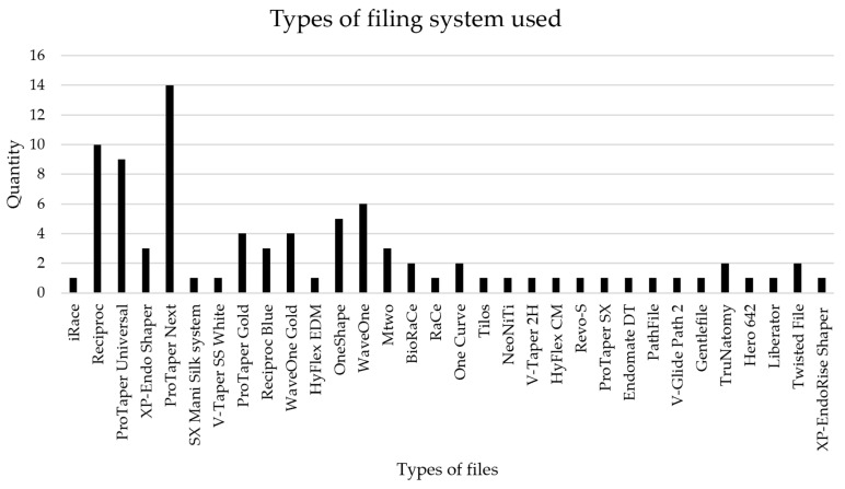 Figure 2
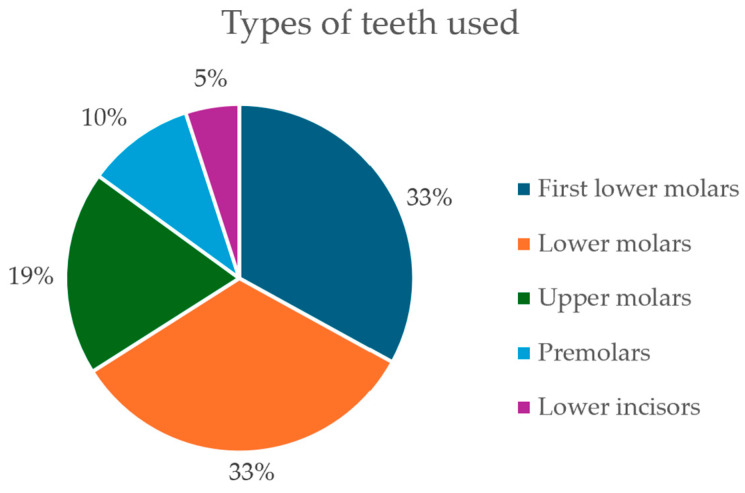 Figure 3
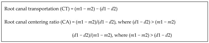 Figure 4
- —FCT—Fundação para a Ciência e Tecnologia, I.P.
- —Centro de Investigação Interdisciplinar em Saúde
Peer Reviews
No public reviews on file for this paper yet. If you reviewed it on a platform where reviews are public (OpenReview, ICLR, NeurIPS, ICML), you can paste yours below so the community can read it here.
Videos
No videos yet. Explain this paper in a talk, walkthrough, or lecture? Add one.
Taxonomy
TopicsEndodontics and Root Canal Treatments · Dental Radiography and Imaging · Dental Trauma and Treatments
1. Introduction
The primary objective of endodontic treatment is to eliminate microorganisms that invade the pulp chambers and root canals, thereby aiding in the preservation of natural teeth [1,2,3]. This treatment protocol includes several critical stages: mechanical preparation and disinfection, root canal preparation, and obturation. During these stages, the root canal is sealed with specific materials to restore the anatomical integrity of the tooth [4]. To achieve optimal outcomes in endodontic procedures, it is essential to ensure meticulous preparation and cleaning, as well as effective disinfection of the root canal.
Root canal preparation plays a crucial role in facilitating the obturation process and enhancing treatment success [5,6]. This phase involves the use of endodontic instruments, commonly known as files, to enlarge the root canals, maintaining their original shape and trajectory [7,8]. Ideally, a tapered morphology is achieved, narrowing towards the apex in both straight/wide and curved/narrow canal configurations [7]. Sodium hypochlorite between each procedural step is vital for the effective elimination of microorganisms and debris [9,10,11,12,13]. Root canal preparation techniques have evolved significantly due to technological advancements and can be performed either manually or mechanically. Manual instrumentation, developed by Edward Maryard, uses stainless steel files, while rotary techniques utilize Nickel-Titanium (Ni-Ti) rotary instruments, enabling faster and more efficient root canal preparation. Systems such as ProTaper Universal and XP-Endo Shaper provide flexibility and better adaptation to canal morphology. Multiple techniques have been explored to enhance quality and reduce operator fatigue: these include the conventional, balanced force, step-back, flared, rotary Ni-Ti, and crown-down techniques [1,4,7,14,15,16].
Cone Beam Computed Tomography (CBCT) has emerged as a sophisticated radiological modality, using cone-shaped X-rays to produce high-resolution images with reduced radiation exposure [17,18,19]. In endodontics, CBCT is instrumental for diagnosing root canal morphology, periapical bone loss, fractures, and resorptions, particularly where standard radiographs are inadequate [20,21,22]. Despite its benefits, CBCT should be considered a complementary rather than a replacement tool for routine digital radiography [23].
Given the rapidly growing and heterogeneous literature on the use of CBCT for evaluating root canal shaping, a scoping review is warranted to systematically map the characteristics, scope, and gaps in current research. This scoping review aims to provide an evidence map of ex vivo studies that have used CBCT to assess root canal and apical preparation with various instrumentation techniques, highlighting trends, evidence clusters, and areas requiring further investigation.
2. Materials and Methods
2.1. Protocol and Registration
This scoping review was conducted and reported in accordance with the Preferred Reporting Items for Systematic Reviews and Meta-analyses for Scoping Reviews (PRISMA-ScR). All procedures, including study selection and data charting, were performed independently by two reviewers; any disagreements between the reviewers were resolved through consensus or, if necessary, by a third reviewer.
2.2. Databases and Search Strategy
A comprehensive literature search was conducted across the databases Medline (via PubMed) and Web of Science (WoS). Search terms incorporated synonyms and free-text terms related to CBCT and root canal preparation. The search question was developed according to the PCC (Population, Concept, Context) framework, in alignment with Scoping Review methodology: Population, extracted teeth (ex vivo studies); Concept, assessment of root canal preparation using CBCT; Context, various instrumentation techniques.
The search strategy for PubMed was: (((CBCT) OR (cone-beam computed tomography)) AND ((“root canal preparation” OR “root canal instrumentation”) OR “mechanical preparation”)) AND ((“cleaning efficiency” OR “shaping ability”) OR “outcome”).
A similar strategy was used for Web of Science: (((CBCT) OR (cone-beam computed tomography)) AND (((“root canal preparation”) OR (“root canal instrumentation”)) OR (“mechanical preparation”))) AND (((“cleaning efficiency”) OR (“shaping ability”)) OR (outcome)).
2.3. Study Selection
Titles and abstracts were screened independently by two reviewers. Full-text articles were obtained for potentially relevant studies. Eligibility was determined against the inclusion criteria, with reasons for exclusion recorded, and discrepancies were resolved by discussion or a third reviewer.
2.4. Inclusion and Exclusion Criteria
The inclusion criteria for this review comprised studies published in English between 2014 and 2024 that provided full-text availability and were conducted as ex vivo studies. The exclusion criteria encompassed non-English articles, literature reviews, editorials, or conference abstracts, and studies focusing on deciduous teeth or artificial tooth replicas.
2.5. Data Charting and Synthesis
Data were charted using a standardized form developed by the team. The following variables were extracted: year, country, tooth type, experimental design, file system, outcome measures, measurement formulas, and main findings. The results were mapped and synthesized descriptively, focusing on the distribution of instrumentation systems, CBCT approaches, and areas of evidence concentration or gaps.
3. Results
This scoping review identified a total of 28 studies meeting the inclusion criteria after screening 70 full texts, following removal of duplicates and title/abstract screening (Figure 1). Included studies were mapped for characteristics including instrumentation techniques, file types, tooth types, and CBCT-based analysis (Table 1). The broad heterogeneity in study design and outcome measures highlights the variability in current research, with a predominance of rotary instrumentation systems.
Most studies focused on rotary instrumentation systems, with ProTaper Next^®^ being the most extensively evaluated (featuring in 14 articles), followed by Reciproc^®^ (10 articles) and ProTaper Universal^®^ (9 articles) (Table 1, Figure 2). Other systems were investigated less frequently, indicating research concentration on a few established rotary systems: the WaveOne^®^ system was assessed in 6 studies, while the OneShape^®^ system was assessed in 5 studies. ProTaper Universal Gold^®^ and WaveOne Gold^®^ were examined in 4 studies each. BioRaCe^®^, One Curve^®^, TruNatomy^®^, and Twisted File^®^ were each the subject of 2 investigations, with other systems being referenced in only 1 article. While no pooled effect sizes are reported given heterogeneity, individual studies frequently report performance metrics such as canal transportation and centering ratios, suggesting rotary files generally maintain canal anatomy more effectively than manual systems. However, based on the data presented in Figure 2, it can be concluded that most systems were analyzed in a single study, highlighting a pressing need for further investigation into their efficacy in root canal preparation.
3.1. Teeth Typology
The majority of studies examined molars, mainly first lower molars [25,30,31,33,41,45,49], followed by both lower molars [27,28,34,38,40,42,47], and upper molars [24,37,43,46], premolars [39,44], and lower incisors [26] (Figure 3). The majority of permanent teeth examined, encompassing both single-rooted and multi-rooted varieties, were extracted due to periodontal issues [24,26,27,32,36,45], the presence of carious lesions [27], prosthetic complications [32], or the degree of curvature of the roots [24,25,28,29,30,31,32,33,34,35,37,38,39,41,43,46,48]. The type of tooth and the number of root canals are variables that influence the results; an increased number of root canals necessitates more meticulous procedures to achieve optimal outcomes.
The anatomical variability and number of root canals across tooth types influenced results, requiring more meticulous shaping in multi-rooted teeth. Subgrouping by tooth type reveals consistent attention to the complexities of curved and narrow canals, although direct quantitative comparison is limited by study design differences.
3.2. Manual Versus Rotary Systems
Only a limited number of studies directly compared manual and rotary instrumentation [44]. The discrepancies in conclusions across various studies may stem from the examination of multiple files and preparation techniques, leading to divergent findings and relationships [30,31,32,33,34,35,36,37,38,39,40,41,42,51].
3.3. Outcome Measures and Calculation Methods
With respect to file performance in terms of transportation and centralization, several studies reported no significant differences that would affect root canal geometry [24,30,37,41,42,44,47]. However, certain files, such as ProTaper Universal^®^ [25], SX Mani Silk^®^ [35], and Gentlefile^®^ [38], exhibited greater transportation, while others, including ProTaper Gold^®^ [52] and XP-Endo Shaper^®^ [36], demonstrated reduced transportation, thereby enhancing instrumentation. In relation to centralization, files such as Revo-S^®^ [31], Patch File^®^ [34], and XP-Endo Shaper^®^ [36,51] displayed superior root canal centralization compared to the predetermined ideal.
Ideally, minimal transportation and improved centralization contribute to more efficient outcomes, thereby reducing treatment duration and associated complications. All of these factors are essential for achieving an optimal root canal shape, which facilitates effective root canal cleaning. It is noteworthy that the studies in question came to varying conclusions.
Although some investigations identified specific files, such as XP-Endo Shaper^®^ [25,36] and V-Taper^®^ [28], as exceptional, others found no significant differences among files, resulting in the absence of a definitive superior option [24,26,30,37,39,40,41,43,44,47]. Overall, the studies suggest that the files tested are suitable for endodontic treatment without inflicting significant issues on root canal shape or cleaning; however, one article indicated that no file excelled in both transportation and centralization [42].
The pre- and post-instrumentation CBCT images were utilized to assess the outcomes and identify any changes. In addition to image analysis, calculations were conducted to ascertain file centralization and transportation. The majority of articles employed the formulas depicted in Figure 4 to calculate file centralization and transportation using CBCT images [24,25,26,27,28,29,30,31,32,33,34,35,36,38,39,40,41,42,43,44,45,47].
In these equations, m1 and m2 denote the shortest distance from the mesial surface to the periphery of the root canal, measured before and after instrumentation, respectively. Meanwhile, d1 and d2 represent the corresponding measurements for the distal surface. Pagliosa et al. [37] employed a formula proposed by Loizides et al., which encompasses the following metrics:
- -Root canal transportation (CT) is calculated as CT = MT − DT,
- -Root canal centering (CA) is expressed as CA = (m total − d total)/CD
In these equations, MT and DT signify the transportation distances for the mesial and distal surfaces, respectively, while CD denotes the diameter of the root canal. All of these criteria were determined based on the average values obtained from each root canal (total m and total d).
4. Discussion
Endodontic treatment aims to preserve the natural teeth and involves multiple phases that can be time-consuming and technically demanding. A growing range of instrumentation techniques has been developed to improve efficiency and safety, and a clear understanding of their impact on canal transportation and centralization is essential for informed clinical decision-making [1,3,7]. This scoping review mapped ex vivo CBCT-based studies evaluating root canal preparation with different instrumentation systems, focusing on how current research is distributed across file types, tooth anatomy, and assessment methods [24,25,26,27,28,29,30,31,32,33,34,35,36,37,38,39,40,41,42,43,44,45,46,47,48,49,50,52].
Manual instrumentation, although historically established and capable of producing acceptable outcomes [44], is under-represented in the available literature when compared with rotary techniques. Rotary and reciprocating systems dominate current research, with ProTaper Next^®^, ProTaper Universal^®^, and ProTaper Gold^®^ among the most frequently investigated file systems, particularly in lower molars. Many studies also incorporated additional parameters such as root canal curvature, dentin removal, and change in canal volume or cross-sectional area, which illustrates the methodological breadth but also contributes to heterogeneity in reported outcomes [46,50,51].
CBCT has become an important imaging tool in endodontics, providing three-dimensional visualization for diagnosis, treatment planning, and pre- and post-instrumentation assessment [18,30,35,40,52,53]. It offers clinically acceptable image quality at relatively low radiation doses and enables non-destructive evaluation of canal geometry before and after preparation. However, its spatial resolution remains a key limitation in the ex vivo research context. Typical clinical CBCT voxel sizes (approximately 75–100 µm) are substantially larger than those achievable with micro-computed tomography, which can reach sub-20 µm resolutions. This limits the precision with which subtle changes in canal morphology, such as small degrees of transportation or centralization, can be detected or quantified. Consequently, CBCT is suitable for comparative, exploratory assessment of shaping outcomes but may not be sensitive enough for definitive evaluation of fine morphological differences, for which micro-CT remains the reference standard in ex vivo research. Interpretations of detailed shaping performance in the included studies should therefore be made with this constraint in mind.
Most included studies assessed shaping outcomes using transportation and centering ratios, frequently calculated with formulas derived from Gambill and colleagues [52], while some used alternative approaches [37]. Despite differences in specific equations and measurement protocols, the general conceptual focus was consistent: quantifying how well each system maintained the original canal trajectory and minimized unwanted deviation. Several systems, such as Revo-S^®^ [31], Patch File^®^ [34], and XP-Endo Shaper^®^ [36,51], were reported to perform favorably in terms of centering, whereas ProTaper Gold^®^ [52], Reciproc Blue^®^ [52], and other modern systems often showed relatively low transportation in the contexts studied. However, given the heterogeneity across studies in tooth type, curvature, operator protocols, and imaging parameters, these findings should be interpreted as indications of trends within specific experimental conditions, rather than as evidence of clear superiority between systems.
Nonetheless, the review had limitations, including the disparity in the volume of studies comparing manual and rotary methods, variations in assessment techniques, and insufficient research concerning specific files and types of teeth. Substantial variability in experimental design, canal morphology, instrumentation protocols, and CBCT acquisition parameters further reduces the comparability of results and precludes reliable estimation or pooling of effect sizes. In addition, the included studies were ex vivo, which restricts the extrapolation of findings to clinical outcomes such as healing, pain, or long-term tooth survival.
Within these constraints, this scoping review highlights clear areas where evidence is clustered, particularly around a small number of popular rotary systems in molar teeth, as well as substantial gaps in the literature. Future research would benefit from more balanced exploration of manual and rotary techniques, broader inclusion of different tooth types and anatomies, and greater standardization of outcome measures and imaging protocols. The integration of higher-resolution modalities, such as micro-CT, in ex vivo work, combined with well-designed clinical studies, will be essential to translate shaping performance metrics into meaningful clinical recommendations. Overall, the findings reinforce the importance of careful instrument selection and protocol design while underscoring the need for more rigorous and standardized research to support evidence-based endodontic practice.
5. Conclusions
The objective of this study was to map, using CBCT, how different instrumentation techniques have been used to assess apical and root canal preparation in ex vivo models. This scoping review identified a heterogeneous body of evidence, with substantial emphasis on rotary systems and a comparatively smaller number of studies addressing manual techniques. The included studies most frequently evaluated transportation and centralization, and highlighted that contemporary NiTi systems, such as ProTaper Next^®^, ProTaper Gold^®^, and XP-Endo Shaper^®^, are commonly investigated with respect to their ability to maintain canal anatomy and limit undesired deviations.
CBCT emerged as a widely adopted tool for three-dimensional assessment of root canal morphology before and after instrumentation, supporting non-destructive visualization of shaping outcomes in ex vivo research. However, important limitations related to spatial resolution and methodological variability across studies restrict the possibility of drawing firm comparative conclusions between specific systems or between manual and rotary techniques. Within these constraints, the available evidence suggests that both manual and rotary approaches can achieve acceptable shaping under controlled experimental conditions, but does not allow robust ranking of their relative clinical performance.
Overall, this review underscores that current research is clustered around a limited number of popular rotary systems and specific tooth types, while many instruments and clinical scenarios remain underrepresented. Future studies should prioritize more standardized protocols, broader inclusion of different tooth anatomies and instrumentation systems, and, where appropriate, higher-resolution imaging methods and clinical outcome measures. Such work will be essential to move from descriptive mapping of CBCT-based shaping studies toward stronger evidence to inform endodontic clinical practice.
The reference list from the paper itself. Each links out to its DOI / PubMed record.
- 1Gaikwad A. Patil R. Bhamare R. Nisa S.U. A CBCT evaluation of the shaping ability of two different rotary instrumentation systems in oval-shaped root canals: An in vitro study Eur. Chem. Bull.202312114126
- 2Löst C. Quality guidelines for endodontic treatment: Consensus report of the European Society of Endodontology Int. Endod. J.20061292193010.1111/j.1365-2591.2006.01180.x 17180780 · doi ↗ · pubmed ↗
- 3Hargreaves K.M. Berman L.H. Cohen’s Pathways of the Pulp Elsevier—Health Sciences Division Amsterdam, The Netherlands 20161419
- 4Antunes M.S. Instrumentação Endodôntica: Instrumentação Mecanizada vs. Instrumentação Manual—Uma Perspetiva Radiográfica Master’s Thesis Universidade de Lisboa Lisboa, Portugal 2021
- 5Pham K.V. A Comparison of Cone Beam Computed Tomography and Periapical Digital Radiography for Evaluation of Root canal Preparation Appl. Sci.202114659910.3390/app 11146599 · doi ↗
- 6Pham K.V. Khuc N.K. The Accuracy of Endodontic Length Measurement Using Cone-Beam Computed Tomography in Comparison with Electronic Apex Locators Iran. Endod. J.202015121710.22037/iej.v 15i 1.2672036704319 PMC 9723209 · doi ↗ · pubmed ↗
- 7Capelas J.A. Instrumentação de Canais Radiculares: Estudo Comparativo Entre Uma Técnica Manual e Três Técnicas Motorizada Ph.D. Thesis Universidade do Porto Porto, Portugal 2001
- 8Pham K. Nguyen N. Cutting efficiency and dentinal defects using two single-file continuous rotary nickel-titanium instruments Saudi Endod. J.20201566010.4103/sej.sej_64_19 · doi ↗