Renoprotection by 5-Methoxytryptophan in Kidney Disease
Jonah P. Gutierrez, Tram N. Diep, Shaona Niu, Liang-Jun Yan

TL;DR
This paper reviews how 5-methoxytryptophan (5-MTP) protects the kidneys from injury and could be a potential treatment for kidney disease.
Contribution
The paper highlights 5-MTP as a novel therapeutic and biomarker for chronic kidney disease.
Findings
5-MTP levels are reduced in chronic kidney disease patients, suggesting its role as a biomarker.
Exogenous 5-MTP shows nephroprotective effects in multiple rodent models of kidney injury.
5-MTP protects kidneys through antioxidative, anti-inflammatory, and anti-fibrotic mechanisms.
Abstract
Kidney disease, be it acute or chronic, has a complex pathology and is a significant human health problem. Increasing interest has been focused on exploring therapeutic targets that can be used to safeguard kidney function under a variety of detrimental conditions. In this article, we review the protective effects of 5-methoxytryptophan (5-MTP), a tryptophan metabolite, on kidney injury. Published studies indicate that serum 5-MTP level is decreased in patients with chronic kidney disease (CKD), suggesting that 5-MTP is a biomarker for CKD and has therapeutic values. Indeed, rodent models of kidney injury induced by folic acid, lipopolysaccharide (LPS), unilateral ureteral obstruction (UUO), and ischemia/reperfusion all demonstrate that exogenous 5-MTP exhibits nephroprotective effects. The underlying mechanisms involve antioxidative damage via activating antioxidant systems such as…
Genes, proteins, chemicals, diseases, species, mutations and cell lines named across the full text — each resolved to its canonical identifier and authoritative record.
Click any figure to enlarge with its caption.
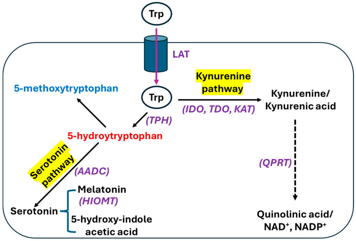 Figure 1
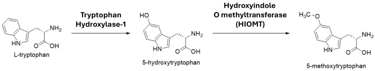 Figure 2
| Model | References |
|---|---|
| Glycerol-induced kidney injury | [ |
| 5/6 Nephrectomy-induced kidney injury | [ |
| Doxorubicin-induced kidney injury | [ |
| Cisplatin-induced kidney injury | [ |
| Cadmium- and other metal-induced kidney injury | [ |
| Adenine-induced CKD | [ |
| Streptozotocin-induced diabetic kidney disease (DKD) | [ |
| Zucker diabetic rats, ZSF-1 rats, db/db mice | [ |
| High-fat diet/streptozotocin-induced DKD | [ |
| High fructose/streptozotocin | [ |
| Nicotinamide/streptozotocin-induced DKD | [ |
| D-galactose-induced kidney injury | [ |
| Cecal ligation and puncture-induced kidney injury | [ |
| Caloric restriction | [ |
| Ketogenic diet and ketone body ingestion | [ |
- —Diabetes Action Research and Education Foundation
- —UNT Health Fort Worth
Peer Reviews
No public reviews on file for this paper yet. If you reviewed it on a platform where reviews are public (OpenReview, ICLR, NeurIPS, ICML), you can paste yours below so the community can read it here.
Videos
No videos yet. Explain this paper in a talk, walkthrough, or lecture? Add one.
Taxonomy
TopicsChronic Kidney Disease and Diabetes · Acute Kidney Injury Research · Chemotherapy-induced organ toxicity mitigation
1. Introduction
The kidney is a vital organ for the maintenance of body function. It excretes water-soluble waste via glomerular filtration and selective tubular reabsorption of nutrients, minerals, and electrolytes [1]. As such, the kidney maintains blood pH within very narrow limits and regulates blood pressure. The kidney also produces and responds to hormones. Examples include erythropoietin production, vitamin D maturation, the aldosterone and parathyroid hormone, the renin–angiotensin system, and the vasopressin or antidiuretic hormone. The kidney can also participate in glucose production via the gluconeogenic pathway under anerobic conditions [2]. All these functions require a large amount of ATP produced by mitochondrial oxidative phosphorylation, a metabolic process that can also generate excessive reactive oxygen species (ROS) that can lead to oxidative damage to nephrons and result in kidney dysfunction [3,4]. Therefore, there is a constant need to search for therapeutic targets that can be used to safeguard kidney function under a variety of harmful conditions. In this review article, we focus on the protective role of 5-methoxytryptophan (5-MTP) in a variety of kidney diseases after a brief overview of kidney injuries such as acute kidney injury (AKI), chronic kidney disease (CKD), and diabetic kidney disease (DKD) as well as well-established underlying mechanisms such as oxidative stress, inflammation, and fibrosis.
1.1. Acute Kidney Injury
Clinically, AKI is a common renal disorder characterized by a sudden decline in kidney function and occurs more frequently in old-aged patients [5]. It is also highly prevalent in hospitalized individuals and is linked with numerous etiologies such as ischemia [6], cardiac surgery [7], sepsis [8], rhabdomyolysis [9], and drug or chemical toxicity [10]. AKI is often associated with increased mortality in severely ill patients. Moreover, even for patients who survive AKI, they could further develop CKD or end-stage renal failure [11]. Despite advances in our understanding of the underlying mechanisms of AKI, few therapeutic options are currently available.
1.2. Chronic Kidney Disease
CKD is characterized by a progressive decline of kidney function caused by various chronic disorders such as hypertension [12], environmental exposure to heavy metals [13,14], and diabetes [15]. It has been estimated that CKD may affect more than 10% of the Earth’s population. CKD can be identified by measuring a glomerular filtration rate (GFR) that is less than 60 mL/min/1.73 m^2^ for a period of more than 3 months while the albumin–creatinine ratio is constantly above 30 mg/g/day [16]. If unmanaged, CKD can become end-stage renal failure [17]. CKD is usually classified into five stages as shown in Table 1 and albuminuria categories are shown in Table 2.
1.3. Diabetic Kidney Disease
As an emerging global health issue, DKD is a major complication of diabetes mellitus. It is caused by persistent exposure of the kidney to hyperglycemia. DKD can develop in about 35% of diabetic patients [19]. It is a renal microvascular disorder of diabetes and is characterized by albuminuria levels greater than 30 mg/day accompanied by a declining GFR [20]. DKD can lead to anemia and vitamin D deficiency given that the kidney is responsible for erythropoietin generation and vitamin D maturation [4]. Redox imbalance, oxidative stress, inflammation, and fibrosis are all implicated in the pathogenesis of DKD [21].
1.4. Oxidative Stress
Oxidative stress occurs when cellular production of ROS overwhelms the cellular antioxidant defense system [22]. ROS can be produced by a variety of systems such as NADPH oxidases [23], the mitochondrial electron transport chain [24], dihydrolipoamide dehydrogenase [25], cyclooxygenase and lipoxygenase [26], and xanthine oxidase [27]. Excessive ROS can cause oxidative damage to proteins, lipids, and DNA, leading to dysfunction of the macromolecules [28]. On the other hand, disease status-caused functional decline of the antioxidant systems such as superoxide dismutase, catalase, glutathione peroxidase and reductase, thioredoxin reductase, and peroxiredoxin reductase can further perturb cellular redox balance toward aggravated oxidative stress. All these can collectively lead to the collapse of cellular function and cell death [29]. Table 3 lists major ROS-generating systems and antioxidant defense systems. With respect to the determination of oxidative stress levels, there are numerous methods that can be used. For example, protein oxidation can be measured as protein carbonyls [22], lipid peroxidation can be measured by malondialdehyde (MDA) using the assay for thiobarbituric acid reactive substances [30,31], and DNA damage can be measured as 8-oxo-deoxyguanosine [32,33]. Moreover, superoxide and hydrogen peroxide can also be measured by the use of a fluorescent probe called 2,7-dicholorofluorescin [34]. In the meantime, cellular antioxidant capacities such as glutathione levels [34], SOD, and catalase activities [35,36] can also be measured to indicate the magnitude of oxidative stress.
1.5. Inflammation
Renal inflammation, as a hallmark of and response to kidney injury [37], involves numerous signaling pathways. Participating cells in renal inflammation include neutrophils, monocytes, macrophages, T cells, B cells, and natural killer cells [38]. Inflammatory mediators can be classified into two family members. One is the chemokine family including SDF-1, CCL-2/MCP-1, and fractalkine; another is the cytokine family including TNF-α, IL-6, and IL-13 [38,39]. If unmanaged, renal inflammation can gradually lead to renal fibrosis [38,39].
1.6. Fibrosis
Renal fibrosis underlies AKI, CKD, and DKD as well as the AKI to CKD transition [40]. It is characterized by accumulation and excessive deposition of extracellular matrix (ECM). The fibrotic process also involves tubulointerstitial fibrosis, tubular atrophy, and glomerulosclerosis, which collectively can lead to renal failure [40]. Key signaling pathways involved in renal fibrosis include NF-kB, Nrf2, NADPH oxidase-4, autophagy activation, and transforming growth factor-β [41]. Additionally, oxidative stress and inflammation are highly involved in renal fibrosis [39], which has been repeatedly demonstrated and confirmed by the use of antioxidants and anti-inflammation molecules in a variety of animal models of kidney disease [42,43,44,45].
2. Tryptophan and Biosynthesis of 5-MTP
Tryptophan, as an essential amino acid, is both ketogenic and glucogenic in nature. It is involved in biosynthesis of proteins, lipids, glucose, and indole acetic acid [2]. Tryptophan is also the precursor of serotonin and melatonin [2,46]. It enters cells via L-amino acid transporters (LATs) and is then catabolized via two pathways (Figure 1) [47]. The catabolic pathway generating serotonin and melatonin is often called the serotonin pathway, while nearly 95% of ingested tryptophan is catabolized by a pathway called the kynurenine pathway, which produces NAD^+^/NADP^+^ and immunomodulatory molecules such as kynurenine and kynurenic acid [46,48,49]. It should be noted that de novo synthesis of NAD^+^/NADP^+^ from tryptophan can only occur in the liver and kidney while all the other tissues may use the salvage pathway as a source for NAD^+^/NADP^+^ [50]. As shown in Figure 2, tryptophan is also a source of 5-hydroxyindole acetic acid [51]. 5-MTP is synthesized via two reactions (Figure 2). In the first step, tryptophan is converted to 5-hydroxytryptophan (5-HTP) by tryptophan hydroxylase (TPH) [52]. This reaction is rate-limiting. In the second step, 5-HTP is converted to 5-MTP by hydroxyindole O-methyltransferase (HIOMT) [52]. 5-MTP, being a 5-methoxyindole metabolite [53,54], can also serve as a precursor for the synthesis of 5-methoxyindole-2-carboxylic acid (MICA), which has been rigorously evaluated by our laboratory for its neuroprotective role in ischemic stroke [55,56,57,58].
The catabolic pathways of tryptophan. Tryptophan enters cells via L-amino acid transporters (LATs) followed by two catabolic pathways: the serotonin pathway and the kynurenine pathway. 5-methoxytryptophan is synthesized from 5-hydroxytryptophan via two-step reactions as shown in Figure 2. LAT: L-amino acid transporter. The enzymes involved in the pathways are TPH, tryptophan hydroxylase; AADC, aromatic amino acid carboxylase; HIOMT, hydroxyindole O-methyltransferase; IDO, indoleamine 2,3-dioxygenase; TDO, tryptophan 2,3-dioxygenase; KAT, kynurenine amino transferase; and QPTR, quinolinate phosphoribosyltransferase.
Synthesis of 5-MTP from 5-hydroxytryptophan. Note: the first step catalyzed by tryptophan hydroxylase is rate-limiting.
3. Potential Biological Functions of 5-MTP
5-MTP was originally named as cytoguardin due to its ability to suppress cyclooxygenase-2 (COX-2) in proliferating fibroblasts [59,60,61,62]. It was later confirmed that 5-MTP can also be made by other types of cells including smooth muscle cells, vascular endothelial cells, bronchial epithelial cells, and renal epithelial cells [63]. Moreover, bacteria in the gut microbiota can also produce 5-MTP [64]. Numerous studies have revealed that 5-MTP is a disease-fighting molecule as it can exhibit protective properties such as antioxidation, anti-inflammation, anti-fibrosis, anti-senescence, and anti-tumor properties [65,66,67,68,69]. In this review, we will focus on the protective effects of 5-MTP on kidney disease.
3.1. 5-MTP and Chronic Kidney Disease
Chen et al. first reported that 5-MTP content was lower in CKD patients than in healthy subjects [70]. Using UPLC-HDMS metabolomics and real-time PCR quantification techniques, the authors compared healthy individuals and CKD patients and analyzed the concentrations of 25 metabolites in serum. 5-MTP content showed an inverse correlation with kidney function, indicating that 5-MTP may be a biomarker of CKD. The same research group further discovered that serum 5-MTP had an anti-fibrotic effect in CKD [71]. These studies signify that exogenous 5-MTP has nephroprotective effects, which indeed have been explored and confirmed by other investigators, as discussed in this article.
Mogos et al. also found that serum 5-MTP level is an important biomarker for early-stage diabetic kidney disease [72]. The authors analyzed urine and serum amino acid metabolites in initial stages of DKD in type 2 diabetic patients. A total of 90 T2D patients were grouped into three subgroups based on their albuminuria stage from normal albuminuria to micro-albuminuria and macro-albuminuria, with each subgroup having 30 patients. Additionally, 20 healthy individuals were recruited as controls. Statistical analysis revealed that 5-MTP, among others, was more significant in terms of discrimination between the subgroups and the controls. This study indicates that 5-MTP not only can serve as a biomarker in early stage of DKD but also plays an active role in the progression of DKD. Nevertheless, whether 5-MTP is lower or higher in rodent models of DKD than in controls remains to be investigated.
3.2. Effect of 5-MTP on Lipopolysaccharide (LPS)-Induced Acute Kidney Injury
Sepsis caused by over-inflammatory response to infections can result in multiple organ failures including the kidney [73]. In animal models, septic AKI can be induced by a simple administration of LPS via intraperitoneal injection [74,75], which has been widely used to investigate septic mechanisms of kidney injury and also serves as a platform for testing the therapeutic values of numerous chemicals, compounds, and natural products [75]. In this septic kidney injury process, many inflammatory mediators such as interleukin-1, interleukin-6, and tumor necrosis factor-α can overload the kidneys and cause inflammation that further leads to tissue hypoxia and damage [76]. Using this LPS-induced AKI mouse model, Sun et al. demonstrated that exogenous 5-MTP exerts protective effects on kidney injury [77]. The authors further demonstrated that 5-MTP activated the Nrf2 transcription factor, leading to upregulation of heme oxygenase-1 (HO-1), which is a major antioxidant protein. This upregulation of the Nrf2 pathway also augmented renal tubular mitophagy reflected by LC3 immunohistochemical staining, thereby helping to maintain mitochondrial homeostasis and decrease mitochondrial oxidative stress. It should be noted that the authors’ interest in studying the effect of 5-MTP on septic AKI was triggered by their observation that serum 5-MTP levels in patients with AKI were significantly elevated when compared with control subjects, an observation seemingly in contrast with that of Chen et al. who reported that 5-MTP content in the serum of CKD patients was lower than in controls [71]. This difference might be due to the nature of the kidney injury in the respective study, that is, AKI vs. CKD. Nevertheless, it is possible that acute stress response in AKI may upregulate tryptophan metabolism, whereas the chronic response in CKD may exhaust tryptophan metabolism.
3.3. Effect of 5-MTP on Renal Ischemia/Reperfusion Injury
Renal ischemia–reperfusion (I/R) injury can occur in many clinical settings such as cardiac surgery, systemic vasodilation, volume depletion, shock, decreased cardiac output, and kidney transplantation [78,79,80]. Decrease in renal blood flow often results in hypoxia and a lack of energy supply that culminate in cell death and kidney functional decline [81,82,83]. Using a mouse model of renal I/R injury, Li et al. tested the protective effects of 5-MTP in I/R kidney injury [84]. 5-MTP was administered to mice 30 min before ischemic surgery. The authors found that exogenous 5-MTP mitigated renal damage and improved kidney function. Hematoxylin–eosin staining clearly showed I/R-induced renal tissue damage that was ameliorated by 5-MTP pretreatment (Figure 3). The underlying mechanisms involved attenuated ER stress and apoptosis. Moreover, the Nrf2 pathway was also activated by 5-MTP with corresponding upregulation of HO-1. When Nrf2 knockout mice were used, the protective effects of 5-MTP were partially suppressed, demonstrating that 5-MTP’s protective effect on renal I/R injury is Nrf2-dependent.
3.4. Effects of 5-MTP on Renal Tissue Inflammation and Fibrosis
Unilateral ureteral obstruction (UUO) is a widely used animal model for induction of kidney inflammation and fibrosis [85,86,87]. Wu et al. used this established animal model to evaluate the effect of 5-MTP on UUO-induced kidney injury [88]. The authors treated mice with 5-MTP prior to UUO surgery and then measured toll-like receptor 2 (TLR2) function and TGF-β signaling pathways. The authors also used TLR2 knockout mice in their study. Results indicated that in wildtype mice, the protective effect of 5-MTP in the UUO model was due to the downregulation of TLR2 and was similar to that in the TLR2-deficient mice, demonstrating that 5-MTP protection against UUO kidney injury involving inflammation and fibrosis is mediated by TLR2. Moreover, renal fibrosis attenuated by 5-MTP was accompanied by decreased macrophage infiltration of the kidney tissue as reflected by decreased TGF-β levels. The authors concluded that 5-MTP is nephroprotective against UUO-induced renal fibrosis and inflammation by blocking TLR2 and TGF-β signaling pathways.
3.5. Effects of 5-MTP on CKD-Induced Cerebrovascular Injury
It is well-known that kidney disease has a negative impact on the brain [89,90]. Indeed, kidney–brain interactions have garnered increasing interest over the years [91,92]. While there are numerous studies on epidemiology and mechanisms regarding the kidney–brain axis [93,94], the role of 5-MTP in this axis remains unknown. To address this unmet need, Zhou et al. used an animal model of kidney injury induced by a high concentration of folic acid and then performed the Morris water maze test on these animals [95]. As expected, the kidneys were injured by folic acid as measured by blood urea nitrogen, and the CKD animals exhibited increased escape latency, indicating a cerebrovascular injury. Interestingly, the authors found that both kidney injury and brain injury were suppressed by 5-MTP, an effect that was partially reversed by NF-kB overexpression. This study demonstrates that CKD-induced cerebrovascular injury can be mitigated by 5-MTP via attenuating the NF-kB pathway.
3.6. Predictive Value of 5-MTP on Clinical Outcomes in Patients Having Septic AKI
As a potential biomarker of kidney injury, does 5-MTP have any predictive value in terms of clinical outcomes of patients with septic AKI? This question was addressed by Sun et al. [96]. The authors recruited 31 healthy individuals and 78 patients who were diagnosed with septic AKI. Serum 5-MTP contents were measured by targeted metabolomics and correlation between serum 5-MTP and kidney function was assessed. The authors found that serum 5-MTP was significantly increased in patients with septic AKI, and this increase was associated with blood urea nitrogen (BUN), serum creatinine, and eGFR values. Additionally, the authors also found that higher content of 5-MTP was linked to a faster recovery of renal function, and a lowered content of 5-MTP was linked to an increased 90-day mortality in septic AKI patients. Therefore, 5-MTP has a predictive value in the development of septic AKI and an early elevation in serum may positively influence the prognosis of septic AKI.
3.7. Does Microbial 5-MTP Have a Nephroprotective Effect?
The gut microbiota also produces 5-MTP [97,98]. Whether microbial 5-MTP can protect the kidney against a variety of kidney injuries has not been explicitly investigated. A recently published study by Gong et al. may shed light on this aspect [99]. The authors studied the role of microbial 5-MTP in the shedding of angiotensin-converting enzyme-2 (ACE2) from the intestinal epithelial cells and sepsis induced by gut leak. They found that ACE2 deficiency was clearly associated with a decrease in microbial production of 5-MTP. When septic mice were given exogenous 5-MTP, the gut leak was ameliorated via enhanced proliferation and repair of epithelial cells. This amelioration was mediated by the PI3-Akt-WEE1 signaling pathway. Therefore, it is conceivable that microbial 5-MTP can also exert a protective effect on kidney injury. In a separate study by Kuo et al. [64], it was found that microbial 5-MTP was low in patients undergoing hemodialysis, further indicating that microbial 5-MTP is involved in kidney function or dysfunction depending on the context of the kidney injury. Future studies need to be performed to evaluate under what disease conditions microbial 5-MTP is beneficial or detrimental.
3.8. Pharmacological Boosting of 5-MTP Levels for Renoprotection
Shenkang injection (SKI), a formula of traditional Chinese medicine, consisting of astragalus, rhubarb, and safflower, has been used to treat chronic kidney failure (CKF) induced by adenine in rats. Zhou et al. [100] investigated the protective mechanisms of SKI and found that when CKF rats were given SKI, renal content of 5-MTP was significantly elevated when compared with CKF rats that were not treated with SKI. Moreover, SKI also attenuated the PI3/Akt and NF-kB signaling pathways. This study strongly suggests that elevation of 5-MTP is one of the mechanisms by which SKI ameliorates CKF. Nonetheless, whether the elevation of 5-MTP is due to increased enzymatic activity of tryptophan hydroxylase remains unknown. This study also suggests that boosting 5-MTP content by pharmacological agents, dietary modulation, or drugs is possible for nephroprotective purposes.
4. Future Perspectives
A PubMed search using the keywords “kidney” and “methoxytryptophan” yielded only 14 articles, which were the basis of this review article. This PubMed search outcome also indicates that more studies need to be performed to widen and deepen our knowledge on the protective effects and mechanisms of 5-MTP on kidney injuries. In our opinion, numerous rodent models of AKI and CKD remain to be explored. These models are listed in Table 4. Table 4 also gives potential approaches that may facilitate or enhance endogenous production of 5-MTP for renoprotective purposes. For example, it would be interesting to know whether caloric restriction including methionine restriction [101] and isoleucine restriction [102] or ketogenic diet can promote 5-MTP production in the context of kidney injury. Targeting delivery of 5-MTP to the kidneys using nanoparticle delivery techniques [103,104,105] is also worth exploring.
Finally, it should also be emphasized that given that 5-MTP acts as an antioxidant that attenuates renal oxidative stress, inflammation, and fibrosis, the underlying mechanisms remain to be comprehensively elucidated. One area would be investigation of the relationship between 5-MTP and NAD^+^/NADH redox balance. Does 5-MTP elevate NAD^+^ levels? If so, how is the NAD^+^-dependent network modulated by this elevation? This would require studies on NAD^+^-dependent enzymes including pyruvate dehydrogenase complex, mitochondrial complex I, NAD kinase, sirtuins, poly-(ADP)-ribosylase, and CD38 [10,106]. It is likely that 5-MTP may boost NAD^+^ content and ameliorate kidney injury caused by a variety of endogenous and exogenous insults. Another area would be the effects of 5-MTP on the infiltration of neutrophils and macrophages into the kidney upon injury. Indeed, how 5-MTP affects NETosis [107] and macrophage release of various inflammatory cytokines such as TNF-α and IL-6 [108] should be investigated.
5. Summary
In this article, we reviewed the protective effects of 5-MTP on kidney injury in various models shown in Table 5. We started with a summary of findings in humans that serum 5-MTP was decreased in CKD patients. This was followed by discussions of the nephroprotective effects of 5-MTP in animal models of AKI or CKD. The rodent models covered include kidney injury induced by folic acid, LPS, UUO, and ischemia–reperfusion. The mechanisms by which 5-MTP protects against kidney injury involve upregulation of antioxidant defense systems, downregulation of oxidative damage, decreased fibrosis, and increased mitophagy (Figure 4). Overall, studies discussed in this article indicate that 5-MTP is not only a potential biomarker in kidney injury but also a renal protectant. Nevertheless, in addition to the above-mentioned potential studies that remain to be performed, future studies regarding endogenous boosting of 5-MTP and its synergistic effects with other therapeutic approaches also remain to be conducted.
The reference list from the paper itself. Each links out to its DOI / PubMed record.
- 1Reed S. Essential Physiological Biochemistry: An Organ-Based Approach Willey-Blackwell Noida, India 2009
- 2Lieberman M. Peet A. Marks’ Basic Medical Biochemistry: A Clinical Approach 6th ed.Wolters Kluwer/Lippincott Williams & Wilkins Philadelphia, PA, USA 2023
- 3Duann P. Lin P.H. Mitochondria Damage and Kidney Disease Adv. Exp. Med. Biol.20179825295512855180510.1007/978-3-319-55330-6_27PMC 8049117 · doi ↗ · pubmed ↗
- 4Kamt S.F. Liu J. Yan L.J. Renal-Protective Roles of Lipoic Acid in Kidney Disease Nutrients 202315173210.3390/nu 1507173237049574 PMC 10097220 · doi ↗ · pubmed ↗
- 5Marquez-Exposito L. Tejedor-Santamaria L. Santos-Sanchez L. Valentijn F.A. Cantero-Navarro E. Rayego-Mateos S. Rodrigues-Diez R.R. Tejera-Munoz A. Marchant V. Sanz A.B. Acute Kidney Injury is Aggravated in Aged Mice by the Exacerbation of Proinflammatory Processes Front. Pharmacol.20211266202010.3389/fphar.2021.66202034239439 PMC 8258347 · doi ↗ · pubmed ↗
- 6Yan Y. Bai J. Zhou X. Tang J. Jiang C. Tolbert E. Bayliss G. Gong R. Zhao T.C. Zhuang S. P 2X 7 receptor inhibition protects against ischemic acute kidney injury in mice Am. J. Physiol. Cell Physiol.2015308 C 463C 47210.1152/ajpcell.00245.201425588875 PMC 4360025 · doi ↗ · pubmed ↗
- 7Rosner M.H. Okusa M.D. Acute kidney injury associated with cardiac surgery Clin. J. Am. Soc. Nephrol.20061193210.2215/CJN.0024060517699187 · doi ↗ · pubmed ↗
- 8Wang Y. Zhu J. Liu Z. Shu S. Fu Y. Liu Y. Cai J. Tang C. Liu Y. Yin X. The PINK 1/PARK 2/optineurin pathway of mitophagy is activated for protection in septic acute kidney injury Redox Biol.20213810176710.1016/j.redox.2020.10176733137712 PMC 7606859 · doi ↗ · pubmed ↗