Carbon Monoxide Therapy: Evidence and Prospects for Preventing and Treating Retinal Diseases
Mathew Reese Land, Marybeth Koepsell, Noah Nussbaum, Edward Gomperts, Andrew Gomperts, Menaka C. Thounaojam, Ravirajsinh N. Jadeja, Pamela M. Martin

TL;DR
Carbon monoxide therapy may help prevent and treat retinal diseases by reducing inflammation and cell death, though more clinical research is needed.
Contribution
This review highlights CO's emerging therapeutic potential in retinal diseases and identifies barriers to clinical translation.
Findings
Low-dose CO therapy shows anti-inflammatory and anti-apoptotic effects in retinal disease models.
Current CO delivery methods lack robust clinical validation and reproducibility.
Improved delivery strategies and mechanism-specific trials are needed for successful translation.
Abstract
In carbon monoxide (CO) therapy, CO is administered at low concentrations as a controlled solution; this approach enables the drug to achieve its cytoprotective properties, including anti-inflammatory, anti-apoptotic, and vasodilatory effects. CO therapy, initially reported to benefit cardiovascular and pulmonary conditions, is now used to treat ocular diseases in preclinical models. Carbon monoxide, a compound most famously known for its deleterious effects, is receiving more attention as a potential therapeutic candidate in ocular medicine. In a few studies, controlled low-dose CO therapy has shown anti-inflammatory and anti-apoptotic effects in various models of retinal disease (such as retinal ischemia-reperfusion injury, optic nerve crush, ocular hypertension, and autoimmune uveitis). We have summarized the clinical and preclinical findings, along with the potential therapeutic…
Genes, proteins, chemicals, diseases, species, mutations and cell lines named across the full text — each resolved to its canonical identifier and authoritative record.
Click any figure to enlarge with its caption.
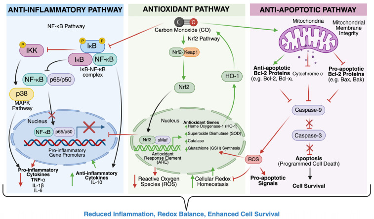 Figure 1
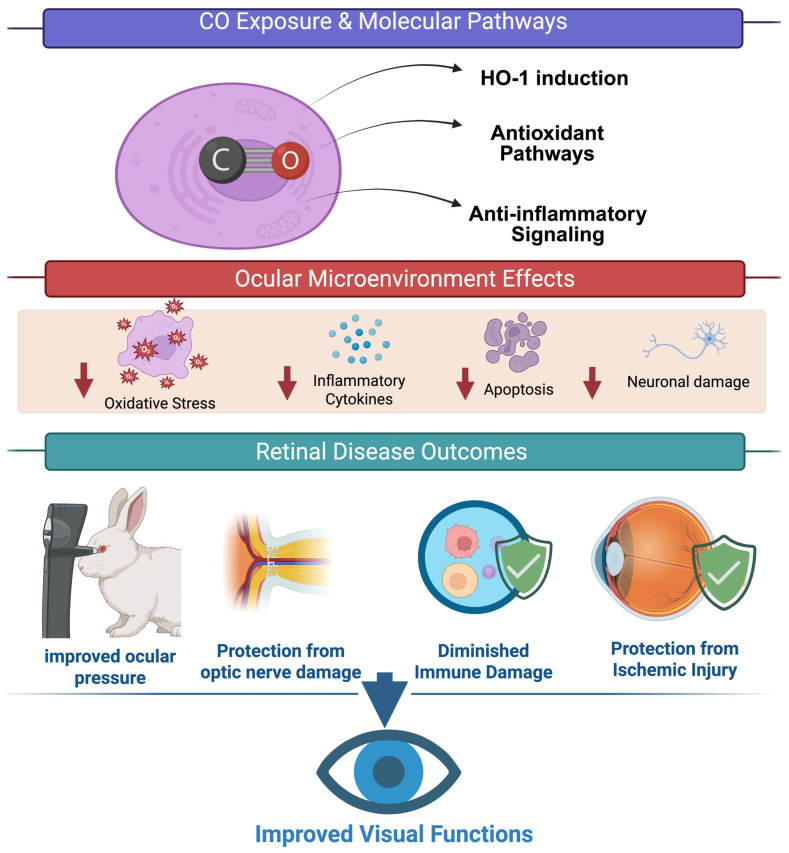 Figure 2
- —NIH
Peer Reviews
No public reviews on file for this paper yet. If you reviewed it on a platform where reviews are public (OpenReview, ICLR, NeurIPS, ICML), you can paste yours below so the community can read it here.
Videos
No videos yet. Explain this paper in a talk, walkthrough, or lecture? Add one.
Taxonomy
TopicsHeme Oxygenase-1 and Carbon Monoxide · Retinal Diseases and Treatments · Neonatal Health and Biochemistry
1. Introduction
Degenerative retinal diseases precipitated by diabetes (diabetic retinopathy), premature birth (retinopathy of prematurity), increased intraocular pressure (glaucoma), and aging (age-related macular degeneration) represent leading causes of visual loss and blindness worldwide [1,2]. Despite significant advances in understanding the molecular mechanisms underlying these conditions, effective early interventions remain limited. Current therapies—including anti-VEGF agents, corticosteroids, laser photocoagulation, and surgical procedures—have improved outcomes for many patients but are often associated with limitations such as incomplete efficacy, recurrence of disease, adverse effects, and the need for repeated invasive treatments [3,4,5].
For example, anti-VEGF therapy has revolutionized the management of neovascular retinal diseases but requires frequent intravitreal injections and may not be effective in all patients [3,4,5]. Corticosteroids, while potent anti-inflammatory agents, carry risks of cataract formation and elevated intraocular pressure. Laser therapy can cause collateral damage to healthy retinal tissue, and surgical interventions are typically reserved for advanced stages of disease [3,4,5].
Moreover, the posterior segment of the eye remains a challenging target for drug delivery due to anatomical and physiological barriers [6,7,8]. Non-invasive yet effective delivery systems are still under development, and many promising compounds fail to reach therapeutic concentrations in retinal tissues when administered systemically or topically [6,7,8].
Given these limitations, there is a pressing need for novel therapeutic strategies that are both effective and minimally invasive. One such emerging approach is the use of carbon monoxide (CO) therapy. Traditionally viewed as a toxic gas, CO has demonstrated cytoprotective properties at low concentrations, including anti-inflammatory, anti-apoptotic, and vasodilatory effects. This review explores the therapeutic potential of CO and heme oxygenase-1 (HO-1) induction in ocular diseases, summarizing current evidence and discussing delivery strategies, safety considerations, and future directions.
CO is classically considered a toxic gas. This is related to its well-known property of binding with high affinity to hemoglobin to generate carboxyhemoglobin (COHb). COHb decreases hemoglobin’s ability to carry oxygen, contributing to hypoxemia, tissue hypoxia, and, if in high concentrations not addressed rapidly, death. CO is also generated physiologically in the body as a byproduct of heme degradation in a reaction catalyzed by heme oxygenase enzymes. Heme oxygenase 1 (HO-1) is essential to endogenous CO production. Several reports have highlighted the benefits of enhanced HO-1 expression and activity in a variety of disease model systems including in animal models of intestinal [9,10,11], hepatic [12,13,14], lung [15,16,17], renal [18,19], cardiac [20,21,22], and ocular ischemic reperfusion injury [23,24,25]. However, the detailed mechanisms to explain these effects, particularly whether or how they relate directly or indirectly to the modulation of CO levels, are unclear.
An increasing number of studies expose the robust and direct benefits of CO in a wide variety of clinical and pre-clinical scenarios, sparking increased interest in CO therapy. This fascinating area of research showcases CO in a light that contrasts starkly with traditional views. The current review focuses on the potential therapeutic effects of CO that could be realized in the eye, providing information on the mechanism(s) of action, available pre-clinical and clinical evidence relevant to the utility of CO in retinal diseases, delivery methods, and safety considerations, as well as prospects and future directions.
Despite extensive preclinical support, translation of CO-based therapeutics to human disease has been challenging. Most early-phase clinical studies have established feasibility and short-term safety but have not demonstrated durable or clinically meaningful efficacy across target indications. This divergence between preclinical promise and clinical outcomes underscores a critical efficacy gap and motivates a more analytic approach to dosing, delivery, and pharmacology, as well as the selection of indications and endpoints that are mechanistically congruent with CO’s modes of action.
2. Literature Search and Selection Criteria
A systematic literature search involving PubMed, Scopus, and Google Scholar was performed to identify peer-reviewed and preprint studies regarding the biological significance of carbon monoxide (CO) and its therapeutic action in ocular diseases. The most current search was conducted on 20 November 2026. The search terms used were: “carbon monoxide,” “CO therapy,” “heme oxygenase-1,” “HO-1,” “cytoprotection,” “retinal disease,” “ischemic retinopathy,” “retinal ischemia-reperfusion,” “uveitis,” and “ocular hypertension.” Inclusion criteria included studies that had evaluated CO signaling, CO-releasing molecules (CORMs), heme oxygenase pathways, or that assessed the therapeutic utility of CO from the perspective of ocular or retinal models, including in vitro, in vivo, and clinical studies. We limited our search of ClinicalTrials.gov, for the following: “carbon monoxide,” “CO therapy,” “CORM,” and included interventions using systemic or localized CO delivery, CO-releasing molecules, and inhaled CO. All articles were excluded for lack of English translation, not available full text references, or for not addressing mechanisms of CO-mediated treatment of ocular pathology. Screening of reference lists of included studies was also performed to identify further relevant literature.
3. Mechanisms of Action for Low-Dose CO
CO is produced during the enzymatic degradation of heme by heme oxygenase-1 (HO-1), a stress-inducible enzyme that responds to various physiological insults. These effects are tightly regulated and dose-dependent, with low-dose CO triggering beneficial responses without inducing toxicity.
The cytoprotective actions of CO begin with the induction of HO-1, which catalyzes the breakdown of heme into biliverdin, free iron (Fe^2+^), and CO. Biliverdin is subsequently converted to bilirubin by biliverdin reductase, and both products contribute to the cellular antioxidant defense system [26] (Figure 1). HO-1 expression is regulated by the transcription factor Nrf2, which is activated via the PI3K/Akt/mTORC1 signaling pathway. Upon activation, Nrf2 translocates to the nucleus and binds to antioxidant response elements (AREs), promoting the transcription of HMOX1 and other genes involved in cytoprotection [27,28]. This pathway has been extensively studied in models of ischemia-reperfusion injury, neurodegeneration, and inflammation, underscoring its central role in CO-mediated cellular resilience.
CO also plays a critical role in modulating inflammation. It suppresses the activation of immune cells such as microglia and macrophages, leading to a reduction in pro-inflammatory cytokines, including TNF-α, IL-1β, and IL-6 [29,30,31]. Simultaneously, CO enhances the production of anti-inflammatory cytokines like IL-10, promoting the resolution of inflammation [32]. In the central nervous system, CO downregulates allograft inflammatory factor-1 (AIF-1), a marker of microglial activation, thereby mitigating neuroinflammatory responses [33]. These effects are mediated through mitogen-activated protein kinase (MAPK) signaling cascades, particularly p38 MAPK and ERK1/2, which regulate cytokine transcription and immune cell behavior (Figure 1). Studies using inhaled-CO and CO-releasing molecules (CORMs) have confirmed these anti-inflammatory effects in various disease models, including sepsis, lung injury, and transplant rejection [34].
In addition to its immunomodulatory properties, CO enhances antioxidant defenses and maintains redox homeostasis [35]. Through HO-1 induction, CO increases intracellular levels of bilirubin, a potent scavenger of reactive oxygen species (ROS). It also modulates the activity of key antioxidant enzymes such as superoxide dismutase (SOD), catalase, and glutathione peroxidase, thereby reducing oxidative damage [35,36]. CO has been shown to inhibit NADPH oxidase, a major source of superoxide in inflammatory cells, further limiting ROS production. Moreover, CO may interact with reactive nitrogen species (RNS) to form carbonate radicals, which modulate redox-sensitive signaling pathways and prevent excessive oxidative injury [35]. These mechanisms have been validated in cardiovascular, renal, and neuroprotective models, demonstrating the role of CO in preserving cellular integrity under stress.
CO’s anti-apoptotic effects are equally significant. It activates soluble guanylate cyclase (sGC), leading to increased cyclic GMP (cGMP) levels and subsequent activation of cAMP response element-binding protein (CREB), a transcription factor that promotes cell survival [37] (Figure 1). CO also influences the expression of Bcl-2 family proteins, inhibiting pro-apoptotic Bax and enhancing anti-apoptotic Bcl-2, thereby stabilizing mitochondrial membranes and preventing cytochrome c release [38,39]. Furthermore, CO inhibits the activation of caspase-3 and caspase-9, key mediators of the intrinsic apoptotic pathway [39,40]. These protective effects have been observed in models of ischemia, neurodegeneration, and organ transplantation, where CO treatment significantly reduced tissue damage and improved functional outcomes.
4. Mechanistic Insights and Controversies from Preclinical Models
The benefits of CO therapy have also been realized in the eye, where the impact of CO therapy has mainly been cytoprotective. Indeed, many ocular disease models have been used to study the effects of CO. The most common model is the rodent model of ocular ischemia-reperfusion injury (IRI). IRI occurs when the blood supply to a tissue, such as the retina, is inadequate; thus, the metabolic and oxygen needs of the tissue are not met [41,42]. Such ischemia can lead to angiogenesis. In the retina, the new vessels that form in response to tissue ischemia are fragile and prone to hemorrhage in the vitreous. The reperfusion of previously under-perfused tissue areas can additionally yield further damage due to the generation of reactive oxygen species (ROS) and pro-inflammatory molecules [43,44,45]. Further, if left untreated, these new vascular tufts can lead to tractional retinal detachment and vision loss [41,42,46]. Tissue ischemia occurs commonly in eye diseases such as glaucoma, diabetic retinopathy, retinopathy of prematurity, and retinal vasculature occlusions [42,47,48]; thus, information gleaned from the ocular IRI model is highly relevant
The efficacy of CO therapy has also been evaluated in models of optic nerve crush (ONC) [49,50] and ocular hypertension [51,52], models that replicate key processes in human glaucoma and traumatic optic neuropathy, and in the rat model of autoimmune uveitis, a model consistent with robust general inflammation in the posterior segment [53]. In these models, CO was found to reduce intraocular pressure and related ocular hypertension, and limit inflammation through the reduction in interleukin-17 (IL-17) expression and increased expression of the anti-inflammatory cytokine IL-10, resulting in improved histological scores.
Ulbrich et al., using a rat model of retinal IRI, found that exogenously delivered CO reduced expression of the pro-inflammatory cytokine interleukin-6 (IL-6); this effect of CO, mediated by soluble guanylate cyclase (sGC), played a key role in protecting retinal ganglion cells (RGCs) from IRI-induced cell death [54,55]. Postconditioning with inhaled CO (250 ppm) in a rat IRI model reduced activation of microglia-neuroglia that can secrete pro-inflammatory cytokines upon activation. Moreover, retinal thickness was preserved in the CO-treated group compared to the IRI + air group [56]. Additional research showed that CO, given post-IRI in a rat retina model, reduces allograft inflammatory factor 1 (AIF-1) expression, a protein implicated in microglial activation; thus, inflammation was reduced while RGCs were protected [57]. Schallner et al. found that postconditioning with 250 ppm inhaled CO reduced NF-kB expression and DNA binding in their rat model of retinal IRI; thus, inflammation and RGC death were reduced [58]. Preconditioning with 250 ppm inhaled CO in a rat model of retinal IRI has been shown to increase heat shock protein 70 (HSP70) expression, a cytoprotective chaperone [57].
Table 1 highlights the findings of these and other preclinical studies on the use of CO delivery to improve various ocular disease conditions.
CO, like HO-1, has extensive anti-apoptotic effects. Much evidence reveals that CO protects RGCs from apoptosis via suppression of caspase-3 and caspase-9 [49,50,56,57,58,59]. The use of CORMs [59] in combination with postconditioning with 250 ppm of inhaled CO [58] reduced expression of Bax and increased expression of Bcl-2 in rat models of retinal IRI. Moreover, preconditioning with inhaled CO (250 ppm) in a rat retinal IRI model increased the activity of cAMP response element-binding protein (CREB), a transcription factor implicated in neuronal plasticity, growth, and survival [57]. The effects of CO on MAPKs have been controversial. For example, evidence suggests that CO stimulates p38 MAPK expression in models of retinal IRI to exert anti-apoptotic effects [56,57,59]. In contrast, Schallner et al. noted less apoptosis secondary to decreased phosphorylation of p38 MAPK with inhaled CO (250 ppm) given post-IRI in a rat model [58]. Several researchers noted that the timing of CO administration could differentially affect the expression of p38 MAPK [57,58]. In studies that showed increased p38MAPK expression, CO treatment, whether via CORM or inhalation, was preconditioned or administered quickly (<3 h) [56,59] after IRI. However, administration of 250 ppm inhaled CO immediately after, 1.5 h, or 3 h post-IRI resulted in decreased p38 MAPK activation [58]. Research on CO’s effect on ERK1/2 MAPK has proven contradictory as well. In their rat model of IRI, Schallner et al. noted increased ERK1/2 phosphorylation after postconditioning with 250 ppm of inhaled CO; however, inhibition of ERK1/2 did not abrogate CO’s protective effects on RGCs [58]. Ulbrich et al., using CORM ALF-186 in a rat model of retinal IRI, found that CO administration reduced ERK1/2 phosphorylation [59]. The apparent differences in CO and CO-induced MAPK responses across studies seem to reflect the context-dependent nature of MAPK signaling, which differs according to cell type, post-injury timing, and experimental model; therefore, these data should be considered indicative of model-specific adaptations and not mechanistic discrepancies.
The aforementioned studies demonstrate the controversy that surrounds CO’s actions on MAPK signaling pathways. Such discrepancies highlight the need for future studies on the effects of CO on MAPKs. Although research on CO’s direct effects on HO-1 in an ocular context is lacking, accumulating evidence suggests that exogenously delivered CO can, in turn, increase HO-1 expression. Yang et al., in their study of STAT3 activation in bovine endothelial cells, discovered that CORM-derived CO increased HO-1 protein levels [60]. Moreover, CO, whether inhaled or from CORMs, increases Nrf2 mRNA and subsequently HO-1 mRNA expression and enzyme activity [61]. Further study in a mouse model of cerebral ischemia revealed that inhaled CO (post-conditioned, 250 ppm) aids Nrf2 translocation into the nucleus, resulting in increased HO-1 expression [62]. Additional evidence from a study of CORM-treated rat pheochromocytoma cells suggests that CO increases phosphorylation of Akt; inhibition of PI3K decreased cell viability [63]. Taken together, CO seems to induce HO-1 expression via a PI3K/Akt/Nrf2 pathway similar to that shown previously in Figure 1 [61,62,63]. Zuckerbraun et al., using a rat model of liver injury, suggested that CO stimulates iNOS, producing nitric oxide (NO) that then stimulates HO-1 [64]. In contrast, Schallner et al. did note that inhaled CO treatment post-IRI in the rat retina resulted in decreased HO-1 mRNA and protein expression. However, they state that this is likely due to a CO-mediated decrease in oxidative stress; since oxidative stress can stimulate HO-1, removing it would naturally decrease HO-1 expression [58]. In any case, further studies on how CO interacts with HO-1 in the eye are needed. CO is known to activate redox-sensitive transcriptional factors and signaling that result in the induction of HO-1 expression. However, exogenous CO can reduce oxidative stress and, hence, also reduce the upstream signals for HO-1 transcription, resulting in decreased HO-1 expression. This reflects reduced oxidative stress rather than the suppression of the pathway per se. This bidirectionality is likely influenced by baseline oxidative stress, timing and duration of CO, tissue compartment, and the specific readout (mRNA, protein, or enzymatic activity).
In a blue light-induced injury model, CORM-2 and CORM-3 demonstrated cytoprotective and anti-inflammatory effects in ARPE-19 cells via NF-kB activation. Compared to CORM-3, CORM-2 was more efficient in increasing levels of GSH synthesis enzymes, resulting in stronger cytoprotective effects in RPE cells. Lastly, this study demonstrated that CORM-2 and CORM-3 inhibit migration in VEGF-induced endothelial cells [65].
As summarized in Figure 2, the mechanistic pathways described in this section converge on key modifications in the ocular microenvironment, which in turn underlie the therapeutic effects observed in preclinical models.
5. Clinical Evidence
Most clinical trials studying the therapeutic efficacy and safety of carbon monoxide are focused on treating respiratory disease using inhaled CO (Table 2). As of right now, there is no therapy for the inflammation that occurs from smoking-related chronic obstructive pulmonary disease (COPD). Researchers found that low-dose CO inhalation in patients with stable COPD was feasible and safe relative to the peak carboxyhemoglobin levels of 3.1–4.5%. CO inhalation also led to reduced sputum eosinophils and improved responsiveness to methacholine in patients with stable COPD. Their findings indicate that CO inhalation might have therapeutic effects in COPD [66].
Several studies have looked at CO inhalation efficacy in treating acute respiratory distress syndrome (ARDS), a prevalent disease for which there is no effective pharmacologic therapy. One study administered bronchoscopic instillation of endotoxin (LPS) in healthy volunteers to model a pulmonary inflammatory response. Although CO did not demonstrate significant anti-inflammatory effects in the pilot study, data analysis for the main study remains ongoing [NCT00094406]. Another study assessed the efficacy of inhaled CO to treat idiopathic pulmonary fibrosis (IPF) in an ambulatory setting. Despite increases in COHb, CO treatment did not significantly raise matrix metalloproteinase-7 (MMP7), a biomarker in IPF. This study demonstrated that inhaled CO is well tolerated and can be safely administered in patients with IPF. However, there were no observed differences in physiologic measures, acute exacerbations, hospitalization, death, or patient-reported outcomes between the subjects treated with CO and the control group [66].
One study conducted a dose escalation trial to determine the feasibility and safety of inhaled CO in patients with sepsis-induced ARDS. By utilizing a personalized inhaled CO dosing algorithm, researchers found that the Coburn-Forster-Kane (CFK) equation was highly accurate at predicting COHb levels, which would ensure COHb levels remain in the target range during future clinical trials [67]. To further this research, an ongoing study is exploring the safety and accuracy of the same CFK equation algorithm to achieve COHb of 6–8% in mechanically ventilated patients with sepsis-induced ARDS [NCT04870125]. This clinical trial has progressed to Phase II to assess the efficacy of low-dose inhaled CO in mechanically ventilated patients with ARDS [NCT03799874].
The negative or inconclusive efficacy seen across inhaled CO trials for COPD, ARDS, and IPF likely reflects several translational barriers rather than an absence of biological activity. First, the strict safety-driven COHb limits used in human studies constrain dosing to levels that may modulate inflammatory biomarkers but remain insufficient for sustained therapeutic engagement of CO-responsive pathways. Second, the PK/PD profile of inhaled CO, characterized by brief systemic exposure and rapid hemoglobin binding, may fail to reproduce the continuous or tissue-localized signaling effects observed in preclinical models. Finally, the complexity and heterogeneity of human lung diseases, including advanced structural damage, comorbidities, and ongoing environmental or infectious insults, can overwhelm modest pathway-specific interventions. Together, these factors help explain why promising preclinical findings have not yet translated into clear clinical efficacy.
An oral liquid CO drug product, HBI-002, is also being studied in the clinic. A Phase 1 clinical trial investigating HBI-002 in healthy adult subjects has been completed [NCT03926819]. In this open-label study, HBI-002 was administered to a total of 20 subjects in a single ascending dose phase followed by a multiple daily dose phase with daily dosing for 7 days. This study demonstrated appropriate safety, with no Serious Adverse Events and only Grade 1 Adverse Events, as well as dose-dependent pharmacokinetics. In addition, a Phase 2a clinical study of HBI-002 in subjects with SCD is ongoing [NCT06144749]. This study is an open-label, ascending multiple-dose study with once daily dosing for 14 days, assessing safety, pharmacokinetics, and proof-of-concept efficacy.
To establish greater transparency concerning the strength and maturity of evidence showing the therapeutic advantage of carbon monoxide (CO) in ocular diseases, we clearly differentiate between cellular, animal, and human data across sections of the manuscript. When moving between levels of evidence, we distinguish between and compare the relative strengths, generalizability, and limitations of different stages—from mechanistic in vitro studies, to in vivo efficacy in animal models, and ultimately to early-phase clinical studies primarily concerned with tolerability. Structuring this evaluation allows for a granular evaluation of how well preclinical results translate to human disease and makes clear just how much of what has been described is the best available evidence for the ocular indications mentioned to date. More critically, although the available clinical evidence indicates the potential feasibility and short-term safety of controlled, low-dose CO inhalation, we do not have robust or reproducible evidence of effectiveness. Despite a variety of clinical trials reporting the presence of benefits that can be varied and often fail to improve clinically. Therefore, no confirmed clinical effectiveness against inhaled CO or CO-releasing molecules has been demonstrated so far. We explicitly identify this ‘efficacy gap’ and treat it as a key limitation to our field rather than as a secondary observation. To further refine the calibration of assertions, we supply a brief evidence map (Table 3) detailing (for each ocular indication) the specific experimental model or type of data, reported biological or clinical effects, the presence or absence of human evidence, and key limitations. In light of these findings, they collectively indicate that future clinical trials should now be structured in relation to PK/PD-informed dosing, mechanistically consistent endpoints, and appropriate patient populations, as opposed to general syndromic outcomes.
6. Delivery Methods
6.1. Inhalation Therapy
Of course, the worry with inhaled CO is the risk of CO poisoning. This is due to the displacement of oxygen (O_2_) from hemoglobin (Hb) by CO, which has a much higher affinity for Hb than O_2_ [68,69,70]. In addition, CO bound to Hb increases Hb’s affinity for O_2_, thus inhibiting O_2_ release to the tissues [69,70,71]. These effects combine to result in tissue hypoxia [70,71]. A carboxyhemoglobin (COHb; fraction of hemoglobin bound to CO) level of 15 to 20% does not seem to have severe side effects; thus, this is presumed to be the threshold for human CO tolerance [72]. More than ten human studies with inhaled CO have shown no adverse effects [73]. Resch et al. found that inhaled CO increased retinal and choroidal blood flow in human volunteers after inhalation of 500 ppm CO for 1 h. The maximum COHb (Dose units (ppm for inhaled gas, mg·kg^−1^ for systemic formulations) are routinely converted to COHb%, the standard readout of systemic CO exposure (Dose unit ppm for inhaled gas is routinely converted to COHb%, the standard readout of systemic CO exposure) level was 8.7%, but CO inhalation was well-tolerated, and no headaches or discomfort were reported [74]. Inhaled CO (100 or 150 ppm) in a clinical trial with COPD patients was well-tolerated. Median COHb was less than 4% with both CO doses, [75]. Ren et al. maintained 10% COHb via inhaled CO (0.4% CO in air) for 8 h in human volunteers without adverse events [76]. Inhaled CO, either 250 ppm for 2 h or 500 ppm for 1 h, was used in a human model of LPS-induced systemic inflammation; COHb levels did not exceed 8%, vital signs remained normal, and no adverse events were noted [77]. In a recent study of interstitial pulmonary fibrosis patients, inhaled CO (100 to 200 ppm) produced maximum COHb levels of 2.24 to 3.82%. The CO treatment was well-tolerated, and no statistical difference in adverse events was noted between the control and CO-treated groups [67]. Based on the above results, CO treatment to achieve low COHb levels is safe and well-tolerated by human subjects. However, human studies with inhaled CO typically fail to produce therapeutic results. Despite CO’s safety in human models, inhaled CO in human trials of COPD [75], LPS-induced systemic inflammation [77], and interstitial pulmonary fibrosis [67] have failed to produce efficacious results. This could be due to underdosing. However, inhaled CO suffers from several disadvantages. Patient compliance is a substantial barrier for inhaled CO. Although inhaled drugs are accepted intraoperatively, there is substantial patient and healthcare provider resistance to inhaled drugs in a nonoperative setting. Dosing accuracy is another substantial issue. Although intraoperative dosing of gases can be well controlled due to the constant monitoring of blood gases and other measures, with nonoperative therapeutic gas dosing there is a substantial risk of a patient not breathing the correct dose, whether due to incorrectly used administration equipment (e.g., incorrectly applied breathing mask), variable inhalation (differences in breathing volume and rate during administration), poor compliance with duration of inhalation, or changes in lung function (e.g., decreased due to infection). Also, inhaled CO lacks specificity, as it is systemically distributed once it reaches the lungs; thus, it is difficult to control its absorption, distribution, and tissue targeting [72,78,79]. Moreover, Hb can serve as a “trap” for inhaled CO. That is, when the CO is bound to Hb, it may be restricted from reaching the site of injury in target tissues [80]. Additionally, CO leaks can be a risk to clinical staff and patients [78].
6.2. Carbon Monoxide-Releasing Molecules (CORMs)
The use of CORMs as a therapeutic has been a fascinating area of research. Indeed, this treatment modality holds the potential for the use of CO as therapy without the downsides of inhaled CO. CORMs are molecules composed of a backbone that release CO under certain conditions and/or stimuli [80]. CORMs are also known as CO prodrugs. The backbone moiety of CORMs can consist of a range of molecules, including organometallic and organic molecules, and both large and small molecules. CORMs have been designed to be dosed orally, intravenously, and through other routes of administration. In fact, CORMS have been designed such that oxidative environments, light, heat, pH changes, etc., can be used to liberate CO from these CORMs, making them attractive candidates for treatment in a variety of ocular diseases [80]. An additional advantage of certain, non-oral CORMs that are tissue-targeted over inhaled CO is minimal COHb elevation with treatment due to the tissue specificity of these CORMs [72,80]. Also, since these targeted CORMs theoretically only release CO when the target tissue is reached, the liberated CO does not fall victim to the “Hb trap” [72]. Perhaps the most attractive aspect of CORMs is the customization offered by the ancillary ligands attached to them; these can be used to “fine-tune” the stability, solubility, pharmacokinetics, and CO-releasing trigger of the CORM [79,80]. Indeed, certain CORMs have been used in animal models of ocular disease with positive results. For example, CORM-A1 had anti-inflammatory effects in a rat model of autoimmune uveitis [53], while ALF-186 has been shown to reduce inflammation and apoptosis [54,59] in rat models of IRI. However, CORMs have their downsides as well. A key concern is the pharmacological, metabolic, and toxic characteristics of the CORM backbone after CO release [72,79,80]. Organometallic CORMs are often made using a variety of transition metals, such as Cr, Mo, Mn, Re, Fe, and Ru [80], all of which can present toxicology concerns. For example, ruthenium-based CORMs (e.g., CORM-2) have been shown to be cytotoxic, and evidence suggests that Mn is neurotoxic, so the fate of the CORM backbone warrants concern [81]. At least in part due to these toxicity concerns, small molecule organometallic CORMs have thus far not been studied in the clinic.
The CORMs that have advanced farthest in development are large molecule organometallic CORMs, the PEGylated COHb drugs. These intravenous drugs typically consist of human or bovine cell-free COHb molecules to which polyethylene glycol (PEG) has been attached. Ten clinical trials have been completed with this class of CORM, including a total of 320 study subjects in both Phase 1 and 2 studies. Studies with these molecules, which have included a total of 320 subjects [73], have reported a toxicology profile similar to that reported for other cell-free hemoglobin (Hb) products (also termed Hb-based oxygen carriers). Reported AEs include cardiovascular issues (hypertension, troponin I increase, and a few myocardial infarction occurrences) as well as hematuria. These AEs are attributed to the effects of circulating free Hb, heme, and ferric iron and/or scavenging of nitric oxide (NO) by the heme moiety [82,83]. The companies developing PEG-COHb drug products focused on acute use of these molecules, rather than chronic use, likely due to the impact of free Hb toxicity. At present, it does not appear that any of these molecules are advancing in development.
There is increasing interest in developing “CO in a pill”, a solid oral CO drug that enables local release in the GI system. The potential to dose orally would provide a substantial advantage over inhaled CO and non-oral CORMs as oral dosing is the preferred modality for patients, providing improved ease of administration and patient compliance. However, as with inhaled CO, oral CORMs present with the downside of systemic exposure. Although oral organometallic CORMs have been developed (e.g., CORM-401) [84], to avoid the abovementioned toxicity of organometallic backbones, organic CORMs (also termed organic CO prodrugs) are also being developed, and drug product candidates have been developed with the goal of minimizing the absorption of the backbone after CO release [85,86]. A number of organic CORMs are in early stage development, for example oxalyl saccharin, which has been studied in initial pharmacokinetic studies in mice, and none so far has advanced to the clinic [87].
The advent of nanomaterials offers another solution to potential organometallic CORM backbone toxicity [88]. These materials, which can be conjugated to the organometallic CORM, may reduce the metal toxicity of the backbone. In addition, nanomaterials can be used to increase the solubility, stability, CO payload, and specificity of organometallic CORMs [88]. Thus, nanomaterials could enhance the therapeutic effects of organometallic CORMs while reducing any potential side effects.
6.3. Oral Liquids Containing CO
Another method of CO administration that holds great promise is that of liquid CO formulations. These CO therapeutics contain CO that is not chemically bound to any molecule, and are designed to circumvent the aforementioned cons of inhaled CO and CORMs. Additionally, oral delivery of the liquid formulation provides a platform for feasibility and compliance, given the preference for oral dosing by patients, especially for chronic administration and for the administration of a therapeutic outside of hospital settings. HBI-002, a liquid CO drug product developed by Hillhurst Biopharmaceuticals, has been shown to be effective, including reducing inflammation, in animal models of SCD [89], Parkinson’s disease [90] and anthracycline-induced cardiotoxicity [91], among other areas. HBI-002 is currently undergoing clinical testing. A Phase 1 clinical trial (NCT03926819) to assess the safety and pharmacokinetics of HBI-002 in healthy volunteers was completed and showed no adverse events of clinical significance. In addition, a Phase 2a clinical trial [NCT 06144749] in subjects with SCD is currently enrolling, and a Phase 2a clinical trial [NCT07005180] in subjects with Parkinson’s disease is reported to begin soon.
We also present a comparative overview of inhaled CO, CORMs, organic prodrugs, and oral CO formulations, as summarized in Table 4, highlighting significant differences among current and emerging CO delivery modalities. These delivery systems vary widely in biodistribution, safety profile, COHb impact, and translational readiness.
Systemic COHb levels may not accurately reflect retinal availability because of hemoglobin buffering and limitations of the blood-retinal barrier. Safe systemic doses may be subtherapeutic for focal diseases such as retinal vein occlusion, whereas localized strategies for these conditions can focus CO at the target with low off-target effects. Future investigations should focus on comparing systemic versus intravitreal, implant, or suprachoroidal routes with similar structural, molecular, and functional endpoints. Direct or surrogate intraocular readouts, in addition to systemic COHb, could be included in future studies. Ocular PBPK (Physiologically Based Pharmacokinetic) may be used to guide dose selection. Optimizing strategies could come from new paradigms—intravitreal CORM nanoparticles and biodegradable depots (to achieve sustained release of this molecule); refillable, port-like reservoirs (to reduce procedure burden); enzyme- or trigger-activated CORMs (to bias release within retinal tissues); suprachoroidal depots (to cover the posterior side region); and liquid CO formulations (oral or intravitreal-ready) to yield accurate titration, rapid activation, adaptive compounding, simplicity of manufacture and scaling, and simple route adaptation. Although evidence is still being collected, these approaches may clearly resolve concerns regarding retinal exposure, disease-applicable targeting, and the systemic dose ceiling, providing a realistic and optimistic approach to CO translation in the eye.
7. Safety Considerations
7.1. Toxicity and Dosage
Studies have shown that COHb levels below 10% are generally well tolerated in humans, with minimal side effects such as headaches or discomfort. For inhaled CO, safe dosage ranges typically fall between 100 and 250 ppm, depending on the duration and frequency of exposure [92]. Clinical trials in patients with COPD, idiopathic pulmonary fibrosis, and sepsis-induced ARDS, among other areas, have used these ranges with good tolerability. Monitoring COHb levels is essential to avoid toxicity. Blood gas machines with co-oximetry that directly measure COHb from venous blood samples are widely used and readily available, and, for inhaled CO, algorithms like the Coburn-Forster-Kane (CFK) equation have proven effective in indirectly predicting safe COHb levels with inhaled CO [93]. Side effects are rare at therapeutic doses but may include mild hypoxia, especially in individuals with pre-existing respiratory conditions. The development of CORMs and oral liquid CO drug products targets improved safety profiles by carefully controlling CO dosing, minimizing systemic COHb elevation, and/or enhancing tissue specificity.
7.2. Regulatory and Ethical Issues
The regulatory landscape for CO therapy is evolving. CO is classified as a medical gas by the FDA and other regulatory agencies [94], and is used widely in pulmonary function testing (diffusion capacity of the lungs). At the same time, the potential toxicity of CO at high exposure is well known. The clinical testing of CO has taken place under the auspices of various regulatory agencies, including the FDA, EMA, and others. Numerous Phase I and II trials have been allowed by these regulatory agencies to assess the safety and, in certain studies, efficacy of inhaled CO, CORMs, and oral liquid CO [73]. As with the development of all drugs, ethical considerations center around the balance between therapeutic benefit and potential toxicity. Informed consent, rigorous safety monitoring, and transparent communication of risks are essential in clinical trials, as is careful ethical oversight. Additionally, as is required in drug development, the potential for off-target effects and long-term consequences of the use of CO must be addressed through the appropriate preclinical and clinical evaluations.
8. Prospects and Future Directions
8.1. Potential for Broader Applications
CO therapy, initially explored for retinal ischemia-reperfusion injury, holds promise for a broader spectrum of ocular diseases. In glaucoma models, CO has demonstrated the ability to lower intraocular pressure and protect retinal ganglion cells from apoptosis and inflammation. Autoimmune uveitis models have benefited from CO’s immunomodulatory effects, including suppression of Th17 responses and enhancement of IL-10 expression. Beyond the eye, CO therapy has shown therapeutic potential in systemic cardiovascular, pulmonary, hepatic, and renal diseases, mainly due to its anti-inflammatory, anti-apoptotic, and vasodilatory properties. These findings suggest that CO-based treatments could be extended to other neurodegenerative and inflammatory conditions, provided delivery methods and safety profiles are optimized for each application.
8.2. Research Gaps and Challenges
Despite encouraging preclinical and early clinical data, several challenges must be addressed before CO therapy can be widely adopted. Mechanistically, the precise molecular pathways through which CO exerts its protective effects in ocular tissues remain incompletely understood, particularly regarding its interactions with MAPK signaling and HO-1 regulation. Delivery methods also pose significant hurdles; inhaled CO lacks tissue specificity, poses dosing issues, and carries risks of systemic toxicity, while CORMs require further refinement in pharmacokinetics, stability, and safety. Oral liquid drug products appear promising, but have yet to be assessed in large, later stage clinicals trials. Clinical trials have demonstrated appropriate safety but limited efficacy, potentially due to subtherapeutic dosing or poor tissue targeting. Regulatory approval is contingent on demonstrating appropriate benefit:risk, along with clinical feasibility. Future research should prioritize mechanistic studies, delivery optimization, and translational trials to fully realize the therapeutic potential of CO in retinal and systemic diseases.
9. Conclusions
The discovery of endogenous CO synthesis via HO-1 has positioned both HO-1 activators and CO as promising therapeutic agents. Both have demonstrated beneficial effects in preclinical models of various diseases, including ocular pathologies such as retinopathy and age-related degeneration. However, the precise molecular mechanisms by which HO-1 and CO exert their protective effects remain incompletely understood. Future research should aim to delineate these pathways more clearly, particularly focusing on the modulation of oxidative stress, inflammation, and apoptosis.
For HO-1, the development of more selective inducers is critical to minimize off-target effects and enhance therapeutic precision. Comparative studies evaluating the efficacy of HO-1 induction versus direct CO administration are also needed to guide clinical translation. Although human studies involving CO inhalation have shown safety, they have largely failed to yield significant therapeutic outcomes-possibly due to subtherapeutic dosing. Future trials should consider higher inhaled CO doses with rigorous monitoring of vital signs and COHb levels to ensure both safety and efficacy.
CORMs offer a delivery strategy that has the potential to circumvent systemic CO exposure and COHb elevation. These compounds can be designed to localize CO release to target tissues, making them attractive candidates for ocular therapy. Comprehensive studies on the pharmacokinetics, pharmacodynamics, and toxicity profiles of CORMs are essential to determine their suitability for human use. Additionally, nanomaterial-based delivery systems may enhance CORM stability and tissue specificity, particularly in the context of retinal disease.
Importantly, HBI-002, an orally bioavailable CO drug product, represents a novel and practical alternative to both inhaled CO and CORMs. Its ease of administration as compared with inhaled CO, favorable safety profile as compared with CORMS, and supportive clinical data thus far make it a compelling candidate for further investigation in ocular disease models. Future research should explore its therapeutic potential, optimal dosing strategies, and long-term effects in retinal pathologies.
In summary, HO-1 activation and CO, whether delivered via inhalation, CORMs, or drug products like HBI-002, hold significant promise as therapeutic agents for retinal diseases. Continued research is essential to refine and translate these approaches into safe, effective treatments for patients suffering from vision-threatening conditions.
The reference list from the paper itself. Each links out to its DOI / PubMed record.
- 1Kovács-Valasek A. Rák T. Pöstyéni E. Csutak A. Gábriel R. Three Major Causes of Metabolic Retinal Degenerations and Three Ways to Avoid Them Int. J. Mol. Sci.202324872810.3390/ijms 2410872837240082 PMC 10218427 · doi ↗ · pubmed ↗
- 2Zhou C. Li S. Ye L. Chen C. Liu S. Yang H. Zhuang P. Liu Z. Jiang H. Han J. Visual impairment and blindness caused by retinal diseases: A nationwide register-based study J. Glob. Health 2023130412610.7189/jogh.13.0412637921040 PMC 10623496 · doi ↗ · pubmed ↗
- 3Wallsh J.O. Gallemore R.P. Anti-VEGF-Resistant Retinal Diseases: A Review of the Latest Treatment Options Cells 202110104910.3390/cells 1005104933946803 PMC 8145407 · doi ↗ · pubmed ↗
- 4Chen Y.T. Radke N.V. Amarasekera S. Park D.H. Chen N. Chhablani J. Wang N.K. Wu W.C. Ng D.S.C. Bhende P. Updates on medical and surgical management of diabetic retinopathy and maculopathy Asia-Pac. J. Ophthalmol.20251410018010.1016/j.apjo.2025.10018040054582 · doi ↗ · pubmed ↗
- 5Bahr T.A. Bakri S.J. Update on the Management of Diabetic Retinopathy: Anti-VEGF Agents for the Prevention of Complications and Progression of Nonproliferative and Proliferative Retinopathy Life 202313109810.3390/life 1305109837240743 PMC 10222751 · doi ↗ · pubmed ↗
- 6Wu K.Y. Joly-Chevrier M. Akbar D. Tran S.D. Overcoming Treatment Challenges in Posterior Segment Diseases with Biodegradable Nano-Based Drug Delivery Systems Pharmaceutics 202315109410.3390/pharmaceutics 1504109437111579 PMC 10142934 · doi ↗ · pubmed ↗
- 7Thrimawithana T.R. Young S. Bunt C.R. Green C. Alany R.G. Drug delivery to the posterior segment of the eye Drug Discov. Today 20111627027710.1016/j.drudis.2010.12.00421167306 · doi ↗ · pubmed ↗
- 8Cabrera F.J. Wang D.C. Reddy K. Acharya G. Shin C.S. Challenges and opportunities for drug delivery to the posterior of the eye Drug Discov. Today 2019241679168410.1016/j.drudis.2019.05.03531175955 PMC 6708448 · doi ↗ · pubmed ↗