Isolated Extra-articular Medial Dislocation of the Long Head of the Biceps Tendon in an Adolescent: A Case Report
Job Alejandro Reyes Jasso, A Lezith Marroquin Rodriguez, Yendi Fernanda Ayala Pérez, Francisco Rafael Espinosa Leal

TL;DR
A 15-year-old male experienced isolated medial dislocation of the biceps tendon without other injuries, managed successfully with conservative treatment.
Contribution
Reports a rare case of isolated extra-articular biceps tendon dislocation in an adolescent without associated injuries.
Findings
MRI showed an empty bicipital groove with intact rotator cuff and labro-bicipital complex.
Conservative management led to a favorable clinical outcome in the patient.
Systematic MRI evaluation of the biceps tendon is emphasized in young patients with shoulder pain.
Abstract
Isolated instability of the long head of the biceps tendon is uncommon and is usually associated with rotator cuff or bicipital pulley injuries. Isolated extra-articular medial dislocation in the absence of associated lesions is particularly rare in the adolescent population. We report the case of a 15-year-old male who presented with anterior shoulder pain after a recreational sports activity. Physical examination revealed localized anterior and anterolateral tenderness, with preserved active and passive range of motion and no clinical signs of instability. Magnetic resonance imaging using MR arthrography demonstrated an isolated extra-articular medial dislocation of the long head of the biceps tendon, characterized by an empty bicipital groove and preserved integrity of the rotator cuff, rotator interval, and labro-bicipital complex. Conservative management with physical therapy…
Genes, proteins, chemicals, diseases, species, mutations and cell lines named across the full text — each resolved to its canonical identifier and authoritative record.
Click any figure to enlarge with its caption.
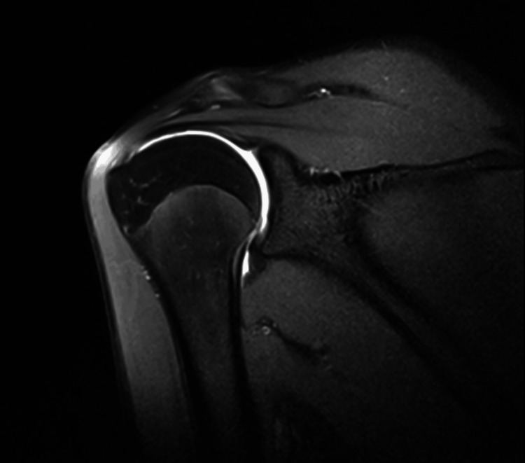 Figure 1
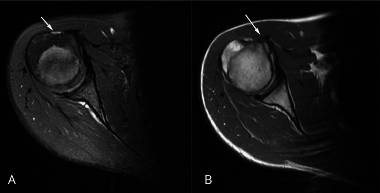 Figure 2
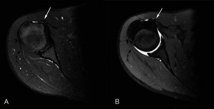 Figure 3
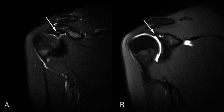 Figure 4
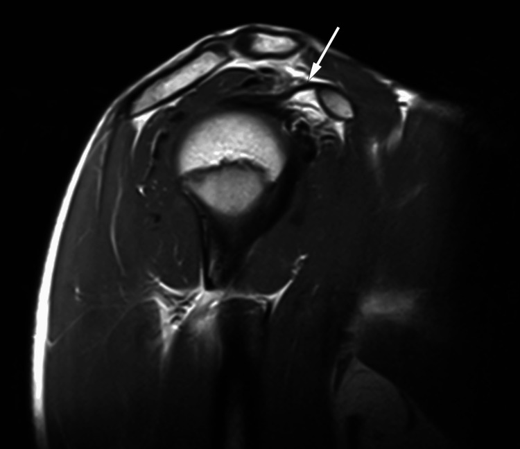 Figure 5
Peer Reviews
No public reviews on file for this paper yet. If you reviewed it on a platform where reviews are public (OpenReview, ICLR, NeurIPS, ICML), you can paste yours below so the community can read it here.
Videos
No videos yet. Explain this paper in a talk, walkthrough, or lecture? Add one.
Taxonomy
TopicsShoulder Injury and Treatment · Elbow and Forearm Trauma Treatment · Tendon Structure and Treatment
Introduction
The bicipital pulley system, formed primarily by the superior glenohumeral and coracohumeral ligaments and reinforced by adjacent fibers of the subscapularis and supraspinatus tendons, plays a key role in the stabilization of the long head of the biceps tendon as it exits the glenohumeral joint through the rotator interval [1-4]. Under normal conditions, the long head of the biceps tendon originates from the supraglenoid tubercle or superior labrum, courses intra-articularly, and then enters the bicipital groove. Medial displacement is biomechanically abnormal and is typically associated with injury to the stabilizing pulley structures [1,5-7]. Instability of the long head of the biceps tendon is uncommon, and medial subluxation or dislocation is most often described in adults with degenerative or traumatic rotator cuff pathology, frequently involving disruption of the pulley complex and or subscapularis tendon abnormalities [1,2,5-8]. In contrast, its occurrence in adolescents is exceptionally rare and has been reported only sporadically in isolated cases [8-11].
The gap in the literature becomes more evident when considering that most reported adolescent cases describe associated structural abnormalities of the subscapularis or the pulley system. The present report is unusual because it demonstrates an isolated extra-articular medial dislocation of the long head of the biceps tendon, with preserved integrity of the rotator cuff, rotator interval, and labro-bicipital complex on MR arthrography. Clinically, this entity may present with nonspecific anterior shoulder pain and preservation of range of motion, making imaging essential for diagnosis and for the exclusion of associated injuries [5-8]. In adolescents, acute rotational stress during recreational sports such as arm wrestling or repetitive overhead activity may contribute to instability even in the absence of obvious structural damage [9-13].
Case presentation
A 15-year-old male patient with no relevant medical history presented with anterior right shoulder pain. One year before the consultation, he had experienced an episode of acute shoulder pain during a recreational arm wrestling match. The pain resolved spontaneously, and no medical evaluation or treatment was sought at that time. The patient remained asymptomatic for several months following the incident. In recent months, after beginning to play paddle tennis occasionally and recreationally, involving repetitive overhead and rotational shoulder movements, he had developed sudden-onset sharp pain in the anterolateral aspect of the right shoulder during play. The pain occurred without direct trauma or radiation.
On physical examination, the anterior and anterolateral areas of the right shoulder were tender to palpation. There were no visible deformities or swelling, and no clinical signs of instability were observed. No neurovascular deficits were identified, and both active and passive ranges of motion remained intact. Given the nonspecific clinical presentation and the need to optimize evaluation of the labro-bicipital complex, rotator interval, and subtle patterns of instability, magnetic resonance imaging was performed as an MR arthrogram. The study employed spin-echo techniques, including T1-, T2-, and inversion recovery sequences acquired in axial, sagittal, and coronal planes, both before and after intra-articular contrast administration (Figure 1).
Coronal T1-weighted fat-suppressed MR arthrography image of the right shoulder demonstrating intra-articular contrast distribution with adequate capsular distension
Adequate capsular distension was achieved after contrast injection, allowing optimal evaluation of the labro-ligamentous structures. Imaging demonstrated extra-articular medial dislocation of the long head of the biceps tendon, superficial to the subscapularis tendon, with an empty bicipital groove (Figure 2).
Axial MRI images of the right shoulderA: proton density fat-suppressed sequence demonstrating an empty bicipital groove (arrow). B: axial T2-weighted fat-suppressed MR arthrogram demonstrates medial extra-articular displacement of the long head of the biceps tendon (LHBT) relative to the bicipital groove, with preservation of the normal morphology of the subscapularis tendon (arrow)
Along its intra-articular course, the tendon appeared thinned from its origin at the supraglenoid tubercle and superior labrum, without discontinuity or abnormal signal intensity suggestive of tearing. Axial proton density fat-suppressed and axial T1-weighted fat-suppressed MR arthrography images confirmed medial displacement of the tendon and its extra-articular position (Figure 3).
Axial MRI images of the right shoulderA: proton density fat-suppressed sequence demonstrating medial displacement of the long head of the biceps tendon outside the bicipital groove (arrow). B: axial T1-weighted fat-suppressed MR arthrography image with intra-articular contrast confirming extra-articular medial dislocation of the long head of the biceps tendon (arrow)
The rotator interval showed normal morphology and signal intensity. The rotator cuff tendons, including the subscapularis, supraspinatus, infraspinatus, and teres minor, demonstrated preserved structural integrity, with no evidence of tendinosis or partial- or full-thickness tears. The coracohumeral ligament demonstrated preserved integrity, without imaging evidence of rotator interval or bicipital pulley disruption (Figures 4, 5).
Coronal MRI images of the right shoulderA: proton density fat-suppressed sequence showing preserved integrity of the coracohumeral ligament and normal rotator interval anatomy. B: T1-weighted fat-suppressed MR arthrography image with intra-articular contrast confirming intact coracohumeral ligament, without evidence of rotator interval or bicipital pulley system injury
Sagittal proton density-weighted MRI of the right shoulderDemonstrating preserved integrity of the coracohumeral ligament, with normal appearance of the rotator interval and no evidence of bicipital pulley system injury
The glenoid labrum and glenohumeral ligaments were normal, with a sublabral recess identified as a normal anatomic variant. Bone marrow signal intensity was normal, without evidence of edema or osseous injury.
Based on the imaging findings, a diagnosis of isolated extra-articular medial dislocation of the long head of the biceps tendon was established. Conservative management with physical therapy focused on pain control and shoulder stabilization was initiated, along with recommendations regarding physical activity modification and injury prevention. The patient demonstrated favorable clinical evolution during follow-up under conservative management.
Discussion
Isolated instability of the long head of the biceps tendon is rare, as medial subluxation or dislocation is most commonly associated with tears of the subscapularis tendon and/or disruption of the biceps pulley system [1,2,5]. Most descriptions involve adult patients with rotator cuff pathology, whereas isolated medial dislocation without concurrent rotator cuff injury has been reported only in a limited number of cases [3,4]. The present case is unusual because an extra-articular medial dislocation occurred in an adolescent patient without detectable structural injury on MR arthrography to the rotator cuff, rotator interval, or labro-bicipital complex. Similar rare cases with intact rotator cuff tendons have been described in the literature, challenging the traditional notion that subscapularis disruption is required for biceps tendon instability [2,3]. Rather than reiterating the imaging findings, the main contribution of this report is to emphasize the importance of careful, systematic assessment of the bicipital groove and the entire course of the long head of the biceps tendon on MRI in young patients with anterior shoulder pain, even when classic associated injuries are absent [3,4,12].
The stabilizing function of the biceps pulley system, composed primarily of the coracohumeral and superior glenohumeral ligaments, has been described in anatomic and imaging studies [1,5,6]. However, MRI primarily assesses structural integrity rather than dynamic or functional competence. One possible explanation for isolated instability in the absence of structural disruption, particularly in adolescents, is transient, microstructural, or functional insufficiency of pulley components; this interpretation should be considered speculative. In this context, subtle microstructural damage or dynamic instability cannot be fully excluded and represents a limitation of static imaging techniques [4,12].
Sports-related mechanisms involving rotational and eccentric loading of the shoulder, such as arm wrestling and repetitive overhead activities, have been linked to a variety of shoulder injuries even without direct trauma [11-13]. In the present case, the initial arm wrestling episode may have predisposed the tendon to instability during subsequent overhead activity. From a clinical perspective, the choice between conservative and surgical management depends on symptoms, associated injuries, and functional impact. In selected patients without associated structural damage, conservative management may be an appropriate initial approach, as demonstrated in this case. The main limitation of this report is its single-case nature, which limits generalization of diagnostic or management recommendations.
Conclusions
Isolated extra-articular medial dislocation of the long head of the biceps tendon is a rare entity, particularly in adolescent patients. This report highlights the importance of a systematic evaluation of the bicipital groove and the entire course of the long head of the biceps tendon on MRI in young patients presenting with anterior shoulder pain, even when associated rotator cuff or pulley injuries are absent. In selected adolescent patients without demonstrable structural damage, conservative management may be an appropriate initial approach. Given the single-case nature of this report, clinical inferences and management recommendations should be interpreted with caution.
The reference list from the paper itself. Each links out to its DOI / PubMed record.
- 1Subluxations and dislocations of the tendon of the long head of the biceps J Shoulder Elbow Surg Walch G Nové-Josserand L Boileau P Levigne C 10010871998959308610.1016/s 1058-2746(98)90218-x · doi ↗ · pubmed ↗
- 2Dislocation of the long head of the biceps tendon with intact subscapularis and supraspinatus tendons J Shoulder Elbow Surg Gambill ML Mologne TS Provencher MT 0215200610.1016/j.jse.2005.09.00817126231 · doi ↗ · pubmed ↗
- 3Isolated medial dislocation of the long head of the biceps without rotator cuff tear: a case report J Korean Orthop Assoc Cho CH Lee KJ Bae KC 655658432008
- 4Medial dislocation of the long head of the biceps without concomitant subscapularis tear: a case report J Orthop Case Rep Vopat ML Yang SY Gregor CM Kallail KJ Saunders BM 610920203254801810.13107/jocr.2019.v 09.i 06.1564 PMC 7276595 · doi ↗ · pubmed ↗
- 5Anterior and posterior instability of the long head of the biceps tendon in rotator cuff tears: a new classification based on arthroscopic observations Arthroscopy Lafosse L Reiland Y Baier GP Toussaint B Jost B 73802320071721043010.1016/j.arthro.2006.08.025 · doi ↗ · pubmed ↗
- 6Instability of the long head of the biceps tendon in patients with rotator cuff tear: evaluation on magnetic resonance arthrography of the shoulder with arthroscopic correlation Skeletal Radiol Kang Y Lee JW Ahn JM Lee E Kang HS 133513424620172852838510.1007/s 00256-017-2669-7 · doi ↗ · pubmed ↗
- 7Hidden long head of the biceps tendon instability and concealed intratendinous subscapularis tears Orthop J Sports Med Chae SH Jung TW Lee SH Kim MJ Park SM Jung JY Yoo JC 468202010.1177/2325967119898123 PMC 733349932656285 · doi ↗ · pubmed ↗
- 8Detour sign in the diagnosis of subluxation of the long head of the biceps tendon with arthroscopic correlation Br J Radiol Khil EK Cha JG Yi JS Kim HJ Min KD Yoon YC Jeon CH 201603759020172788585010.1259/bjr.20160375 PMC 5685103 · doi ↗ · pubmed ↗