Commentary to: Lymphocytic UV autofluorescence: A novel UV-induced fluorescence dermoscopy finding in lichen nitidus: A series of two cases
Paweł Pietkiewicz, Marian Voloshynovych, Julien Anriot, Cristian Navarrete-Dechent

Abstract
Genes, proteins, chemicals, diseases, species, mutations and cell lines named across the full text — each resolved to its canonical identifier and authoritative record.
Click any figure to enlarge with its caption.
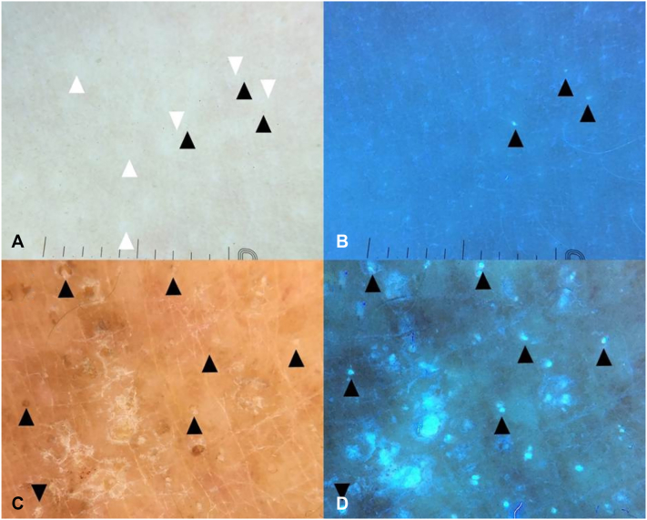 Figure 1
Peer Reviews
No public reviews on file for this paper yet. If you reviewed it on a platform where reviews are public (OpenReview, ICLR, NeurIPS, ICML), you can paste yours below so the community can read it here.
Videos
No videos yet. Explain this paper in a talk, walkthrough, or lecture? Add one.
Taxonomy
TopicsOral Health Pathology and Treatment · Cutaneous lymphoproliferative disorders research · Autoimmune Bullous Skin Diseases
To the Editor: We read with interest the case report by Varun et al1 published in the November issue of JAAD Case Reports, describing lymphocytic UV autofluorescence in generalized lichen nitidus. The authors correlated clinical, dermatoscopic, UV-induced fluorescence dermoscopy (UVFD), and histopathologic findings in 2 pediatric cases. Both demonstrated well-defined hypopigmented clods with occasional peripheral scale and brownish dots that exhibited blue-white fluorescence under UVFD. The authors attributed this fluorescence to autofluorescence of the dermal lymphocytic infiltrate, referencing studies using fluorescence microscopy,2 spectrophotometric microscopy,3 and flow cytometry.4
Although the proposed mechanism is intriguing, we note that other conditions with dense lymphocytic infiltrates—such as lichen planus or folliculotropic mycosis fungoides—do not typically display blue fluorescence on UVFD in our experience. We, therefore, suggest considering an alternative fluorophore that was not discussed: subtle serous crusts rich in bilirubin. Brown dots may represent healing erosions that produce blue-to-green fluorescence depending on their stage and sun exposure.5 Such subtle erosions may result from intentional scratching or friction over lichen nitidus papules by both young patients, leading to minimal exudation that can be inconspicuous on conventional dermoscopy but nonetheless fluoresces under UVFD (Fig 1).Fig 1A, Polarized dermoscopy of a localized lichen nitidus in a young boy featuring subtle tan clods (serous erosions; black arrowheads) occasionally superimposed over white clods of parakeratosis/acanthosis (white arrowheads). B, UV-induced fluorescence dermoscopy of lichen nitidus demonstrates bright bluish clods corresponding to serous erosions (black arrowheads). C, Polarized dermoscopy of eczematous dermatitis in elderly woman featuring brown, orange and tan serokeratotic crusts (black arrowheads). D, UV-induced fluorescence dermoscopy of eczematous dermatitis in elderly woman demonstrating blue-greenish fluorescence of the areas corresponding to serokeratotic crusts (black arrowheads).
We commend the authors for highlighting these uncommon presentations and hope this additional consideration supports future interpretation of UVFD features in lichen nitidus and related dermatoses.
Sincerely,
Paweł Pietkiewicz (on behalf of all authors)
Conflicts of interest
None disclosed.
The reference list from the paper itself. Each links out to its DOI / PubMed record.
- 1Varun H, Singh A, Madke B, Chandak M, Nitya VN, Vadera H. Lymphocytic UV autofluorescence: a novel ultraviolet-induced fluorescence dermoscopy finding in lichen nitidus – a series of two cases. JAAD Case Rep. 2026; 67;178-182. https://doi.org/10.1016/j.jdcr.2025.10.06410.1016/j.jdcr.2025.10.064PMC 1274429541466615 · doi ↗ · pubmed ↗
- 2Hawkins E.P.Hawkins H.K.Armstrong D.Lymphocyte autofluorescence: a screening procedure for neurodegenerative diseases Pediatr Neurol 231986160166285473910.1016/0887-8994(86)90010-x · doi ↗ · pubmed ↗
- 3Monici M.Cell and tissue autofluorescence research and diagnostic applications Biotechnol Annu Rev 1120052272561621677910.1016/S 1387-2656(05)11007-2 · doi ↗ · pubmed ↗
- 4More S.Shastri M.Kotru M.Gupta M.Autofluorescence in flow cytometry: a diagnostic conundrum secondary to drugs J Microsc Ultrastruct 2023 https://journals.lww.com/jmcu/abstract/9000/autofluorescence_in_flow_cytometry__a_diagnostic.99949.aspx
- 5Pietkiewicz P.Navarrete-Dechent C.Togawa Y.Applications of ultraviolet and sub-ultraviolet dermatoscopy in neoplastic and non-neoplastic dermatoses: a systematic review Dermatol Ther 142202436139010.1007/s 13555-024-01104-4PMC 1089099038358617 · doi ↗ · pubmed ↗