Increased atherosclerosis and expression of inflammarafts in macrophage foam cells in AIBP-deficient mice
Shenglin Li, Nicolaus Nazarenkov, Elena Alekseeva, Soo-Ho Choi, Juliana Maria Navia-Pelaez, Aakash Patel, Patrick Secrest, Philip L.S.M. Gordts, Sven Heinz, Yury I. Miller

TL;DR
AIBP deficiency in mice leads to more severe atherosclerosis and increased inflammation in macrophage foam cells.
Contribution
This study reveals that AIBP deficiency alters foam cell characteristics and promotes advanced atherosclerosis.
Findings
AIBP-deficient mice had more lipid-laden foam cells and larger atherosclerotic lesions.
AIBP deficiency increased inflammaraft markers like TLR4 dimers in foam cells.
AIBP-deficient macrophages showed higher expression of inflammation-related genes.
Abstract
Atherosclerotic lesions comprise different populations of macrophages, including lipid-laden macrophage foam cells and non-foamy, inflammatory macrophages, which play distinct roles in disease progression. Non-foamy macrophages express higher levels of inflammarafts – enlarged, cholesterol-rich lipid rafts hosting assemblies of inflammatory receptors – compared to foam cells in atherosclerotic lesions of Ldlr−/− mice. Apolipoprotein A-I binding protein (AIBP) has been shown to control lipid raft dynamics. This study investigated the effect of systemic AIBP deficiency on inflammaraft expression in foam cells and non-foamy macrophages in atherosclerotic lesions of hypercholesterolemic mice. A larger number of foam cells, with increased neutral lipid accumulation, populated atherosclerotic lesions in Apoa1bp−/−Ldlr−/− mice compared to Ldlr−/− mice. Importantly, AIBP-deficient foam cells…
Genes, proteins, chemicals, diseases, species, mutations and cell lines named across the full text — each resolved to its canonical identifier and authoritative record.
Click any figure to enlarge with its caption.
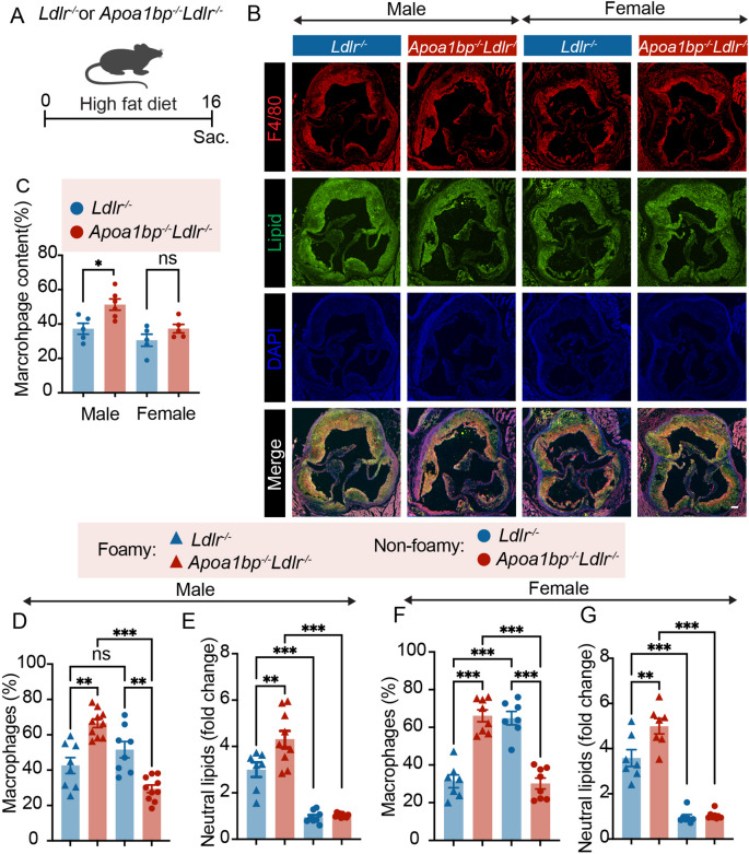 Figure 1
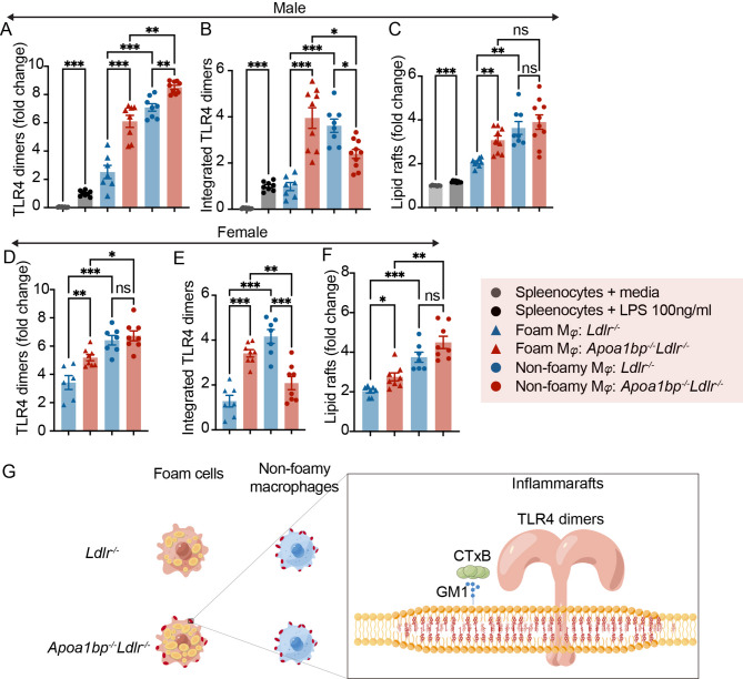 Figure 2
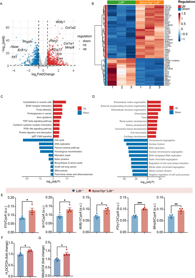 Figure 3
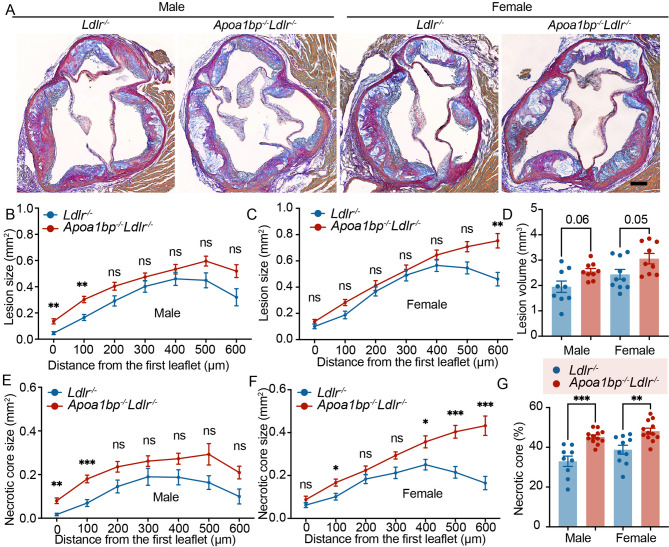 Figure 4
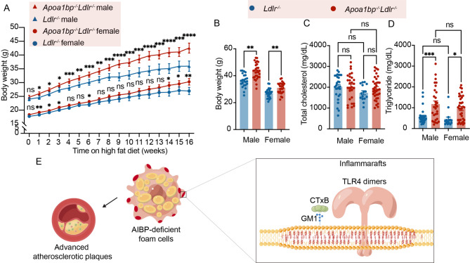 Figure 5
- —NIH
Peer Reviews
No public reviews on file for this paper yet. If you reviewed it on a platform where reviews are public (OpenReview, ICLR, NeurIPS, ICML), you can paste yours below so the community can read it here.
Videos
No videos yet. Explain this paper in a talk, walkthrough, or lecture? Add one.
Taxonomy
TopicsCholesterol and Lipid Metabolism · Inflammasome and immune disorders · Atherosclerosis and Cardiovascular Diseases
Introduction
The advances in flow cytometry and sequencing techniques have helped identify distinct populations of macrophages in human and mouse atherosclerotic lesions^1,2^. One differentiating feature in lesional macrophages is the handling of excess cholesterol. Lipid-laden macrophage foam cells accumulate large deposits of esterified cholesterol in lipid droplets. In contrast, non-foamy macrophages display increased levels of unesterified cholesterol-rich lipid rafts in the plasma membrane. Lipid rafts, due to low diffusion rates and the presence of cholesterol and sphingolipid binding domains in many proteins, serve as a platform for the assembly of functional receptor complexes^3^. Enlarged lipid rafts that host assembled complexes of inflammatory receptors have been designated as inflammarafts^4^. In atherosclerotic lesions, non-foamy macrophages, which express inflammatory genes^1^, retain high levels of inflammarafts, particularly inflammarafts rich in Toll-like receptor (TLR) complexes^3^. In contrast, macrophage foam cells express lipid storage genes, have low expression of inflammatory genes^1^ and lower abundance of inflammarafts^3^. However, a recent single-cell RNA-seq study identified in human carotid lesions a population of macrophages expressing high levels of PLIN2, a lipid droplet protein, and TLR-driven inflammatory genes^5^. The mechanisms regulating the emergence of inflammatory lipid-laden macrophages remain to be fully elucidated.
One possible mechanism may involve the function of apolipoprotein A-I binding protein (AIBP, gene name Apoa1bp or Naxe), which facilitates cholesterol efflux by binding to apoA-I and stabilizing ATP-binding cassette transporter A1 (ABCA1)^6,7^. The AIBP-mediated removal of cholesterol from the plasma membrane reduces the abundance of inflammarafts^7^. AIBP is ubiquitously expressed and can be secreted in response to apoA-I or oxidized LDL (OxLDL)^8,10^. Human and mouse atherosclerotic lesions express high levels of AIBP protein^9,10^. Apoa1bp^−/−^ mice fed a Western-type diet exhibit rapid weight gain and impaired glucose tolerance, hallmarks of metabolic disease^11^. Intracellular AIBP localizes to mitochondria and regulates mitophagy^9,10^. Apoa1bp^−/−^Ldlr^−/−^ mice fed a high-cholesterol, high-fat diet have more atherosclerotic lesions compared to Ldlr^−/−^ mice^11^. In mouse models of neuropathic pain and Alzheimer’s disease, Apoa1bp^−/−^ mice have chronic, increased expression of inflammarafts in spinal cord or brain microglia, respectively, accompanied by oxidative stress and mitochondrial dysfunction^12,13^.
Here, we demonstrate that systemic AIBP deficiency in hyperlipidemic mice is associated with a phenotype shift of macrophage foam cells, characterized by increased lipid accumulation and increased expression of inflammarafts, as well as with larger necrotic cores in atherosclerotic lesions.
Results
Increased inflammarafts in lesional macrophage foam cells in AIBP-deficient mice
In cross-sections of the aortic root of Apoa1bp^−/−^Ldlr^−/−^ and Ldlr^−/−^ mice fed a 16-week high-fat diet (Fig. 1A), AIBP-deficient male mice showed a higher percentage of F4/80^+^ area within the LipidTOX^+^ neutral lipid positive lesion area, than Ldlr^−/−^ male mice (Fig. 1B and C, Supplemental Figure S1), indicating that more macrophages populated the atherosclerotic lesions in AIBP-deficient mice. We then measured by flow cytometry the percentages of foamy and non-foamy macrophages in lesions from the entire aorta. Aortic single-cell suspensions were gated for BODIPY^high^ (foamy) and BODIPY^low^ (non-foamy), CD45^+^F4/80^+^ macrophages (Supplemental Figures S2 and S3). The proportion of macrophage foam cells was significantly increased in Apoa1bp^−/−^Ldlr^−/−^ mice (foamy/non-foamy = 7:3) when compared to Ldlr^−/−^ mice (4:6) (Fig. 1D and F). The macrophage foam cells from Apoa1bp^−/−^Ldlr^−/−^ mice had more neutral lipid accumulated, measured by the BODIPY intensity, than Ldlr^−/−^ foam cells (Fig. 1E and G).
Fig. 1. Higher numbers of foam cells with higher levels of neutral lipids in AIBP-deficient mice. A Experimental design of a high-fat diet feeding experiment. **B **Representative images of immunostaining of aortic root. The representative sections were at the same distance from the aortic valve origin for each group. Scale bar = 100 μm. C Quantitative analysis of F4/80^+^ area in **B, **D and E. Data from male mice (n = 8–10). F and G Data from female mice (n = 7–8). D/F Percentages of foam cells and non-foamy macrophages in atherosclerotic lesions from Apoa1bp^-/-^Ldlr^-/-^ and Ldlr^-/-^ mice. E/G Relative content of neutral lipids in foam cells based on BODIPY fluorescence intensity; BODIPY readings in non-foamy macrophages from the same aorta were set to 1. One-way ANOVA with Tukey’s. Mean ± SEM; ns, nonsignificant; *, p ≤ 0.05; **, p ≤ 0.01; ***, p ≤ 0.001.
We then measured the degree of TLR4 dimerization in lesional macrophages as a key parameter of inflammaraft expression. The expression of TLR4 dimers in aortic macrophages was significantly higher than that in splenic macrophages stimulated with lipopolysaccharide (LPS), used as an intra-assay positive control (Fig. 2A). Further, macrophage foam cells from both male and female Apoa1bp^−/−^Ldlr^−/−^ mice expressed significantly higher TLR4 dimers, when compared to those from corresponding Ldlr^−/−^ mice (Fig. 2A and D). TLR4 dimers in Apoa1bp^−/−^Ldlr^−/−^ non-foamy macrophages were higher in male but not female mice (Fig. 2A and D). The difference was further exacerbated when TLR4 dimers were integrated over the number (percentage) of each macrophage population, showing that macrophage foam cells from both Apoa1bp^−/−^Ldlr^−/−^ male and female mice were the predominant population expressing TLR4 dimers (Fig. 2B and E). In contrast, in Ldlr^−/−^ mice, the major TLR4 dimer-expressing cells were non-foamy macrophages (Fig. 2B and E). Furthermore, Apoa1bp^−/−^Ldlr^−/−^ macrophage foam cells exhibited increased levels of lipid rafts, equal to the lipid raft levels in non-foamy macrophages in both Apoa1bp^−/−^Ldlr^−/−^ and Ldlr^−/−^ male mice (Fig. 2C and F). These results indicate that AIBP deficiency leads to increased inflammaraft expression in macrophage foam cells, making them the major inflammaraft-expressing subpopulation in atherosclerotic plaques in AIBP-deficient mice (Fig. 2G).
Fig. 2. Foam cells are the major macrophage population expressing TLR4 inflammarafts in AIBP-deficient mice. A-C Data from male mice (n = 8–10). D-F Data from female mice (n = 7–8). A/D Expression of TLR4 dimers in foam cells and non-foamy macrophages in Apoa1bp^-/-^Ldlr^-/-^ and Ldlr^-/-^ mice. Splenocytes stimulated with vehicle or LPS (100 ng/ml, 15 min) served as negative and positive controls in A-F. B/E Integrated TLR4 dimers (TLR4 dimers in each cell type in A/D multiplied by cell type percentage in Fig. 1D and F. C/F Expression of lipid rafts in lesional macrophages. G Illustration of a foam cell phenotype in Apoa1bp^-/-^Ldlr^-/-^ mice characterized by higher lipid accumulation and increased expression of TLR4 inflammarafts. GM1: monosialotetrahexosylganglioside. CTxB: cholera toxin subunit B. One-way ANOVA with Tukey’s. Mean ± SEM; ns, nonsignificant; *, p ≤ 0.05; **, p ≤ 0.01; ***, p ≤ 0.001.
To model macrophage foam cells, we incubated bone marrow-derived macrophages (BMDM), obtained from Apoa1bp^−/−^Ldlr^−/−^ or Ldlr^−/−^ mice, with 20 µg/mL OxLDL for 24 h and subjected them to RNA-seq analysis. BMDMs incubated with OxLDL were confirmed to accumulate neutral lipid (Supplemental Figure S4). A total of 604 genes were upregulated, and 660 genes were downregulated in OxLDL-treated Apoa1bp^−/−^Ldlr^−/−^ BMDMs compared to those from Ldlr^−/−^ mice (Fig. 3A). Among the top differentially expressed genes were Wdfy1, suggesting a predominant TRIF signaling downstream of TLR4^14^, P2ry12, a major purinergic receptor in macrophages mediating vascular inflammation^15^, Mmp9 and several genes encoding extracellular matrix proteins, suggesting vulnerable lesion remodeling^16^, as well as Il1b, a proinflammatory cytokine downstream of Tlr4, and its receptor Il1r1 (Fig. 3B). KEGG and GO pathway and enrichment analyses showed upregulated extracellular matrix organization, chemotaxis, and focal adhesion (Fig. 3C and D). We confirmed the increased expression of Wdfy1, P2ry12, Il1b, Il1r1, and Mmp9 with real-time qPCR (Fig. 3E).
Fig. 3. Transcriptional changes in foamy macrophage cell models of Apoa1bp^-/-^Ldlr^-/-^ and Ldlr^-/-^ mice. Bone marrow-derived macrophages (BMDM) from Apoa1bp^-/-^Ldlr^-/-^ and Ldlr^-/-^ male mice were incubated with 20 µg/mL OxLDL for 24 h to mimic macrophage foam cells. A Volcano plot. B Heatmap showing the top 50 differentially expressed genes selected based on a combined rank of adjusted p-value (ascending) and absolute log2 fold-change (descending).C KEGG and D GO Enrichment analysis showing the top 10 upregulated and downregulated pathways. E-F In a separate set of experiments, BMDMs from Apoa1bp^-/-^ and WT mice were incubated with 20 µg/mL OxLDL for 24 h and subjected to qPCR analysis of selected genes expression (E), or flow cytometry detection of oxidative stress as measured with H_2_DCFDA, a general reactive oxidative species (ROS) probe (F), or MitoSOX, a mitochondrial ROS probe (G). Mean ± SEM; t-test, n = 4 biological replicates; *, p ≤ 0.05; **, p ≤ ≦ 0.01.
We next tested whether the AIBP-deficiency in OxLDL-stimulated BMDMs was accompanied by increased oxidative stress. We incubated cells with 2’,7’-dichlorodihydrofluorescein diacetate (H_2_DCFDA), a general reactive oxidative species (ROS) probe, or MitoSOX, a mitochondrial ROS probe. We found that AIBP-deficient BMDMs showed higher intensity of H_2_DCFDA (Fig. 3F) and MitoSOX (Fig. 3G) than control cells, indicating increased oxidative stress in general and the oxidative stress associated with mitochondrial dysfunction in particular in AIBP-deficient foam cells.
Advanced atherosclerosis in AIBP-deficient mice
To assess the extent of atherosclerosis, consecutive cross-sections were collected from the aortic root for quantification. Larger atherosclerotic lesions were observed in the proximal region of the aortic valve in Apoa1bp^−/−^Ldlr^−/−^ male mice when compared to Ldlr^−/−^ mice (Fig. 4A and B). AIBP-deficient female mice displayed a larger distal (500 to 600 μm from valve origin) lesion size (Fig. 4A and C). The analysis of total lesion volume indicated a tendency for increased atherosclerotic plaque burden (Fig. 4D). We observed sex differences in the atherosclerotic lesion morphology in Apoa1bp^−/−^Ldlr^−/−^ mice (Fig. 4B and C). In Ldlr^−/−^ mice, the lesion size curves in both males and females were bell-shaped, peaking at 400 μm from the aortic root. In Apoa1bp^−/−^Ldlr^−/−^ mice, the lesion size curve in female mice continuously ascended all the way from the valve origin to the 600 μm distance, while the curve in male mice remained bell-shaped, peaking at 500 μm.
Fig. 4. Advanced atherosclerotic lesions in AIBP-deficient mice. A Representative images of the aortic root stained with modified Van Gieson staining. The representative sections were at the same distance from the aortic valve origin for each group. Scale bar = 100 μm. Quantitative analysis of lesion size in male mice (B) and female mice (C). D Lesion volume measured by area under the curve from B and C. Quantitative analysis of necrotic core size in male mice (E) and female mice (F). G Quantitative analysis of necrotic core percentage over the total lesion area. N = 9–10; two-way ANOVA with Tukey’s or Dunnett’s multiple comparison tests (B, C, E, F), and t-test (D and G). Mean ± SEM; ns, nonsignificant; *, p ≤ 0.05; **, p ≤ ≦ 0.01; ***, p ≤ 0.001.
Formation and enlargement of a necrotic core imply plaque destabilization, associated with vascular inflammation, and indicate advanced atherosclerotic plaques^17,18^. Consistent with the lesion size measurements, a larger necrotic core size was observed in the proximal region of the aortic valve in Apoa1bp^−/−^Ldlr^−/−^ male mice (Fig. 4A and E) and at the distal region of the aortic valve in Apoa1bp^−/−^Ldlr^−/−^ female mice (Fig. 4A and F) when compared to Ldlr^−/−^ mice. Of note, the percentage of necrotic core in lesion area was significantly higher in Apoa1bp^−/−^Ldlr^−/−^ mice than in Ldlr^−/−^ mice, in both male and female, suggesting a more vulnerable advanced plaque in AIBP-deficient mice (Fig. 4G).
Apoa1bp^−/−^Ldlr^−/−^ mice gained body weight faster than Ldlr^−/−^ mice (Fig. 5A and B) and had higher plasma triglyceride but not cholesterol levels (Fig. 5C and D). Overall, male and female mice showed similar results. These results indicate that AIBP deficiency affects lipid homeostasis and leads to a larger lesion burden and advanced atherosclerotic plaques.
Fig. 5. Weight gain and plasma lipid levels. A Body weight gain of Apoa1bp^-/-^Ldlr^-/-^ and Ldlr^-/-^ mice during HFD feeding. B End point body weight. **C **Plasma total cholesterol levels. D Plasma triglyceride levels. E Diagram illustrating this paper’s findings: AIBP deficiency leads to marked expression of inflammarafts and increased lipid accumulation in lesional macrophage foam cells. Advanced atherosclerotic plaques accompany the transition of foam cells to an inflammaraft-expressing phenotype. N = 23–29; two-way and one-way ANOVA with Tukey’s or Dunnett’s multiple comparison tests for statistical analysis. Results are presented as Mean ± SEM; ns, nonsignificant; *, p ≤ 0.05; **, p ≤ 0.01; ***, p ≤ 0.001.
Discussion
In this study, we demonstrated that the atheroprotective effect of AIBP is strongly associated with the attenuation of inflammarafts in macrophage foam cells. AIBP deficiency leads to marked expression of inflammarafts and increased lipid accumulation in lesional macrophage foam cells. Advanced atherosclerotic plaques accompanied the transition of foam cells to an inflammaraft-expressing phenotype.
Multiple lines of evidence support the concept that macrophage foam cells express lipid storage genes but exhibit suppressed expression of inflammatory-response genes, leading to the reduced contribution of foam cells to inflammation in atherosclerosis compared to non-foamy macrophages. Macrophage foam cells from Ldlr^−/−^ mice exhibit altered cholesterol metabolism and a marked suppression of proinflammatory mediators that are normally characteristic of the inflammatory responses associated with atherosclerotic lesions^19^. Similarly, transcriptome data from Apoe^−/−^ mice show that lipid-rich macrophage foam cells express few inflammatory genes but many lipid-processing genes, while non-foamy macrophages are the primary population expressing IL-1β and other inflammatory mediators in the atherosclerotic aorta^1^. Moreover, we previously established that non-foamy macrophages in atherosclerotic lesions of Ldlr^−/−^ mice express higher levels of inflammarafts than macrophage foam cells, correlating with increased plasma levels of inflammatory cytokines such as monocyte chemoattractant protein-1 (MCP-1), interleukin (IL)−6, and IL-1β^3^.
Despite the evidence for a non-inflammatory character of macrophage foam cells, the mechanisms by which macrophage foam cells in atherosclerotic lesions exhibit less inflammatory response than other macrophage populations remain poorly understood. The most notable difference between foam cells and non-foamy macrophages is the content of intracellular lipid droplets, as well as distinct lipid metabolism. High levels of desmosterol, the last intermediate in the Bloch pathway of cholesterol biosynthesis, have been reported to be associated with suppression of proinflammatory mediators in macrophage foam cells via several homeostatic pathways, including activation of LXR target genes, inhibition of SREBP target genes, and selective reprogramming of fatty acid metabolism^19^. These findings indicate that lipid metabolism is involved in the suppression of inflammatory mediators. On the other hand, a recent publication found the TLR-dependent pathogenic macrophage transition to an inflammatory lipid-associated phenotype in human intraplaque immune cell communities^5^.
AIBP not only facilitates cholesterol efflux from the cell membrane to regulate lipid metabolism, but AIBP-mediated disruption of lipid rafts also protects microglia from inflammaraft-associated oxidative stress and mitochondrial dysfunction^12^. In this study, we demonstrated that AIBP-deficient macrophage foam cells also have increased oxidative stress and increased levels of mitochondria-associated ROS. In our study, we observed that AIBP deficiency leads to enhanced expression of TLR4 dimers and lipid rafts in macrophage foam cells, as well as increased lipid accumulation in lesional macrophages, resulting in macrophage foam cells being the major inflammaraft-expressing macrophage subpopulation in atherosclerotic lesions. The inflammaraft-expressing phenotype of foam cells was accompanied by advanced atherosclerotic plaques. In this study, we examined gene expression in AIBP-deficient and wild-type foam cells using BMDMs incubated with OxLDL. This cellular model only partially replicates the in vivo environment surrounding vascular macrophages in mice with systemic AIBP expression or deficiency, where AIBP is secreted by macrophages and has an autocrine effect^9,10^ and also secreted by other cell types for a paracrine effect. In fact, the choice of a systemic Apoa1bp^−/−^Ldlr^−/−^ mouse for this study was dictated by the secreted nature of ubiquitously expressed AIBP, which exerts its major effect on lipid rafts extracellularly. Among the upregulated genes in AIBP-deficient BMDMs were the genes broadly associated with atherosclerosis progression, including the pathways not directly downstream of TLR4 signaling. These findings suggest the assembly of other receptor complexes in inflammarafts, as we reported earlier^3^. So far, flow cytometry-based detection of TLR4 dimers and lipid rafts is the most robust measure of inflammarafts, which host other, diverse receptor assemblies. Development of more comprehensive methods to characterize inflammarafts in macrophages and other vascular cells will advance the understanding of the inflammatory milieu in atherosclerosis.
Interestingly, our analysis of aortic root lesions showed a sex difference in lesion distribution in response to AIBP deficiency. Female Apoa1bp^−/−^Ldlr^−/−^ mice exhibited a continuous increase in lesion size and necrotic core size starting from the origin of the aortic valve, while lesions in male Apoa1bp^−/−^Ldlr^−/−^ mice and both sexes of Ldlr^−/−^ mice reached the maximum size at 400–500 μm from the origin. These findings indicate that AIBP contributes to the sex difference observed in atherosclerosis^20,21^. Future studies will investigate whether this might be due to sex-specific differences in shear stress response, hormonal modulation of AIBP-mediated cholesterol efflux, or other factors.
In our previous study with Apoa1bp^−/−^Ldlr^−/−^ mice, we observed elevated plasma cholesterol and triglyceride levels^11^, in contrast to the results of this work, which showed increased triglyceride but not cholesterol plasma levels. The likely reason for this difference is the difference in the high-fat diets used in these two studies. The Western diet used in the previous study contained 1.25% cholesterol^11^, which is higher than the 0.2% cholesterol diet in this study. This study did not investigate if increased triglyceride levels may have accounted for the increased size of necrotic core in atherosclerotic lesions. Increased triglycerides is a viable alternative to macrophage inflammarafts as a primary cause for necrotic lesions. Another limitation of the present study is that the analyses were conducted at one time point, 16 weeks after the start of diet intervention. Without earlier time points, it is unclear whether the inflammaraft-expressing foam cell phenotype represents a stable identity or a transitional stage from non-foamy to foamy macrophages.
The major finding of this study – that AIBP suppresses inflammaraft expression specifically in macrophage foam cells – warrants a detailed, genome-wide analysis of gene expression in foamy and non-foamy macrophages from atherosclerotic lesions of Apoa1bp^−/−^Ldlr^−/−^ and Ldlr^−/−^ mice. Future studies will help elucidate the mechanisms by which AIBP regulates inflammarafts and the signaling pathways in macrophage foam cells that prevent them from assuming an inflammatory phenotype.
Methods
Study approval
All experiments involving animals were conducted according to a protocol approved by the Institutional Animal Care and Use Committee at the University of California, San Diego, and in compliance with the ARRIVE guidelines.
Animals
Apoa1bp ^-/-^ Ldlr ^-/-^ mice^11^ and Ldlr^-/-^ mice were bred in-house and housed at a maximum of five animals of the same sex per cage at room temperature and a 12:12 light-dark cycle. Age- and weight-matched mice were used. Both male and female mice were included. Starting at eight weeks of age, mice were fed a 16-week Western-style diet containing 42% kcal from fat and 0.2% cholesterol (TD.88137, Envigo Teklad). Mice were fasted for 8 h before euthanasia. Carbon dioxide inhalation in a closed chamber was used to induce euthanasia according to the American Veterinary Medical Association recommendations. Blood was drawn through a cardiac puncture and collected in EDTA-containing vials. Mice were then perfused with 10 ml ice-cold PBS containing 2 mM EDTA; aortae were dissected and prepared for flow cytometry analyses; hearts were fixed, dehydrated, and embedded in paraffin or O.C.T. Compound (4583, Sakura Finetek USA) for histological analysis.
Aorta single-cell suspension
The entire aorta from the aortic root to the iliac bifurcation (including ascending, arch, thoracic, and abdominal aorta) were used for flow cytometry analysis. Aortae were perfused with 10 mL cold PBS containing 2 mM EDTA. After removal of the surrounding fat and connective tissue, aortae were cut into several segments and digested in the aorta digestion enzyme solution^1^ [HBSS containing 250U/mL collagenase type XI (C7657, Sigma Aldrich), 120 U/mL hyaluronidase type I-s (H3506, Sigma Aldrich), 120 U/mL DNase I (DN25, Sigma Aldrich), and 450 U/mL collagenase type I (SCR103, Sigma Aldrich)] for 60 min at 37 °C. The digested aortae were then passed through a 70 µM cell strainer (25–376, Olympus Plastics), and cells were pelleted by centrifugation and resuspended in RPMI containing 0.5% Poloxamer 188 (24040032, ThermoFisher Scientific).
Flow cytometry-based detection of inflammarafts
Aortic single-cell suspensions were incubated at 37 °C for 30 min for cell surface recovery following enzymatic digestion and stained with Ghost Dye Red 780 Fixable Viability Dye (18452, Cell Signaling Technology). After neutral lipid staining with 1 µg/mL BODIPY 505/515 (D3921, Thermo Fisher Scientific), the single-cell suspensions were fixed with 4% PFA at 4 °C for 20 min. Cells were blocked with an anti-CD16/CD32 antibody (553142, BD Biosciences) in PBS containing 2% BSA on ice for 30 min and then incubated with a mixture of BV650-conjugated anti-CD45 antibody (103151, Biolegend), PerCP/Cy5.5-conjugated anti-F4/80 antibody (123128, Biolegend), AF594-Conjugated anti-Cholera Toxin Subunit B antibody (C22842, Invitrogen), APC-conjugated anti-TLR4 (145406, BioLegend; detecting total TLR4 expression) antibody, and PE-conjugated anti-TLR4/MD-2 (Invitrogen, 12–9924-81; detecting TLR4 monomers) antibody for 60 min on ice. Antibody-matched isotype controls were used to support antibody specificity. Cells were analyzed on a CytoFLEX Flow Cytometer (Beckman Coulter). Splenic F4/80-positive macrophages obtained from Ldlr^−/−^ mice fed a standard laboratory diet were treated for 15 min with 100 ng/ml LPS (to induce transient TLR4 dimerization) and served as a positive control for the TLR4 dimerization assay. Non-stimulated splenic macrophages served as a negative control to standardize data obtained over time. The expression of TLR4 dimers was calculated from geometric mean fluorescence intensities of PE-conjugated TLR4/MD2 antibody (monomers) and APC-conjugated TLR4 antibody (total) as the percentage of dimers of total TLR4. TLR4 dimers and lipid rafts were then normalized to those in LPS-treated splenic F4/80-positive macrophages in each experiment to account for the inter-day assay variation.
Immunofluorescence staining of the aortic root
Briefly, consecutive 10 μm-thick aortic cross-sections were collected with a Dakewe CT520 Research Cryostat. Sections were collected at 100 μm increments starting from the aortic valve origin, blocked with 3% FBS in PBS for 1 h at room temperature and incubated with rat anti-F4/80 antibody (MF48000, Invitrogen) at 4 °C overnight. The sections were then incubated with anti-rat secondary antibody (A78947, Invitrogen) at room temperature for 2 h, and counterstained with LipidTOX neutral lipid stain (H34476, ThermoFisher) for 30 min. Sections were mounted with DAPI-containing mounting media (P36931, ThermoFisher). No-primary antibody/no-LipidTox negative controls were imaged to account for auto-fluorescence (Supplemental Figure S1). Images were captured using a Leica Sp8 confocal microscope. Quantification of LipidTOX^+^ and F4/80^+^ area was analyzed on split channels, respectively, using the tracing function in ImageJ (Version 2.16.0)^22^.
LDL oxidation
OxLDL was produced in vitro as previously reported.^3^ Briefly, human native LDL (36010, Lee BioSolutions) was extensively dialyzed against PBS to remove EDTA, and 0.1 mg/mL of LDL was incubated with 10 µM CuSO_4_ for 18 h at 37 °C. Thiobarbituric acid reactive substances (typically, > 30 nmol/mg in OxLDL) were measured to confirm LDL oxidation. OxLDL was concentrated to 1 mg/mL using a 100 kDa cutoff centrifugal concentrator (UFC810024, Millipore) and sterile filtered (0.22 μm). Endotoxin contamination was tested using a Pierce Chromogenic Endotoxin Quant Kit (A39553, Thermo Fisher), and only LDL preparations with endotoxin levels below 0.025 EU/mg protein were used in this study.
Bone marrow-derived macrophages
BMDMs were cultured as previously described^3^. Briefly, bone marrow cells were isolated from femurs and tibias of 8-week-old Apoa1bp^−/−^Ldlr^11^ and Ldlr^−/−^ male mice and incubated in L929-conditioned medium for one week. Cells were then replated in 6-well plates at a density of 1 million cells per well for a 24-hour resting period. Cells were incubated with 20 µg/mL OxLDL in DMEM supplemented with 5% lipoprotein-deficient serum for 24 h. Total RNA for RNA-seq and qPCR analyses was extracted from OxLDL-stimulated BMDM using the NucleoSpin RNA purification kit (740955.250, MACHEREY-NAGEL). To validate lipid accumulation, BMDMs were cultured on cover slips, incubated for 24 h with 20 µg/mL OxLDL, stained with Oil Red O and counterstained with hematoxylin.
RNA-seq
RNA samples were quantified and qualified using a high-sensitivity RNA screen tape kit (5067–5579, Agilent Technologies). Libraries of RNA samples were prepared using a Stranded mRNA Prep kit (20040534, Illumina) and sequenced on a NovaSeq XPlus sequencer (Illumina). Sequencing reads were quality-checked by fastqc^23^, cleaned by trim-galore^24^, mapped to the GRCm39 mouse genome using STAR^25^, and counted using featureCounts^26^. Differential gene expression analysis was performed using the DESeq2 R package^27^. Raw read counts were normalized using the DESeq2, and differential expression between conditions was estimated with the Wald test, incorporating Benjamini–Hochberg correction to control the false discovery rate (FDR). Genes were considered significantly differentially expressed if they exhibited an adjusted p-value < 0.05 and an absolute log2 fold-change greater than log2(1.5). To prioritize genes with both strong statistical support and substantial effect size, significant genes were independently ranked by adjusted p-value (ascending) and by absolute log2 fold-change (descending). Genes were subsequently ordered according to a composite score of the sum of these two rank values.
qPCR
RNA concentration and quality were assessed using NanoDrop. Next, 1 µg of RNA was reversely transcribed to cDNA using RNA to cDNA EcoDry™ Premix kit (Takara, 639543) according to the manufacturers’ protocol. Real-time qPCR was carried out to determine gene expression. All reactions were performed in the Rotor-Gene Q cycler (Qiagen) in triplicate using 250 ng of cDNA, qPCR Master Mix (Azura Genomics, AZ-2301), and primers in a total reaction volume of 20 µl. Relative quantities of mRNA were calculated using the ΔΔCt formula and two standard curves relative quantitation using Rotor-Gene Q Software 1.7 (Qiagen) with cyclophilin A as the reference gene. Primers used for qPCR are listed in Supplemental Table T2.
Measurement of reactive oxygen species
Intracellular ROS in the BMDMs incubated with OxLDL was measured by incubating cells with 1 µM 2’,7’-dichlorodihydrofluorescein diacetate (H_2_DCFDA, Invitrogen, D399) at 37 °C for 30 min. In a separate panel, mitochondrial-specific ROS was measured by incubating cells with 1 µM MitoSOX (Invitrogen, M36008) at 37 °C for 30 min. Cells were analyzed with a CytoFLEX Flow Cytometer (Beckman Coulter). Unstained OxLDL-stimulated BMDMs were used as negative controls and for gating – to account for an increased autofluorescence of lipid-laden cells.
Quantification of atherosclerotic lesions
Hearts were fixed and embedded in paraffin for the assessment of atherosclerosis as described previously^3^. Briefly, consecutive 10 μm-thick aortic cross-sections were collected in 100 μm increments starting from the first appearance of the first leaflet of the aortic valve until the last leaflet. Sections were stained with modified Van Gieson staining to enhance the contrast between the intima and surrounding tissues. Images were acquired using a Nano Zoomer Slide Scanner. At each 100 μm increment, the lesion size and necrotic core size in each mouse were analyzed by computer-assisted morphometry (Image-Pro Plus 6.3, Media Cybernetics) by two investigators blinded to the study protocol. Lesion volumes were analyzed by calculating the area under the curve (AUC) in each mouse. Necrotic core percentages were calculated as necrotic core/lesion size at each distance from the valve origin.
Measurements of cholesterol and triglycerides
Mice were fasted for 8 h before euthanasia. Total cholesterol and triglyceride levels in plasma samples were quantified using a cholesterol/cholesteryl ester assay kit (ab65359, Abcam) and a triglyceride assay kit (ab65336, Abcam), respectively, according to the manufacturer’s protocol.
Statistical analyses
Data are presented as mean ± SEM. N represents biological replicates. Data were analyzed for normality and equal variance using the Kolmogorov-Smirnov test. A two-sided Student’s t-test was employed to compare two groups. For multiple-group comparisons with one or two independent factors, one-way or two-way ANOVA was conducted with Tukey’s or Dunnett’s multiple comparison tests for normally distributed variables. All statistical analyses were performed using GraphPad Prism version 9.4.1. A p-value of less than 0.05 was considered statistically significant.
Supplementary Information
Below is the link to the electronic supplementary material.
Supplementary Material 1
Supplementary Material 2
Supplementary Material 3
Supplementary Material 4
Supplementary Material 5
Supplementary Material 6
Supplementary Material 7
The reference list from the paper itself. Each links out to its DOI / PubMed record.
- 1Navia-Pelaez, J. M. et al. Differential Expression of Inflammarafts in Macrophage Foam Cells and in Nonfoamy Macrophages in Atherosclerotic Lesions—Brief Report. Arteriosclerosis, Thrombosis, and Vascular Biology 43, 323–329 (2023). 10.1161/ATVBAHA.122.31800610.1161/ATVBAHA.122.318006 PMC 987714936453276 · doi ↗ · pubmed ↗
- 2Andrews, S. & Fast QC: A Quality Control Tool for High Throughput Sequence Data. (2010). http://www.bioinformatics.babraham.ac.uk/projects/fastqc/