In vitro evaluation of PEI-coated microbubble– neutrophil conjugates for ultrasound-guided cell delivery
Hossein Razmi Bagtash, Roshni Gandhi, Ghazal Rastegar, Amine Azizi, Aparna Priyadarshani Jha, Shuai Shao, Emma Salari, Shashank R. Sirsi, Caroline N. Jones

TL;DR
This study explores using microbubble-coated immune cells for ultrasound-guided delivery to improve cancer immunotherapy effectiveness.
Contribution
The novelty lies in creating PEI-coated microbubble–neutrophil conjugates and evaluating their viability and migration under ultrasound.
Findings
PEI microbubble-conjugated dHL-60 cells retained over 88% viability four hours post-conjugation.
Cell migration rates were higher at 1:1 and 1:2 cell-to-microbubble ratios compared to higher ratios.
Ultrasound exposure did not significantly alter cell migration toward chemoattractants.
Abstract
Immunotherapies have advanced cancer treatment; however, their clinical efficacy remains limited for solid tumors due to challenges associated with effectively directing immune cells into the complex tumor microenvironment. Recent developments in Ultrasound Contrast Agent (UCA — also known as “microbubble”) technology have provided novel opportunities to enhance targeted therapeutic delivery. In this study, we introduce an innovative approach of leveraging microbubbles to enhance immune cell targeting by directly attaching microbubbles to immune cells, establishing an in vitro platform to evaluate microbubble–immune cell conjugation with potential applicability to ultrasound-guided delivery strategies. To create novel microbubble-immune cell conjugates, we created polyethyleneimine (PEI) coated microbubbles and attached them to differentiated HL-60 (dHL-60) cells. These positively…
Genes, proteins, chemicals, diseases, species, mutations and cell lines named across the full text — each resolved to its canonical identifier and authoritative record.
Click any figure to enlarge with its caption.
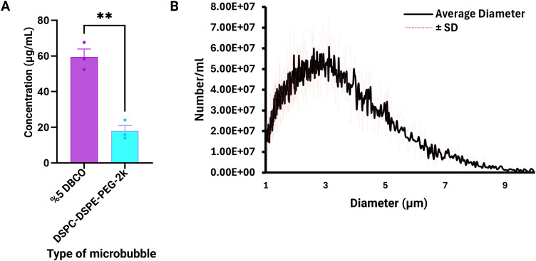 Figure 1
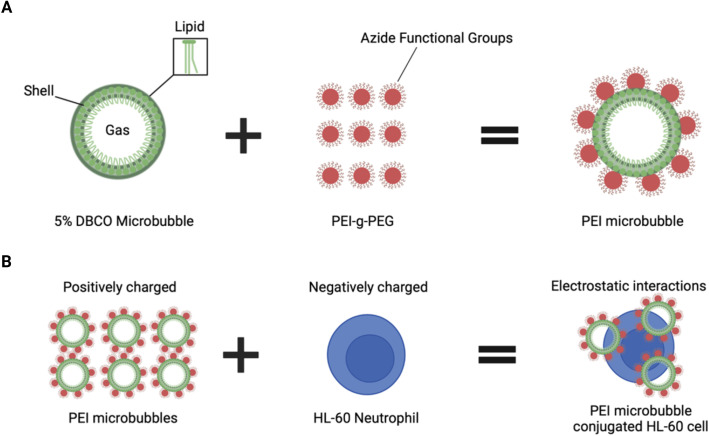 Figure 2
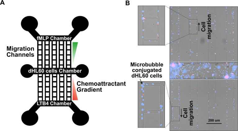 Figure 3
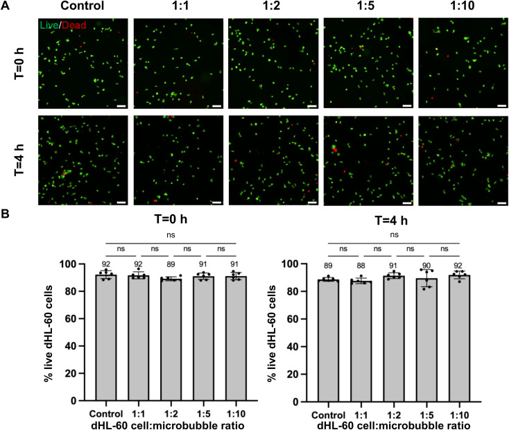 Figure 4
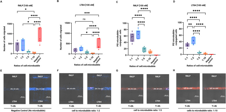 Figure 5
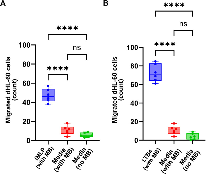 Figure 6
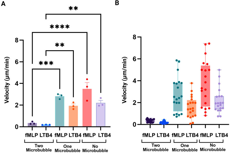 Figure 7
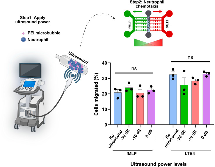 Figure 8
- —National Institute of General Medical Sciences10.13039/100000057
Peer Reviews
No public reviews on file for this paper yet. If you reviewed it on a platform where reviews are public (OpenReview, ICLR, NeurIPS, ICML), you can paste yours below so the community can read it here.
Videos
No videos yet. Explain this paper in a talk, walkthrough, or lecture? Add one.
Taxonomy
TopicsUltrasound and Hyperthermia Applications · Nanoplatforms for cancer theranostics · Nanoparticle-Based Drug Delivery
Introduction
The tumor microenvironment (TME) plays a pivotal role in determining the efficacy of cancer immunotherapies (1). Its complex and dynamic landscape establishes diverse physical, chemical, and immunological barriers that obstruct the effective infiltration and activity of immune cells within tumor tissues (1, 2). Specifically, the pronounced heterogeneity of the TME, its dense extracellular matrix (ECM), and abnormal vasculature impede immune cell migration and limit their cytotoxic capabilities upon reaching the tumor core (3, 4). In addition to these structural challenges, the TME exerts active immunosuppressive effects through factors such as hypoxia, acidosis, and the accumulation of immunosuppressive cell types, including regulatory T cells and myeloid-derived suppressor cells (1, 3). To overcome this limitation, strategies that deliver pre-activated or pre-treated immune cells directly into the tumor hold promise for enhancing their functional activity and persistence in these hostile environments (1, 2, 5–7). Cancer immunotherapy relies on using the natural killing mechanisms of the immune system to eliminate tumor cells (2, 8) by either activating an antigen on the tumor or enhancing the immune system itself. Given the current limitations of immunotherapy, especially in solid tumors, a method of delivering immune cells to the complex tumor microenvironment can help improve efficacy of this therapy. Many immunotherapy strategies aim to enhance immune cell infiltration into tumors by manipulating chemokines that attract effector cells to the tumor microenvironment (4, 9). Directly introducing specific subpopulations of immune cells into solid tumors, rather than relying on tumor-driven immune cell recruitment, has the potential to revolutionize the clinical application of immunotherapy. However, instead of conventional intratumoral injection approaches, employing focused ultrasound (FUS) for targeted delivery offers several distinct advantages (10). First, ultrasound enables broader spatial coverage within the tumor and surrounding stroma, ensuring more uniform modulation and minimizing regions that remain inaccessible to injected cells (11–13). Second, it reduces procedural risks associated with needle-based delivery, such as bleeding, infection, tumor seeding, and patient discomfort, making it safer and less invasive, particularly for deep or delicate anatomical sites (14, 15). Third, FUS provides noninvasive access to multifocal or anatomically challenging lesions, guided by real-time imaging for high precision (10, 16). Together, these advantages position focused ultrasound as a powerful alternative for enhancing the safety, efficacy, and translational potential of intratumoral immunotherapy.
Microbubbles are composed of an exterior shell that can be made up of lipids, polymers, or proteins that encapsulate a gas core (17). These microbubbles are similar in size to red blood cells and stay in circulation for in vivo applications, they can be employed for a variety of therapeutic purposes (17). Moreover, they are acoustically responsive and can move in the direction of a propagating ultrasound wave; this phenomenon is known as primary radiation force application (18). This unique aspect of microbubbles makes them versatile cargo delivery vehicles, meaning therapeutic drugs and genes can be delivered to specific areas of the body (17, 19–21). Cells can be delivered using microbubbles for in vitro and in vivo applications as a way to improve upon immunotherapy (22–24). Previous studies have demonstrated the ability to conjugate Sonazoid microbubbles to natural killer cells for cell therapy and imaging (22). Other studies have demonstrated the ability to deliver stem cells using microbubbles to treat post myocardial infarction atherosclerosis (23). Further research is needed to explore different types of microbubble and cell conjugates, identify optimal ratios of these compounds, and translate them into clinical trials for immunotherapy. We have developed a novel microbubble and cell conjugate for immunotherapy purposes using polyethyleneimine (PEI) microbubbles and neutrophils. By pre-conjugating our microbubbles to immune cells, we propose to enable their site-specific delivery to the solid tumor using low-intensity radiation force ultrasound. As a crucial initial step, it is necessary to show that the microbubble conjugation process onto immune cells does not affect their function or activity, which is the main focus of this study.
To evaluate the effects of microbubble conjugation on immune cell viablility and function, a microfluidic platform to monitor cell migration was used. Such platforms enable the quantification of immune cell migration function in physiologically appropriate, controlled settings (25–28). Microfluidics technologies have been used to quantify immune cell- function (25, 29–32). These platforms have been used to define dynamic processes including chemotaxis (33, 34), cell-cell interactions (35, 36), and immunological responses (29, 35–38). Microfluidic systems can measure immune cell migration in linear gradients of chemoattractants (31, 39, 40).
In this study, a microfluidic system was utilized to explore the migratory patterns of immune cells conjugated with microbubbles, allowing for real-time observation of how microbubble conjugation and ultrasound power influences cell migration to chemotactic signals. Neutrophil-like differentiated HL-60 (dHL-60) cells have been used in this study, as HL-60 cells are frequently used as model systems to investigate neutrophil function. DHL-60 cells exhibit characteristics of neutrophils, including chemotaxis, bactericidal activity, neutrophil extracellular trap (NET) creation, and reactive oxygen species (ROS) generation (41, 42). dHL-60 cells provide a reproducible, genetically tractable, and scalable system for studying neutrophil behavior, including migration, all of which are critical to our microbubble-conjugation and ultrasound-based targeting strategy (43, 44). To validate our findings in a physiologically relevant context, we also performed key functional experiments using primary human neutrophils. These primary cells were used for migration studies under ultrasound stimulation. Importantly, the results observed in primary neutrophils were consistent with those obtained using dHL-60 cells, supporting the relevance of the cell line as a model system while ensuring that our conclusions are applicable to actual human immune cells. This approach not only supports the development of targeted delivery strategies but also deepens our understanding of immune cell function within engineered microenvironments. We validate the formulation of a novel type of polyethyleneimine (PEI) microbubbles which is a branched cationic polymer that has the ability to carry more cargo due to its chemical structure (45). Moreover, because of the click chemistry, PEI microbubbles are fast and stable (46). We confirmed that the attachment of PEI microbubbles to immune cells does not have a detrimental impact on viability and migration.
Materials and methods
dHL-60 cell culture
HL-60 cells (ATCC CCL- 240) were purchased from the American Type Culture Collection (ATCC, Rockville, MD, USA). Cells were cultured in Iscove’s Modified Dulbecco’s Medium (ATCC) and supplemented with 20% fetal bovine serum. Cells were cultured at 37°C and at 5% CO_2_. In order to bring the cells to a differentiated state, dimethyl sulfoxide (DMSO, Sigma-Aldrich, St. Louis, MO) was added and the cells were incubated for 4–5 days. dHL-60 cells were used for preliminary viability and conjugation experiments due to their reproducibility and ease of culture.
Primary human neutrophils
Five milliliters of peripheral blood were collected from healthy donors into heparinized tubes. Human neutrophils were isolated using the Human Neutrophil Isolation Kit (Stemcell Technologies) following the manufacturer’s protocol. After isolation, the cells were adjusted to the desired concentration and mixed with PEI-coated microbubbles at specific ratios. Following exposure to defined ultrasound power, the neutrophils were injected into the microfluidic system to assess chemotaxis and migratory behavior over a 4-hour period using time-lapse imaging. Primary neutrophils were used for migration studies to ensure physiologically relevant functional responses.
Preparation of microbubbles
Lipid microbubbles for PEI conjugation were prepared using a similar protocol that has been described previously (47). Notably, in this study we have changed the conjugation chemistry from maleimide-thiol to copper free Strain-Promoted Azide-Alkyne Cycloaddition (SPAAC) click chemistry. Lipid films were prepared with 1,2-distearoyl-sn-glycero-3-phosphocholine (DSPC), N-(methylpolyoxyethyleneoxycarbonyl)-1,2-distearoyl-sn-glycero-3-phosphoethanolamin (DSPE-PEG2000), and 1,2-distearoyl-sn-glycero-3-phosphoethanolamine-N-poly (ethylene glycol)-dibenzocycolctyne (DSPE-PEG-DBCO) using a molar ratio of 90:5:5 respectively. Each lipid was dissolved in a chloroform (Sigma-Aldrich, St. Louis, MO) stock solution and mixed into a single 20 mL glass vial to achieve the desired lipid ratios. This mixture was nitrogen evaporated and stored at -20°C. The stored 5% DBCO film was suspended in 10 mL of 1X phosphate buffer saline (PBS, Fisher Scientific, Waltham, MA), 10% propane-1,2,3-triol (glycerol, 92.09 FW) (v/v), and 10% propane-1,2-diol (propylene glycol, 76.1 FW) (v/v), creating a 2 mg/mL solution. An Isotemp Heating Block was used to heat the solution up to 65°C. In order to completely suspend the lipid, the solution was bath sonicated using an Ultrasonic Bath Sonicator (Fisher Scientific, Waltham, MA). A probe tip sonicator with a microtip attachment at maximum amplitude for 10 seconds was used to emulsify the lipid solution with Decafluorobutane (PFB, 238 MW, FluoroMed LP, Round Rock, TX) gas to form the microbubble suspension. The microbubbles are then cooled in an ice bath and centrifuged three times using a Bucket Centrifuge Model 5804R (Eppendorf, Hauppauge, NY) at 300 relative centrifugal force (RCF) for 3 min to separate the microbubbles from free lipid. The size distribution and concentration of the final bubble suspension was measured by a Multisizer 4e Coulter Counter (MS4, Beckman Coulter, Brea, CA) and shown in Figure 1B.
*Characterization of PEI microbubbles. (A) PEI microbubbles were synthesized using azide-DBCO click chemistry. Results show that there is a greater concentration of PEI detected using the fluorescamine assay for the 5% DBCO microbubbles than DSPC-DSPE-PEG-2k microbubbles since more amine groups were detected after the click-chemistry reaction. (B) Size distribution of PEI microbubbles from the Coulter Counter. The PEI microbubbles have an average concentration of 8.532E9 microbubbles per mL and a mean diameter of 2.568 µm. Data are presented as mean ± SD from three independent experiments. Statistical significance was assessed using an unpaired two-tailed Student’s t-test. *p < 0.01.
Azide-PEI conjugation to DBCO-microbubbles
Azide-labeled branch polyethylene glycol polyethylenimine (N3-PEI-PEG) with a PEI molecular weight of 10k, PEG molecular weight of 2k, and a 20% substitution ratio (Nanosoft Polymers, Winston-Salem, NC) was conjugated to the microbubbuble surface using a protocol similar to our previous work (48). The suspension volume for 10 mg/mL of PEI-g-PEG is determined using the molecular weight and size distribution data of the 5% DBCO microbubbles. Then, 100 uL of microbubbles are slowly mixed in the solution consisting of PEI-g-PEG and 1XPBS. The microbubbles and PEI-g-PEG solution were stored in a 3 mL Luer tip syringe. This solution is then mixed thoroughly for 60 minutes using a Laboratory Tube Rotator to allow sufficient time for the azide-DBCO covalent bonds to form (Figure 1A). This solution is then washed three times using the same settings described above for the centrifuge and characterized again using the Coulter Counter.
PEI microbubble conjugated dHL-60 cell preparation
The HL-60 cells (CCL-240, ATCC) were mixed with PEI microbubbles, shown in Figure 2A, along with the microbubble generation process, at ratios of 1:1,1:2, 1:5, and 1:10. These ratios were calculated using the concentration and volume of the HL-60 cells and PEI microbubbles. Schematic design of the formulation of PEI microbubbles using 5% DBCO microbubbles and polyethyleneimine (PEI). Due to electrostatic interactions, the negatively charged HL-60 cells will bind to the positively charged PEI microbubbles (Figure 2B).
Preparation and conjugation process of PEI microbubbles with HL-60 cells. (A) Schematic design of the formulation of PEI microbubbles using 5% DBCO microbubbles and polyethyleneimine (PEI). (B) PEI microbubble conjugated HL-60 cells were made by utilizing the electrostatic forces between the positively charged PEI microbubbles and negatively charged HL-60 neutrophils. Created with BioRender.com.
Fluorescamine detection assay
Dilutions of 10 ug/mL to 1000 ug/mL were made using polyethyleneimine (PEI) (Sigma-Aldrich, St. Louis, MO) and phosphate buffered saline (PBS). A fluorescamine stock solution with a concentration of 3 mg/mL was made (Sigma-Aldrich, St. Louis, MO) with dimethyl sulfoxide (DMSO) (Sigma-Aldrich, St. Louis, MO). Aliquots of 90 µL of the polymer stocks were pipetted into a 96 well microplate. PEI microbubbles and DSPC-DSPE microbubbles (acting as a negative control) were also pipetted into the microplate and diluted with 1xPBS to reach a final volume of 90 µL and concentration of 3E9 #/mL. Each well was mixed with 30 µL of fluorescamine. The fluorescence was detected using a fluorescence plate reader with an emission filter of 460 nm and an excitation filter of 400 nm.
Live/dead assay of dHL-60 cells
After treatment with microbubbles, dHL-60 cells were seeded on a glass-bottom 96-well plate (Cellvis, P961.5HN) at a concentration of 8 × 10^5^ cells/mL in 100 µL of complete IMDM. To assess the effect of microbubble treatment on the viability of dHL-60 cells, dHL-60 cells were stained with the LIVE/DEADTM Viability/Cytotoxicity Assay Kit (Invitrogen, L32250) based on the manufacturer’s protocol. Briefly, a 2X dye solution was prepared in complete IMDM and 100 µL of the dye solution was added to each well to reach a working concentration of 1X at t=0 h and 4 h after microbubble treatment. Cells were imaged on a Nikon ECLIPSE Ti2-E microscope using a Plan Apo 20X objective (NA = 0.80) at 37°C to capture live and dead cells at t=0 h and t=4 h. Images were acquired using NIS-elements (Nikon Inc.) software and recorded using FITC (live cells) and Cy5 (dead cells) channels. Cells were stored in the incubator at 37°C with 5% CO2 between the two time points of imaging. Image analysis was performed in ImageJ. Cell viability was quantified as the percentage of live cells per image, i.e., the number of live cells divided by the sum of the number of live cells and the number of dead cells. The number of cells was measured using Thresholding and Analyze Particles functions. The Watershed function was used to segment clumps of cells. Only objects with a larger area than 15 µm^2^ were classified as cells to exclude spurious spots and debris.
PEI microbubble formulation
PEI microbubbles were created using azide-DBCO click chemistry. A fluorescamine assay was used to detect the amine groups present in the azide-DBCO reaction. We determined that more PEI was coated on the 5% DBCO microbubbles than the DSPC-DSPE-PEG2k microbubbles since the 5% DBCO microbubbles had an average concentration of 59.5 µg/mL while the DSPC-DSPE-PEG2k microbubbles only had an average concentration of 18.05 µg/mL (Figure 1A). The DSPC-DSPE-PEG2k microbubbles did not have any DBCO amine groups present, and therefore a click chemistry reaction did not occur; any residual PEI left over after the centrifugation steps could have led to the values detected for the control group. Size distribution of PEI microbubbles was normal with a final concentration of 8.5E9 #/mL and mean diameter of 2.568 µm (Figure 1B).
Microfluidic device preparation along with chemotactic migration of dHL-60 cells conjugated with PEI microbubbles
Microfluidic devices were fabricated using traditional photolithography techniques. Silicon mold was used to create devices with two chemoattractant reservoirs for generating linear gradients toward a central reservoir, where dHL-60 cells were cultured to study their migratory behavior. Each device also included ten migration channels (10 µm width × 10 µm height) connecting the central reservoir to the left and right chemoattractant reservoirs (Figures 3A, B). This process also utilizes replication molding, in which a PDMS prepolymer is created by mixing PDMS and a curing agent. The PDMS prepolymer is poured into the silicon mold of desired structure; it is then cured and later punched with a biopsy puncher. Oxygen plasma bonding (Harrick Plasma) was used to attach a glass slide to the bottom of the device. The migration patterns of the HL-60 cells were determined using microfluidic chemotaxis assays. Fibronectin, a glycoprotein in the extracellular matrix, was used to coat the microfluidic channels in order to increase cell adhesion. The PEI microbubbles and HL-60 cells were mixed at ratios of 1:1, 1:2, 1:5, and 1:10. These conditions were loaded onto the dHL-60 cells chamber of multiple devices (Figure 3A). Chemoattractants N-Formylmethionine-leucyl-phenylalanine (fMLP, Sigma-Aldrich, St. Louis, MO) and Leukotriene B4 (LTB4, Cayman Chemical, Ann Arbor, MI) were loaded into two chemoattractant chambers at a concentration of 100 nM. Time lapse images were taken using the Nikon TiE fully automated microscope (Nikon Inc., Melville NY). Images were recorded at 2:30 minute intervals for 4 hours. The number of cells migrating towards fMLP and LTB4 and the number of PEI microbubble conjugated HL-60 cells were quantified. Figure 3B shows a real-time microscopic image of the microfluidic system, highlighting dHL-60 cells within the cell loading chamber and migration channels.
Design of a microfluidic device, chemotactic movement of dHL-60 cells conjugated with PEI microbubbles. (A) The microfluidic device’s schematic illustration. An array of migration channels (10 µm width, 10 µm height) connects the device’s central reservoir to two chemoattractant reservoirs (fMLP and LTB4). Cells migrate along the channels in response to chemoattractant gradients created by the fMLP and LTB4 reservoirs, while the central reservoir acts as the loading point for cells. (B) Microscopic image of the microfluidic system showing PEI microbubble-conjugated dHL-60 cells migrating toward the fMLP and LTB4 chemoattractant reservoirs in the upper and lower chambers. 200 µm is represented by the scale bar.
Ultrasound treatment of microbubble-conjugated neutrophils
To evaluate the effect of ultrasound power on primary neutrophil migratory behavior, microbubble-conjugated neutrophils were exposed to ultrasound at different power levels prior to microfluidic migration assays. While most experiments in this study were conducted using differentiated HL-60 (dHL-60) cells, human primary neutrophils were employed in this section to validate the observed ultrasound effects in physiologically relevant cells.
Ultrasound exposure was performed using the Siemens Sequoia ultrasound with the 15L8 Linear transducer (Dr. Lux’s laboratory, UTSW Medical Center). Cells were treated under four conditions: (1) control (no ultrasound), (2) low-power ultrasound, (3) medium-power ultrasound, and (4) high-power ultrasound. The specific acoustic power settings for the low, medium, and high-power conditions were -30 dB, -10 dB, and 0 dB, respectively. Supplementary Video 1 demonstrates the visual response of microbubble-conjugated cells to 0 dB ultrasound application. The ultrasound acoustic radiation force was done using continuous pulse wave doppler mode to emit radiation force at different outputs.
Acoustic radiation force was generated by pulsed-wave (PW) Doppler on a Siemens Sequoia ultrasound system using a methodology adapted from Gessner et al (49). PW Doppler was operated at a center frequency of 7.0 MHz. Power output was adjusted between −30 dB (MI ≈ 0.03), −10 dB (MI = 0.29), and 0 dB (MI = 0.93). B-mode imaging for anatomical guidance was performed at 8.0 MHz. PW Doppler settings included a gain of 10 dB, gate length of 18 mm, sample volume depth of 10 mm, and a sweep speed of 50 mm/s."
For each condition, neutrophils were conjugated to microbubbles at a 1:2 cell-to-microbubble ratio. The cell–microbubble suspensions were transferred to sterile Eppendorf tubes and subjected to ultrasound exposure for 10 seconds at room temperature. Following ultrasound treatment, the cells were immediately introduced into the microfluidic migration assay platform.
Neutrophil migration was quantified as described in the migration assay section. The percentage of migrating cells was compared across all ultrasound conditions to assess the impact of ultrasound power on neutrophil motility.
Ethics statement
Primary human neutrophils were isolated from peripheral blood obtained from healthy adult donors under a protocol approved by the Institutional Review Board (IRB) at the University of Texas at Dallas. All donors provided written informed consent prior to participation.
Results
Microbubble treatment does not impact the viability of dHL-60 cells in 4 h
We investigated the viability to determine the effect of PEI microbubble conjugation process on the survival of dHL-60s (Figures 4A, B). Previous literature states that microbubbles are stable, specifically polymer-based microbubbles (17). The viability of the PEI conjugated dHL-60 cells was compared with the control dHL-60 cells. A LIVE/DEAD™ Viability/Cytotoxicity assay was used to determine the viable and nonviable cell count, and the percent viability. The percent viability of the PEI microbubble conjugated HL-60 cells was compared to that of the control dHL-60 cells. There was no significant difference between the percentage viability of the PEI microbubble conjugated dHL-60 cells and that of the control dHL-60 cells which have been shown in Figures 4A, B.
Microbubble treatment does not impact the viability of dHL-60 cells in 4 h. (A) 20X representative images showing live (green) and dead (red) dHL-60 cells on a glass-bottom 96-well plate at t= 0 h and 4 h after treatment with microbubbles at 1:1, 1:2, 1:5, and 1:10 ratios or no treatment (control). Cells were stained with the LIVE/DEADTM Viability/Cytotoxicity Assay Kit at two separate time points. Scale bar, 50 μm. (B) Bar plots showing the viability or the percentage of live dHL-60 cells in specified conditions at t=0 h and 4 h. Each data point represents a ROI and n = 6 ROIs per condition (technical replicates). Bars show mean ± SD with the mean values written above the points. ns: p ≥ 0.05, ordinary one-way ANOVA with Tukey multiple comparisons test.
Migration patterns of HL-60 cells and PEI microbubble conjugated HL-60 cells
The migration patterns of HL-60 cells and PEI microbubble conjugated dHL-60 cells were determined using microfluidic chemotaxis assays. We tested cells to microbubble ratios of 1:1, 1:2, 1:5, 1:10, and control with no microbubbles conjugated with cells. fMLP and LTB4 chemoattractants were used in this assay. Cells migratory behavior was different for different cell: microbubble ratios. As shown in Figures 5A, B, dHL-60 cells showed the highest migration for 1:1 and 1:2 ratio considering this fact that conjugating cells with microbubbles significantly decreased their migratory behavior. For 1:5 and 1:10 cell: microbubble ratio we observed the lowest migration toward both fMLP and LTB4 chemoattractants. Interestingly, PEI microbubble conjugated dHL-60 cells migration was the highest for 1:2, 1:1, 1:5 and 1:10 respectively. We observed that more of the PEI microbubble conjugated HL-60 cells migrated towards the fMLP and LTB4 chemoattractant reservoir for certain ratios. We found that there was a significant difference in the number of cells that migrated towards fMLP and LTB4 when the cell to microbubble ratio increased (Figures 5C, D). Figures 5E–H show representative images illustrating the migration behavior of dHL-60 cells conjugated with varying amounts of microbubbles in response to fMLP (top chamber) and LTB4 (bottom chamber).
*dHL-60 cells showed peak migration at a 1:2 cell-to-microbubble ratio, with reduced migration at higher ratios (1:5 and 1:10) due to impaired adhesion and mobility. (A, B) In comparison to the control (no microbubble conjugation), the number of dHL-60 cells migrated in response to (A) 100 nM fMLP and (B) 100 nM LTB4 at various cell-to-microbubble ratios (1:1, 1:2, 1:5, 1:10). (C, D) The proportion of dHL-60 cells that responded to (C) fMLP and (D) LTB4 by conjugating to PEI microbubbles at the specified ratios. (E-H) Representative images display the migration behavior of dHL-60 cells conjugated with varying quantities of microbubbles in response to fMLP (top chamber) and LTB4 (bottom chamber). In panel (E) dHL-60 cells (no microbubble conjugation) show strong and directed migration toward both chemoattractants, serving as a negative control. In panel (F), cells conjugated with microbubbles at a 1:2 cell-to-microbubble ratio still demonstrate effective chemotaxis. As shown in panel (G), increasing the microbubble load to a 1:5 cell-to-microbubble ratio results in a noticeable reduction in chemotactic migration. In panel (H), where the ratio reaches 1:10, dHL-60 cells completely lose their ability to migrate toward either fMLP or LTB4, suggesting that excessive microbubble conjugation severely inhibits migratory capacity. Individual data points are displayed in box plots. Data represents mean ± SD from six technical replicates (n = 6). Statistical analysis was performed using one-way ANOVA with Tukey’s post-hoc test. *p < 0.05, **p < 0.01, ***p < 0.0001; ns: not significant. Individual data points are shown as box plots.
PEI microbubbles do not independently induce migration
After identifying the migration patterns of different ratios of dHL-60: PEI microbubble conjugates towards two different chemoattractants (fMLP and LTB4), we aimed to demonstrate that the microbubbles do not induce migration independently. To test this, we conducted a chemotaxis assay using a 1:2 ratio of dHL-60: PEI microbubbles in the presence of chemoattractants and without any chemoattractant (Figures 6A, B). The 1:2 dHL-60: PEI microbubble ratio without chemoattractant served as a negative control. The results showed that in the absence of chemoattractants, the conjugates exhibited no significant migration. This lack of migration in the negative control indicates that microbubbles do not independently induce. The minimal migration observed was attributed to the random movement of the conjugates within the microfluidic chip, further confirming that the directed migration was specifically due to the presence of chemoattractants. This experiment underscores that the microbubbles themselves do not have an inherent tendency to migrate.
*Microbubbles alone do not induce migration. Quantification of dHL-60 cell migration in response to (A) fMLP and (B) LTB4 compared to control conditions (no chemoattractant). The lack of migration in cells exposed to microbubbles in the absence of a chemoattractant (negative control) suggests that microbubbles by themselves do not promote migration. Technical replicates (n = 6) were performed for each condition. Data are presented as mean ± SD. Statistical significance was assessed using an unpaired two-tailed Student’s t-test. ***p < 0.0001.
Increased ratio of microbubble conjugation to dHL-60 cells reduces their velocity
The velocity of dHL-60 cells in response to two distinct chemoattractants, fMLP and LTB4, under various microbubble (MB) conjugation settings (0 MB, 1 MB, and 2 MB per cell) is shown in Figures 7A, B. The data clearly show that, under both fMLP and LTB4 circumstances, dHL-60 cells without microbubbles (0 MB) had the highest velocities. These findings suggest that when the ratio of microbubbles to cells exceeds a certain threshold, the conjugated microbubbles impair the cells’ ability to adhere to the substrate, thereby limiting their migration toward chemotactic signals. Cells conjugated with 1 MB demonstrate a moderate drop in velocity in comparison to the 0 MB group in both chemoattractant conditions, but cells conjugated with 2 MB show the most noticeable decrease in velocity. This dose-dependent decrease in velocity suggests that adding more microbubbles further limits cellular mobility, possibly as a result of cytoskeletal dynamics being impacted by greater physical forces. Moreover, in all microbubble groups, cells exposed to fMLP exhibit somewhat higher velocities than those under LTB4 circumstances. This finding is consistent with earlier research showing that fMLP is a strong chemoattractant for neutrophil-like cells that causes robust migration reactions (50). Figure 7A presents the average velocity of dHL-60 cells conjugated with varying microbubble to cell ratios, while Figure 7B illustrates the single-cell velocity distribution across these different ratios.
*Higher microbubble binding reduces cell velocity. (A) The velocity of dHL-60 cells conjugated with varying quantities of microbubbles (2 MB, 1 MB, or no microbubbles) is shown in the right panel. Increased microbubble binding (2 MB) dramatically decreases motility, indicating a restricting effect on cell movement, whereas cells lacking microbubbles (0 MB) show the maximum velocity. (B) Average velocities under each condition confirm that increased microbubble loading correlates with decreased cell speed, with the lowest velocity observed in the 2 MB group and the highest in cells without microbubbles. Technical replicates (n = 3) were performed for each condition. Data are presented as mean ± SD. Statistical significance was assessed using one-way ANOVA. **p < 0.01, ***p < 0.001, ***p < 0.0001.
Effect of ultrasound and microbubble conjugation on neutrophil cell migration in response to chemotactic signals
To investigate whether ultrasound exposure alters neutrophil migratory behavior, the percentage of cells migrating toward the chemoattractants fMLP and LTB4 was quantified under four ultrasound power conditions (negative control: no ultrasound, low, medium, and high power). As shown in Figure 8, neutrophil migration toward both fMLP and LTB4 remained comparable across all ultrasound power levels. For fMLP stimulation, no statistically significant differences were detected between control and ultrasound-treated groups. Similarly, migration toward LTB4 was consistently higher but again showed no significant differences among the different ultrasound conditions.
Effect of ultrasound power on neutrophil migration toward fMLP and LTB4. The percentage of migrated neutrophils was quantified under four conditions: negative control (no ultrasound), low, medium, and high ultrasound power. Each bar represents the mean ± SD of three independent experiments (black dots). No significant (ns) differences were observed among ultrasound power levels for either chemoattractant, indicating that ultrasound exposure did not alter neutrophil chemotactic activity toward fMLP or LTB4.
These results indicate that the applied ultrasound power levels did not significantly influence neutrophil chemotactic responsiveness to either fMLP or LTB4. Thus, within the tested range, ultrasound exposure appears to have minimal or no modulatory effect on neutrophil migratory capacity.
Discussion and conclusion
There is established research on improving immune cell infiltration into tumors using focused ultrasound and bubbles. However, this relies on increasing permeability and allowing uncontrolled immune cell infiltration (1, 51). In solid tumors, where immune infiltration is frequently inadequate or spatially limited, chemotactic migration is a crucial step in the efficient utilization of immune cells for cancer therapy (52, 53). The natural recruitment of immune effector cells, including neutrophils, is restricted by the complex and frequently immunosuppressive microenvironments created by tumors (54, 55). In this study, we sought to develop and evaluate an in vitro platform to examine whether microbubble conjugation is compatible with immune cell viability and chemotactic migration under conditions relevant to ultrasound-assisted delivery strategies.
Immune cells can more effectively perform cytotoxic, pro-inflammatory, or remodeling actions when they are able to locate precisely within tumor tissues because of efficient chemotaxis (52). Translationally significant, improved chemotactic guidance can decrease off-target activity in healthy tissue, increase local immune responses, and increase immune cell density at the tumor site (53, 56). It is well recognized that solid tumors have immunosuppressive barriers, thick extracellular matrix, and aberrant vasculature, all of which prevent immune cells like neutrophils from infiltrating (57, 58). Normal tissues usually do not have these structural and functional defects. Sonoporation can temporarily increase vascular and tissue permeability during ultrasound exposure. Because of the increased permeability and retention effect, this is more likely to occur in tumor vasculature (59). These tumor-specific characteristics motivate continued investigation of ultrasound-responsive platforms; however, direct tissue accumulation or targeting was not evaluated in the present study. Focused ultrasound has the potential to enable spatially controlled activation in future studies, but such targeting effects were not examined here. Accordingly, the current work establishes foundational in vitro evidence supporting further investigation of this approach in tissue-mimicking and in vivo models. Importantly, the absence of in vivo experimentation in the present study precludes direct validation of radiation-force-assisted guidance of microbubble–immune cell conjugates within solid tumors, and such validation remains a critical next step for translational development.
While acoustic radiation force is discussed as a motivating physical mechanism, this study did not directly assess radiation-force-driven steering or localization, focusing instead on the in vitro compatibility of microbubble conjugation and ultrasound exposure with immune cell migratory function. In the present study, PEI-coated microbubbles were shown to form stable conjugates with immune cells under standard in vitro culture and microfluidic chemotaxis conditions; however, stability was not evaluated in plasma or other protein-rich environments that more closely mimic the tumor microenvironment. In such settings, electrostatic interactions may be partially attenuated due to charge screening effects and competitive protein adsorption at the microbubble and cell surfaces, potentially influencing conjugate stability (60–62). Protein corona formation on microbubble surfaces has been reported in serum-containing environments and may alter surface charge and binding behavior (60, 63, 64). Accordingly, the absence of stability testing under plasma or tumor-mimicking conditions represents a limitation of the present study. Future work will focus on evaluating conjugate stability in protein-rich media and plasma, as well as exploring surface modification strategies to enhance binding robustness under physiologically relevant conditions. While short-term viability and chemotactic migration were preserved following PEI-mediated microbubble conjugation, longer-term neutrophil functions such as ROS production, NETosis, and phagocytosis were not assessed in this study and remain important areas for future investigation.
Mechanical interactions associated with microbubble conjugation and ultrasound exposure may influence cytoskeletal organization and membrane-associated signaling pathways that regulate immune cell migration (65–67). Prior studies have shown that mechanotransduction cues can modulate actin remodeling, cell polarity, and chemotactic responsiveness in immune cells (68, 69). Although these effects were not directly assessed in the present study, the preservation of chemotactic migration following PEI-mediated microbubble attachment and ultrasound exposure suggests that key cytoskeletal functions required for directed motility remain intact under the tested conditions. Compared to existing immune cell modification strategies, the PEI-based conjugation approach offers several distinct advantages (70, 71). The electrostatic, non-covalent nature of PEI-mediated attachment enables rapid microbubble conjugation without permanent chemical modification or genetic manipulation of immune cells (72, 73). This strategy allows controllable microbubble loading per cell, compatibility with primary human neutrophils, and preservation of short-term viability and migratory function. These characteristics suggest that PEI-based conjugation may serve as a practical platform for ultrasound-compatible immune cell delivery studies, with further optimization needed to extend the temporal window of cell viability and function.
The use of microbubble conjugation and ultrasound introduces a physical and controllable approach to enhance immune cell infiltration into tumors, potentially overcoming the limitations of natural chemotaxis. According to earlier research, ultrasound can promote immune cell infiltration through improved mechanical transport, such as NK cell penetration into tumors (74). Another study by Joiner et al. have demonstrated that application of low intensity focused ultrasound with microbubbles can help activate the innate immune response for treatment of pancreatic cancer in murine models. However, the progression of disease impacted the treatment outcomes, with larger tumors being more difficult to treat due to the immunosuppressive microenvironment (75). Other studies have shown the ability to use ultrasound and microbubbles for sonopermeation, causing subsequent T-cell infiltration to the tumor (76, 77). While these studies have promising results, the ability to control the type of immune cells delivered and to preserve immune cell function during delivery remains an important challenge. In this context, our study focuses on evaluating whether pre-conjugation of immune cells to microbubbles preserves viability and chemotactic behavior, rather than demonstrating selective delivery to tumors. To test the feasibility of this approach, we evaluated whether PEI-coated microbubble attachment affects the viability or migration of dHL-60 cells. The LIVE/DEAD cytotoxicity assay showed no significant difference in viability between microbubble-conjugated and control cells, indicating that conjugation did not compromise cell health. Next, we characterized the migration of different cell-to-microbubble ratios under the influence of chemoattractants (fMLP and LTB4) on a microfluidic device. The purpose of performing the chemotaxis assay was to determine if the PEI microbubble-conjugated dHL-60 cells followed normal migration patterns. We found that a ratio of 1:2 showed the highest migration, followed by a 1:1 ratio, out of all the experimental ratios for both the chemoattractants. On the contrary, ratios of 1:5 and 1:10 inhibited the migration of PEI microbubble-conjugated dHL-60 cells due to the overloading of dHL-60 cells with the microbubbles which inhibited the migration towards both the chemoattractant. After identifying the migration pattern, we also validated if the microbubbles did not induce migration by themselves. We validated it by visualizing the migration pattern 1:2 ratio of dHL-60: PEI microbubbles in the presence and absence of chemoattractants. We observed that, in the absence of the chemoattractants, the conjugates showed no significant migration which underlines that the microbubbles themselves do not have any tendency to migrate. Lastly, we studied whether the PEI microbubbles adhered to the cells in the expected proportions. We quantified the number of PEI microbubbles (one, two, and no bubbles) and the speed of the conjugates adhering to dHL-60. It was observed that the speed of the conjugates depended on the number of microbubbles attached to the dHL-60 cells. This concluded that the more the PEI microbubbles attached to the cells lowered their migration speed towards the chemoattractants.
In addition to these experiments, we also examined the impact of ultrasound exposure on neutrophil migration behavior. Four different ultrasound power levels were tested, including a negative control with no ultrasound, low power (–30 dB), medium power (–10 dB), and high power (0 dB). The cells were exposed to these ultrasound conditions in the presence of fMLP and LTB4 gradients to evaluate potential changes in chemotactic behavior. Interestingly, we observed that ultrasound exposure across these power levels did not significantly alter the motility or directional migration of neutrophils toward either chemoattractant. These findings suggest that, within the tested range, ultrasound stimulation does not impair neutrophil migratory function, further supporting the compatibility of ultrasound-assisted delivery approaches with immune cell behavior.
Future explorations should aim to deepen our understanding of the immune response cascade initiated by these interactions. Investigating how different immune cells react to PEI microbubbles and deciphering the subsequent effects on the tumor microenvironment will provide valuable insights. These studies will facilitate the refinement of ultrasound-assisted delivery techniques, ensuring a more robust and comprehensive approach to treating solid tumors effectively. This study presents a proof-of-concept platform demonstrating that microbubble conjugation to neutrophil-like dHL-60 cells is safe, non-cytotoxic, and compatible with essential immune cell functions. We showed that cell viability remained high (above 88%) after conjugation and that, when the microbubble-to-cell ratio is appropriately optimized, the chemotactic migration of these cells toward distinct chemoattractants (e.g., fMLP and LTB4) remains largely unaffected. To further validate the physiological relevance of our platform, we conducted additional experiments using primary human neutrophils, demonstrating that ultrasound exposure does not impair their migratory behavior under the tested conditions. These results indicate that microbubble conjugation preserves critical aspects of cell health and directed migration in both model and primary immune cells, supporting the potential for immune cell delivery applications. Future studies will focus on extending this framework to tissue-mimicking systems and in vivo models to evaluate radiation-force-assisted delivery performance.
In conclusion, this study sets a precedent in the field of targeted cancer therapy, opening doors to revolutionary treatment methodologies using immune cell specific delivery to tumors. By addressing its current limitations and delving deeper into the unexplored facets, we could witness transformative advances in the precision and efficacy of cancer treatment.
The reference list from the paper itself. Each links out to its DOI / PubMed record.
- 1Tiwari A Trivedi R Lin SY . Tumor microenvironment: barrier or opportunity towards effective cancer therapy. J Bio Med Sci. (2022) 29:83. doi: 10.1186/s 12929-022-00866-3, PMID: 36253762 PMC 9575280 · doi ↗ · pubmed ↗
- 2Sahu M Suryawanshi H . Immunotherapy: The future of cancer treatment. J Oral Maxillofac Pathol. (2021) 25:371. doi: 10.4103/0973-029X.325257, PMID: 34703141 PMC 8491352 · doi ↗ · pubmed ↗
- 3Tang T Huang X Zhang G Hong Z Bai X Liang T . Advantages of targeting the tumor immune microenvironment over blocking immune checkpoint in cancer immunotherapy. Sig Transduct Target Ther. (2021) 6:1–13. doi: 10.1038/s 41392-020-00449-4, PMID: 33608497 PMC 7896069 · doi ↗ · pubmed ↗
- 4Tang H Qiao J Fu YX . Immunotherapy and tumor microenvironment. Cancer Letters. (2016) 370:85–90. doi: 10.1016/j.canlet.2015.10.009, PMID: 26477683 PMC 4725050 · doi ↗ · pubmed ↗
- 5Mishra AK Ali A Dutta S Banday S Malonia SK . Emerging trends in immunotherapy for cancer. Diseases. (2022) 10:60. doi: 10.3390/diseases 10030060, PMID: 36135216 PMC 9498256 · doi ↗ · pubmed ↗
- 6Anderson NM Simon MC . Tumor microenvironment. Curr Biol. (2020) 30:R 921–5. doi: 10.1016/j.cub.2020.06.081, PMID: 32810447 PMC 8194051 · doi ↗ · pubmed ↗
- 7Dhar R Seethy A Singh S Pethusamy K Srivastava T Talukdar J . Cancer immunotherapy: Recent advances and challenges. J Cancer Res Ther. (2021) 17:834–44. doi: 10.4103/jcrt.JCRT_1241_20, PMID: 34528529 · doi ↗ · pubmed ↗
- 8Liu C Yang M Zhang D Chen M Zhu D . Clinical cancer immunotherapy: Current progress and prospects. Front Immunol. (2022) 13:961805. doi: 10.3389/fimmu.2022.961805, PMID: 36304470 PMC 9592930 · doi ↗ · pubmed ↗