Single-arm robot-assisted endoscopic mucosal resection for oxyntic gland adenoma in the gastric fundus
Zichuang Hao, Suhuan Liao, Erzhen Zhong, Guifa He, Longbin Huang, Jialin Yang, Silin Huang

Abstract
Genes, proteins, chemicals, diseases, species, mutations and cell lines named across the full text — each resolved to its canonical identifier and authoritative record.
Click any figure to enlarge with its caption.
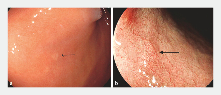 Fig. 1
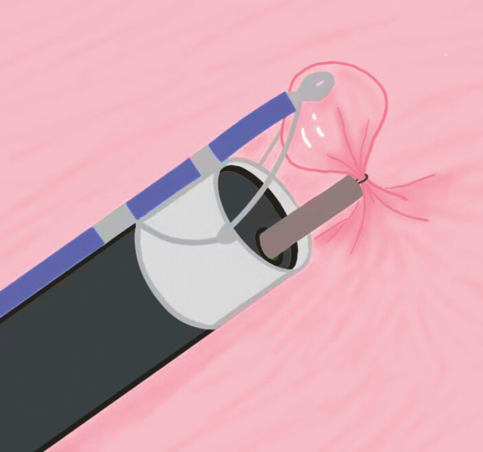 Fig. 2
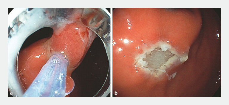 Fig. 3
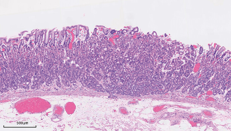 Fig. 4
Peer Reviews
No public reviews on file for this paper yet. If you reviewed it on a platform where reviews are public (OpenReview, ICLR, NeurIPS, ICML), you can paste yours below so the community can read it here.
Videos
No videos yet. Explain this paper in a talk, walkthrough, or lecture? Add one.
Taxonomy
TopicsGastrointestinal Tumor Research and Treatment · Gastric Cancer Management and Outcomes · Helicobacter pylori-related gastroenterology studies
An oxyntic gland adenoma (OGA) is a benign epithelial neoplasm defined by cellular differentiation into chief and/or parietal cells 1 . Due to its potential for submucosal infiltration, endoscopic submucosal dissection (ESD) is frequently selected in clinical practice to secure a complete excision 2 . However, the technical difficulty of ESD is often increased when such lesions are located in the gastric fundus or fornix of the upper stomach 3 . To address this challenge, we modified the resection technique by incorporating robotic assistance, enabling complete lesion removal via endoscopic mucosal resection (EMR).
A 53-year-old man was found to have a 3-mm flat, pale lesion in the gastric fundus during a routine endoscopic examination ( Fig. 1 a, b ). EMR was successfully performed for the patient with the assistance of a single-arm transluminal endoscopic robot ( Fig. 2 ) (EndoFaster, Robo Medical Technology Co., Ltd, Shenzhen, China; Video 1 ). After marking around the lesion, a single-arm transluminal endoscopic robot was mounted to the tip of the gastroscope via a soft hood. Under external joystick control, the robotic grasping forceps were placed at the 1-o’clock position. With the snare positioned around the lesion, upward traction was applied by grasping the lesionʼs center with forceps ( Fig. 3 a ). While maintaining traction, the snare was tightened around the lesion base, including the surrounding normal mucosa, followed by electrocautery resection ( Fig. 3 b ). The total resection time was approximately 3 minutes, which is significantly shorter than ESD. The wound was closed with metallic clips, and postoperative pathology confirmed a OGA with negative margins ( Fig. 4 ).
a A flat and pale lesion measuring 3 mm was found in the gastric fundus. b Magnified observation of the lesion.
Illustration of single-arm robotic-assisted endoscopic mucosal resection of the oxyntic gland adenoma in the gastric fundus.
Single-arm robot-assisted endoscopic mucosal resection for oxyntic gland adenoma in the gastric fundus.Video 1
a Under robotic-assisted traction, the lesion was completely snared. b The lesion was entirely removed.
Hematoxylin and eosin (H&E) staining revealed that the tumor was composed of numerous irregular and fused glandular tubules.
The use of the single-arm transluminal endoscopic robot appears to simplify the resection of small gastric lesions while enhancing procedural safety, representing a promising alternative strategy for the management of such cases. Further studies involving larger case series and longer follow-up periods are warranted to validate the clinical benefits of this technique.
Endoscopy_UCTN_Code_TTT_1AO_2AC
The reference list from the paper itself. Each links out to its DOI / PubMed record.
- 1Kim GH Lee JS Lee JH Oxyntic Gland Neoplasms- From Adenoma to Advanced Gastric Cancer: A Review of 29 Cases J Gastric Cancer 20242437839039375054 10.5230/jgc.2024.24.e 30PMC 11471317 · doi ↗ · pubmed ↗
- 2Miyazawa M Matsuda M Yano M Gastric adenocarcinoma of the fundic gland (chief cell-predominant type): A review of endoscopic and clinicopathological features World J Gastroenterol 201622105231053110.3748/wjg.v 22.i 48.1052328082804 PMC 5192263 · doi ↗ · pubmed ↗
- 3Kim JH Nam HS Choi CW Risk factors associated with difficult gastric endoscopic submucosal dissection: predicting difficult ESD Surg Endosc 2017311617162627495343 10.1007/s 00464-016-5149-6 · doi ↗ · pubmed ↗