Enhanced endoscopic transillumination enables precise tumor delineation in laparoscopic-endoscopic cooperative surgery for duodenal neuroendocrine tumor
Huan He, Yuting Tian, Mingxin Ye, Xiaowei Tang, Wenguang Fu, Muhan Lü, Lei Shi

Abstract
Genes, proteins, chemicals, diseases, species, mutations and cell lines named across the full text — each resolved to its canonical identifier and authoritative record.
Click any figure to enlarge with its caption.
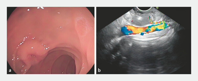 Fig. 1
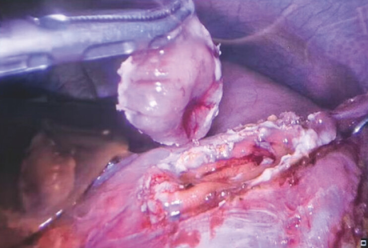 Fig. 2
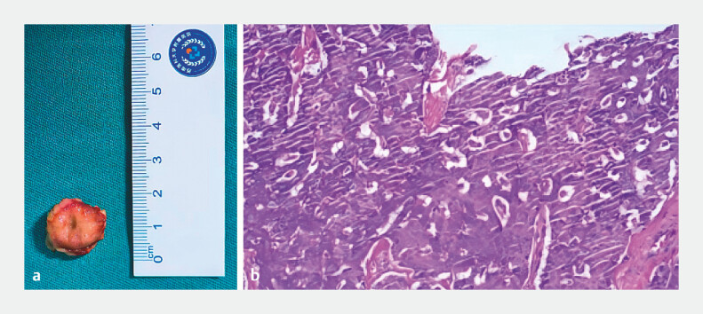 Fig. 3
Peer Reviews
No public reviews on file for this paper yet. If you reviewed it on a platform where reviews are public (OpenReview, ICLR, NeurIPS, ICML), you can paste yours below so the community can read it here.
Videos
No videos yet. Explain this paper in a talk, walkthrough, or lecture? Add one.
Taxonomy
TopicsNeuroendocrine Tumor Research Advances · Gastrointestinal Tumor Research and Treatment · Minimally Invasive Surgical Techniques
A 21-year-old woman presented with melena. Gastroscopy revealed a protruding lesion in the descending duodenum. Endoscopic ultrasonography showed a ~2.0 × 1.9 cm hypoechoic mass originating from the muscularis propria ( Fig. 1 ). Given the deep location, laparoscopic-endoscopic cooperative surgery (LECS) was selected for full-thickness resection.
a Gastroscopy identified a protruding lesion in the descending duodenum. b Endoscopic ultrasound revealed a hypoechoic mass, measuring approximately 2.0 × 1.9 cm, within the muscularis propria.
After Kocher maneuver, the gastroscope was positioned at the tumor. Endoscopic enhanced transillumination (Olympus, TRANSILLUM Mode) clearly illuminated the intestinal segment, outlining the entire tumor contour and margins ( Video 1 ), enabling precise co-localization. The resection area was marked 3 mm outside the border. Following laparoscopic resection ( Fig. 2 ), the duodenum was sutured in layers. Final gastroscopy confirmed no bleeding, normal papilla, and no stenosis. The operative time was 40 minutes with minimal blood loss. Pathology confirmed an R0 resection of a 19 × 16 × 6 mm NET G1 ( Fig. 3 ). The patient recovered uneventfully.
Enhanced endoscopic transillumination provides clear delineation of tumor margins in a duodenal NET, facilitating a conservative and precise resection via the LECS approach. LECS, laparoscopic-endoscopic cooperative surgery; NET, neuroendocrine tumor.Video 1
The tumor was completely resected laparoscopically.
a A macroscopic view of the 19 × 16 × 6 mm specimen. b H&E staining reveals nests of epithelioid cells in the lamina propria, consistent with a neuroendocrine tumor, G1.
LECS is a feasible, safe approach for duodenal tumors 1 . However, determining serosal-side resection margins is challenging, and excessive resection risks stenosis. Although existing transillumination techniques have been documented 2 , they often provide suboptimal illumination for defining the complete tumor margins. This case demonstrates that the novel application of enhanced endoscopic transillumination enabled precise tumor delineation without compromising laparoscopic lighting, thereby facilitating a conservative yet precise resection and establishing a safe and effective visualization strategy for duodenal neuroendocrine tumors.
Endoscopy_UCTN_Code_TTT_1AT_2AD
The reference list from the paper itself. Each links out to its DOI / PubMed record.
- 1Nunobe S Ri M Yamazaki K Safety and feasibility of laparoscopic and endoscopic cooperative surgery for duodenal neoplasm: a retrospective multicenter study Endoscopy 2021531065106810.1055/a-1327-593933264810 · doi ↗ · pubmed ↗
- 2Kato M Nakajima K Nishida T Local resection by combined laparoendoscopic surgery for duodenal gastrointestinal stromal tumor Diagn Ther Endosc 2011201164560910.1155/2011/64560921808595 PMC 3145349 · doi ↗ · pubmed ↗