Case Report: Vogt–Koyanagi–Harada syndrome complicated by secondary glaucoma: diagnostic insights and mechanistic correlations in uveitic ocular hypertension
Fenglong Li, Dongshen Wu, Xiaowei Zhu

TL;DR
A rare case of Vogt–Koyanagi–Harada syndrome presented as acute angle-closure glaucoma, emphasizing the need for accurate diagnosis and anti-inflammatory treatment.
Contribution
The case emphasizes the diagnostic value of ciliary body imaging and the mechanistic link between inflammation and secondary angle closure.
Findings
VKH syndrome can present with acute angle-closure glaucoma as an initial manifestation.
Ciliary body detachment and serous retinal detachments were identified through imaging.
Early anti-inflammatory treatment improved visual acuity and resolved subretinal fluid.
Abstract
This case highlights an uncommon presentation of Vogt–Koyanagi–Harada (VKH) syndrome, in which acute angle-closure glaucoma (AACG) served as the initial ocular manifestation. This atypical onset may lead to misdiagnosis as primary angle-closure glaucoma. This report adds to the existing literature by emphasizing the diagnostic value of ciliary body imaging and the mechanistic link between inflammatory ciliary body detachment and secondary angle closure. A 27-year-old woman presented with acute ocular pain, vision loss, and markedly elevated intraocular pressure (IOP). Important clinical findings included conjunctival congestion, corneal edema, medium-depth anterior chambers, and the absence of keratic precipitates or aqueous flare. Ultrasound biomicroscopy revealed a ciliary body detachment, whereas optical coherence tomography revealed multiple serous retinal detachments. The patient…
Genes, proteins, chemicals, diseases, species, mutations and cell lines named across the full text — each resolved to its canonical identifier and authoritative record.
Click any figure to enlarge with its caption.
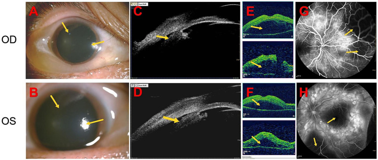 Figure 1
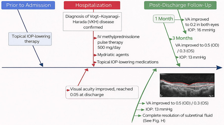 Figure 2
- —2024 Zhongshan City Traditional Chinese Medicine Inheritance and Innovation Development Research Special Fund
- —2024 Zhongshan City First Batch of Social Welfare and Basic Research Project
Peer Reviews
No public reviews on file for this paper yet. If you reviewed it on a platform where reviews are public (OpenReview, ICLR, NeurIPS, ICML), you can paste yours below so the community can read it here.
Videos
No videos yet. Explain this paper in a talk, walkthrough, or lecture? Add one.
Taxonomy
TopicsOcular Diseases and Behçet’s Syndrome · Retinal Diseases and Treatments · Retinal and Optic Conditions
Introduction
1
Vogt–Koyanagi–Harada (VKH) syndrome is an autoimmune disease that primarily affects melanocytes and predominantly affects individuals aged 20–50 years. The hallmark of this syndrome is bilateral granulomatous panuveitis, often accompanied by systemic manifestations, including decreased hearing and vision, headache, tinnitus, and vitiligo (1, 2). The course of VKH syndrome is complex, with a high rate of misdiagnosis and missed diagnosis in the early stages. This disease is easily misdiagnosed as sympathetic ophthalmia, birdshot retinochoroidopathy, and Behçet’s disease (3). Moreover, complications are commonly mistaken for primary disease (4). In some cases, VKH syndrome can be misdiagnosed as primary acute angle-closure glaucoma (AACG), presenting with peripheral anterior synechiae and increased intraocular pressure (IOP). However, AACG, as the first ocular manifestation of VKH syndrome, is uncommon (1). Its pathogenesis may involve acute inflammatory edema, disruption of the blood–aqueous humor barrier in the ciliary body, or forward rotation of the iris–lens diaphragm (5, 6). Herein, we present a case of VKH syndrome with AACG as the initial manifestation and investigate its underlying pathogenesis.
Case presentation
2
A 27-year-old woman was admitted for treatment of red eyes, ocular pain, increased IOP, and self-perceived decreased vision (near-absent light perception) for 2 days. The patient had been diagnosed with glaucoma at another hospital 1 day before admission and underwent glaucoma treatment, after which IOP decreased; however, vision continued to deteriorate progressively. After admission, relevant examinations were performed, and the findings suggested that the initial manifestation was VKH syndrome with AACG. Anterior segment photography (after pupil dilation) showed conjunctival congestion with edema, corneal opacity, absence of keratic precipitates, medium-depth anterior chambers, and absence of aqueous flare (−) (Figures 1A,B). Notably, ultrasound biomicroscopy revealed ciliary body detachment (Figures 1C,D). Secondary angle-closure glaucoma in eyes with uveitis can result from several mechanisms, including angle closure with pupillary block, which occurs when anterior chamber inflammation leads to 360° posterior synechiae, as well as inflammation and edema causing forward rotation of the ciliary body and angle closure. Optical coherence tomography of both eyes revealed multiple serous retinal detachments at the posterior pole (Figures 1E,F). Fundus fluorescein angiography revealed diffuse fluorescein leakage and staining (Figures 1G,H). Before admission, the patient had received topical IOP-lowering therapy. After admission, following the confirmation of a diagnosis of VKH syndrome, intravenous methylprednisolone pulse therapy (500 mg/day) was initiated in combination with mydriatic agents and topical IOP-lowering medications. Visual acuity gradually improved and reached 0.05 at discharge. At the 1-month follow-up, visual acuity had improved to 0.2 in both eyes, with an IOP of 16 mmHg. At the 3-month follow-up, visual acuity further improved to 0.5 in the right eye and 0.3 in the left eye, with an IOP of 13 mmHg, and complete resolution of subretinal fluid was observed. The patient’s diagnostic and therapeutic course is shown in Figure 2.
After outpatient pupil dilation, anterior segment photography shows conjunctival congestion with edema, corneal opacity, absence of keratic precipitates, medium-deep anterior chambers, and absence of aqueous flare (A,B). Ultrasound biomicroscopy reveals ciliary body detachment (C,D). Optical coherence tomography demonstrates multiple serous retinal detachments involving the posterior pole in both eyes (E,F). Fundus fluorescein angiography shows diffuse fluorescein leakage and staining (G,H).
Diagnostic and therapeutic course of the patient from pre-admission to follow-up, illustrating the sequence of management and disease progression. IOP, intraocular pressure.
Diagnosis of VKH syndrome
3
The diagnosis of VKH syndrome was established according to the revised diagnostic criteria of the International Committee on VKH Disease. This patient exhibited the following features:
Bilateral ocular involvement.Serous retinal detachments on optical coherence tomography.Ciliary body detachment, suggesting choroidal inflammation.Systemic features, including headache.Absence of keratic precipitates, infectious symptoms, or a history of trauma.
HLA-DR4/DRB104 testing, while supportive, was not performed because the diagnosis was sufficiently supported by clinical findings and multimodal imaging.
Discussion
4
Pathophysiological interplay
4.1
The exact pathophysiological mechanisms underlying the association between VKH syndrome and AACG remain incompletely understood (7, 8). Current evidence suggests that the primary mechanisms include posterior iris adhesion with pupillary block, anterior chamber angle occlusion, trabecular meshwork inflammation, accumulation of inflammatory cells near the trabecular meshwork, and long-term use of glucocorticoids (9). Notably, this patient did not exhibit angle recession but instead demonstrated ciliary body detachment, which we believe was the primary cause of elevated IOP. This finding is consistent with that of previous reports (10). Detachment of the ciliary body leads to zonular laxity, allowing anterior displacement of the lens and subsequent compression of the iris, resulting in pupillary block. This obstruction impairs aqueous humor flow from the posterior to the anterior chamber, ultimately leading to the development of malignant glaucoma. Differential diagnoses, including sympathetic ophthalmia, infectious uveitis, central serous chorioretinopathy, and primary AACG, were excluded based on clinical features and imaging findings.
Treatment considerations
4.2
Management of VKH syndrome-associated AACG requires a comprehensive approach that addresses both the underlying inflammatory and mechanical components of the disease (11, 12). In this case, the patient underwent the following interventions.
Systemic corticosteroids
4.3
Systemic corticosteroids (intravenous methylprednisolone, 500 mg/day for 5 days) were administered to treat the acute inflammatory process. This high-dose therapy aims to reduce inflammation, alleviate pupillary block, and prevent further complications, such as retinal detachment. The dosage was gradually tapered over several weeks to minimize adverse effects associated with prolonged steroid use, in accordance with standard management strategies for autoimmune uveitis (13, 14).
Topical corticosteroids
4.4
Topical corticosteroid eye drops were administered to reduce anterior segment inflammation. The dosage and frequency were adjusted according to the severity of anterior chamber inflammation and corneal edema and were gradually tapered once inflammation was adequately controlled.
Mydriatic therapy
4.5
Mydriatic agents were used to induce pupil dilation and relieve pupillary block caused by anterior displacement of the lens. Mydriatic therapy helped maintain pupillary dilation and reduce elevated IOP secondary to forward displacement of the lens–iris diaphragm resulting from ciliary body detachment.
Changes in interventions
4.6
During the course of treatment, the initial priority was controlling elevated IOP, a critical concern in secondary angle-closure glaucoma. As the patient’s condition improved, corticosteroid therapy was gradually tapered to reduce potential adverse effects. Visual acuity and IOP normalized, and subretinal fluid progressively resolved during follow-up visits at 1 and 3 months.
Rationale for treatment changes
4.7
The initial use of systemic corticosteroids was essential for controlling inflammation and secondary angle closure caused by ciliary body detachment and pupillary block.
Corticosteroids were tapered as the patient’s condition stabilized, following established protocols to minimize complications associated with prolonged steroid use while maintaining inflammatory control.
The use of mydriatic agents was critical for preventing further IOP elevation and improving patient comfort by alleviating pupillary block.
These treatment strategies are consistent with established clinical guidelines for VKH syndrome-associated AACG and were selected to address both the acute and chronic aspects of the disease while accounting for the patient’s individual response (7, 14, 15).
Conclusion
5
This case report provides important insights into the pathogenesis and clinical decision-making involved in VKH syndrome-associated AACG. Early recognition of systemic symptoms, appropriate imaging evaluation, and a multidisciplinary treatment approach are essential for optimizing outcomes and preventing irreversible visual loss.
The reference list from the paper itself. Each links out to its DOI / PubMed record.
- 1Bai X Hua R. Case Report: Vogt–Koyanagi–Harada syndrome mimicking acute angle-closure glaucoma in a patient infected with human immunodeficiency virus. Front Med. (2022) 8:752002. doi: 10.3389/fmed.2021.752002, 35096859 PMC 8789872 · doi ↗ · pubmed ↗
- 2Urzua CA Herbort CP Takeuchi M Schlaen A Concha-del-Rio LE Usui Y . Vogt–Koyanagi–Harada disease: the step-by-step approach to a better understanding of clinicopathology, immunopathology, diagnosis, and management: a brief review. J Ophthalmic Inflamm Infect. (2022) 12:17. doi: 10.1186/s 12348-022-00293-3, 35553272 PMC 9098759 · doi ↗ · pubmed ↗
- 3See WS Sawri Rajan R Alexander SM. Diagnostic and management challenges in atypical central serous chorioretinopathy mimicking Vogt–Koyanagi–Harada disease: a case report. Cureus. (2025) 17:e 91080. doi: 10.7759/cureus.91080, 41018434 PMC 12464508 · doi ↗ · pubmed ↗
- 4Patyal S Narula R Thulasidas M. Vogt–Koyanagi–Harada disease associated with anterior ischemic optic neuropathy in a young woman presenting as acute angle closure glaucoma. Indian J Ophthalmol. (2020) 68:1937. doi: 10.4103/ijo.IJO_793_20, 32823420 PMC 7690515 · doi ↗ · pubmed ↗
- 5Iwahashi C Fukuda M Makita S Takahashi A Kurihara T Sugioka K . A rare case of membrane pupillary-block glaucoma in a phakic eye with uveitis. BMC Ophthalmol. (2025) 25:358. doi: 10.1186/s 12886-025-04191-9, 40597887 PMC 12220562 · doi ↗ · pubmed ↗
- 6Yao J Chen Y Shao T Ling Z Wang W Qian S. Bilateral acute angle closure glaucoma as a presentation of Vogt–Koyanagi–Harada syndrome in four Chinese patients: a small case series. Ocul Immunol Inflamm. (2013) 21:286–91. doi: 10.3109/09273948.2013.792937, 23718571 · doi ↗ · pubmed ↗
- 7Abu El-Asrar AM Van Damme J Struyf S Opdenakker G. New perspectives on the immunopathogenesis and treatment of uveitis associated with Vogt–Koyanagi–Harada disease. Front Med. (2021) 8:705796. doi: 10.3389/fmed.2021.705796, 34869409 PMC 8632721 · doi ↗ · pubmed ↗
- 8Baltmr A Lightman S Tomkins-Netzer O. Vogt–Koyanagi–Harada syndrome—current perspectives. Clin Ophthalmol. (2016) 10:2345–61. doi: 10.2147/OPTH.S 94866, 27932857 PMC 5135404 · doi ↗ · pubmed ↗