A novel PTEN variant causing hemimegalencephaly and focal nodular heterotopias in the developing human brain
Franziska Fazekas, Amit Haboosheh, Bernhard Hennebichler, Thomas Roetzer‐Pejrimovsky, Julia Binder, Theresa Reischer, Mateja Pfeifer, Anke Scharrer, Christof Worda, Tina Linder, Alex Farr, Romana Höftberger, Ellen Gelpi, Christian Mitter, Gregor Kasprian, Christine Haberler

TL;DR
A new PTEN gene variant is found to cause brain overgrowth and seizures in a fetus, offering insights into how this mutation leads to neurological issues.
Contribution
The study identifies a novel PTEN variant and its role in hemimegalencephaly, clarifying its non-dominant-negative nature and cellular effects.
Findings
A somatic second hit in PTEN causes biallelic alteration in the affected brain hemisphere.
Nodular heterotopias composed of SATB2+ glutamatergic neurons are linked to focal seizures.
The PTEN variant is not dominant-negative, as the unaffected hemisphere carries the same germline mutation.
Abstract
Brain development and subsequent brain function are highly sensitive to genetic mutations, which can result in severe neurodevelopmental malformations. Alterations in PTEN signaling cause a spectrum of developmental malformations and neurological diseases including epilepsy. To date, a detailed understanding of the neuropathological underpinnings of PTEN‐associated brain malformations, particularly in fetuses, is missing. We have thus investigated a fetal case of hemimegalencephaly (HME), which is a rare disorder characterized by hemispheric overgrowth, developmental delay, and epileptic seizures. Our assessment of the male fetus includes genetic, radiologic, and histologic features and provides a comprehensive characterization of the cellular alterations in HME together with a genotypic correlation. Genetic analyses uncovered that hemispheric overgrowth was caused by a somatic second…
Genes, proteins, chemicals, diseases, species, mutations and cell lines named across the full text — each resolved to its canonical identifier and authoritative record.
Click any figure to enlarge with its caption.
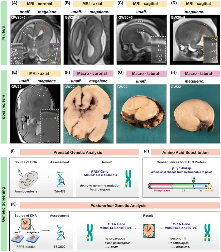 FIGURE 1
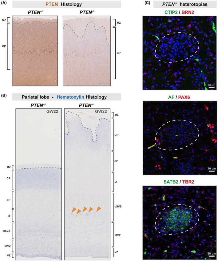 FIGURE 2
- —Austrian Science Fund10.13039/501100002428
Peer Reviews
No public reviews on file for this paper yet. If you reviewed it on a platform where reviews are public (OpenReview, ICLR, NeurIPS, ICML), you can paste yours below so the community can read it here.
Videos
No videos yet. Explain this paper in a talk, walkthrough, or lecture? Add one.
Taxonomy
TopicsPI3K/AKT/mTOR signaling in cancer · Tuberous Sclerosis Complex Research · Hedgehog Signaling Pathway Studies
Key points
- Upon biallelic alteration, a novel PTEN variant is associated with hemispheric overgrowth during fetal brain development.
- The affected hemisphere presents with focal nodular heterotopias within the outer subventricular zone.
- PTEN variant‐associated heterotopias consist of SATB2^+^ neurons.
INTRODUCTION
1
The brain comprises a remarkable cell‐type diversity, which arises during embryonic and fetal development and provides the basis for adequate brain function. Errors in the tightly orchestrated process of brain development, for example, by generating distinct subtypes of neurons in wrong ratios, can result in neurodevelopmental malformations and neurological diseases including epilepsy.1
Alterations in PTEN signaling, an essential regulator of cell growth, proliferation, survival, and metabolic activity, are known to cause brain malformations and neurological diseases of developmental origin. Depending on the time point/cell type in which respective mutations occur, altered PTEN signaling results in a spectrum of defects ranging from focal cortical dysplasia to hemimegalencephaly (HME) and macrocephaly.2, 3, 4 HME is a rare PTEN‐associated neurodevelopmental disorder,5, 6 characterized by overgrowth of one hemisphere, developmental delay, and epileptic seizures.7 However, a detailed cellular and molecular understanding of the neuropathological underpinnings of HME and onset of neurological symptoms, particularly in fetuses, is still missing to date.
We have thus assessed the genetic, radiologic, and histologic features of a pronounced HME in a male fetus (gestational week [GW] 22) and provide a comprehensive characterization of the cellular alterations in this malformation, focusing on the cortex and the locally produced glutamatergic neurons. HME provides the unique opportunity to directly compare the unaffected and megalencephalic hemispheres derived from the same developmental time point, harboring the same gross genetic background, and that have undergone identical postmortem processing for magnetic resonance imaging (MRI), genetic, and histological analysis.
RESULTS
2
Hemisphere‐specific loss of heterozygosity of a novel
PTEN variant
2.1
In utero fetal MRI (1.5 T) was performed at GW 20 + 5 to confirm the ultrasonographic suspicion of a unilateral and asymmetric enlargement of the entire right hemisphere with cortical dyslamination, as determined by loss of the normal T2‐high signal within the subplate (SP) zone (Figure 1A–D).8 Pregnancy was terminated at 22 weeks of gestation. A postmortem brain MRI (3 T) showed similar findings as in utero MRI (Figure 1E). In addition, subtle areas of irregular and undulating cortical plate (CP) surface, mainly along the medial frontobasal and medial occipital lobes, were noticed (Figure 1E). Both MRI examinations correlated with the gross macroscopic appearance of the brain, showing a similar pronounced enlargement and irregular surface of the right hemisphere as compared to the smooth and normal‐sized left hemisphere (Figure 1F–H).
Novel pathological PTEN variant in a fetus with hemimegalencephaly. (A–D) In utero evaluation. Selected coronal (A, B) and sagittal (C, D) T2‐weighted images from 1.5‐T in utero MRI examination of a fetus with hemimegalencephaly at gestational week (GW) 20 + 5 are shown. Distinct cortical zones can be discriminated based on signal intensity in the zoom‐ins. The enlarged right hemisphere shows an almost homogeneous signal across all cortical zones, thus not allowing discrimination between particular zones such as the ventricular zone (VZ), the subventricular zone (SVZ), the subplate (SP), and the cortical plate (CP). (E–H) Postmortem evaluation. (E) T2‐weighted axial image of 3‐T postmortem magnetic resonance imaging (MRI) of the fetal brain at GW 22, correlating with the observations gained from (F) macroscopic coronal section of the same brain. (G, H) Postmortem whole‐mount photographs of the lateral view of the fetal brain, correlating well with the in utero MRI findings shown in panels C and D. (I–K) Genetic evaluation. (I) Results from trio exome sequencing (ES) following amniocentesis identified a heterozygous de novo point variant in PTEN (c.1036 T>G). (J) Schematic of the PTEN protein, indicating the location of the amino acid substitution in the PTEN protein caused by the c.1036 T>G variant. (K) Results from TruSight Oncology 500 (TSO500) panel sequencing of DNA isolated from the formalin‐fixed hemispheres. FFPE, formalin‐fixed paraffin‐embedded; megalenc., megalencephaly; unaff., unaffected; PDZ‐BD, PDZ binding domain motif.
To perform a phenotype–genotype correlation, we first turned to the prenatal trio exome sequencing (ES) results, which were obtained after amniocentesis (Figure 1I). Trio ES identified a novel heterozygous de novo point variant in the phosphatase and tensin homolog (PTEN) gene (NM_000314.8: c.1036 T>G; Figure 1I), causing an amino acid substitution in the C2 domain of the PTEN protein (p.Tyr346Asp; Figure 1J). Because PTEN serves as a negative regulator of mTOR signaling, PTEN loss‐of‐function variants typically result in overgrowth syndromes.3, 4, 5, 6 However, a dominant‐negative germline variant in PTEN should result in a whole‐body overgrowth. Instead, the fetus showed no signs of peripheral organ abnormalities and displayed normal body length as compared to healthy fetuses of the same developmental stage (Supplemental Material) but had a unilateral HME phenotype where only one hemisphere overgrew, thus suggesting the presence of a second somatic hit or synergizing mutations in the affected hemisphere.9
To uncover hemisphere‐specific somatic mutations, we isolated DNA from formalin‐fixed paraffin‐embedded brain tissue of the unaffected and the affected hemisphere and performed next generation sequencing using TruSight Oncology 500 panel sequencing (Figure 1K, Table S1). In the unaffected hemisphere, we found an allelic frequency of 49.31% for the PTEN c.1036 T>G variant, thus reflecting the heterozygous germline mutation that was already discovered by trio ES (Figure 1K). Importantly, the megalencephalic hemisphere showed an allelic frequency of 88.06% for the PTEN c.1036 T>G variant, consistent with loss of heterozygosity in the neuronal lineage (Figure 1K, Table S1). As a result, we assume that the neuronal lineage of the megalencephalic hemisphere carries the PTEN c.1036 T>G variant in a biallelic manner, and that other lineages, such as endothelial cells and immune cells, carry the heterozygous germline variant. We thus refer to the unaffected hemisphere as PTEN ^ +/− ^ and to the megalencephalic hemisphere as PTEN ^ −/− ^.
Biallelic
PTEN variant causes reduced PTEN signaling and nodular heterotopias in the overgrown hemisphere
2.2
To validate our hypothesis of the biallelic PTEN c.1036 T>G variant resulting in reduced PTEN levels/activity, we performed PTEN immunostaining. The biallelic variant resulted in strongly diminished PTEN immunoreactivity (Figure 2A), which correlated with tissue enlargement (Figures 1 and 2B) and was in line with the known role of PTEN loss of function in overgrowth syndromes.9 Based on these results we infer that the c.1036 T>G variant is pathogenic upon biallelic alteration. According to the American College of Medical Genetics and Genomics criteria, we thus classify the variant as “likely pathogenic” (Table S1).10
*Histological appearance of PTEN‐associated hemimegalencephaly. (A) PTEN staining of the unaffected (PTEN
+/− ) and the affected hemisphere (PTEN
−/− ). (B) Hematoxylin staining of the unaffected and the PTEN
−/− hemisphere and identification of the different zones of the fetal cortex. Orange arrowheads highlight nodular heterotopias localized in the outer subventricular zone (oSVZ) of the PTEN
−/− hemisphere. (C) High‐resolution images of immunofluorescent staining of representative nodular heterotopia, showing (top panel) layer V neuron marker CTIP2 (green) and layer II–III neuron marker BRN2 (red), (middle panel) apical radial glial progenitor marker PAX6 (red) and autofluorescence (AF; green), and (lower panel) intermediate progenitor marker TBR2 (green) and layer II–IV neuron marker SATB2 (red), together with nuclear stain (4,6‐diamidino‐2‐phenylindole, blue). Scale bars for panels A and B: 250 μm; scale bars for panel C: 20 μm. CP, cortical plate; GW, gestational week; iSVZ, inner subventricular zone; IZ, intermediate zone; MZ, marginal zone; SP, subplate; VZ, ventricular zone.*
Next, we used nuclear staining with hematoxylin on histological sections to discriminate between the different zones of the developing fetal neocortex based on cell density (according to the developing human brain atlas; Figure 2B).11, 12 We detected nodular heterotopias specifically located in the outer subventricular zone (oSVZ) of the megalencephalic hemisphere (arrowheads, Figure 2B). The heterotopias were not visible on MRI, likely reflecting limited sensitivity at this developmental stage. Additionally, the CP of the PTEN ^ −/− ^ hemisphere showed features of cortical dyslamination and an irregular surface with many fingerlike protrusions, which correlated well with the MRI observations (Figures 1E and 2B).
To gain a deeper understanding of the nodular heterotopias occurring in PTEN‐associated HME, we stained for cell type‐specific markers (Figure 2C), using anti‐PAX6 antibody to identify apical radial glial progenitors and anti‐TBR2 antibody for intermediate progenitors.13, 14 Cortical projection neurons were discriminated based on CTIP2 (early born subcerebral neurons located in layer V), SATB2 (late born callosal neurons in layers II–IV), and BRN2 (late born corticocortical neurons in layer II–III) immunostaining.15, 16 We could not detect any progenitor cell subtypes within the heterotopias as determined by absence of PAX6^+^ or TBR2^+^ cells, suggesting that these ectopic cells consisted of neurons. We found the heterotopias to almost exclusively harbor upper layer glutamatergic SATB2^+^ cells and not lower layer CTIP2^+^ or upper layer BRN2^+^ cells (Figure 2C). Further immunohistochemistry stainings with interneuron markers (calbindin, calretinin, GAD67, somatostatin; Figure S1)17 showed that the heterotopias did not display interneuron enrichment, thus strengthening the evidence of a glutamatergic lineage phenotype in the *PTEN^−/−^
- hemisphere.
DISCUSSION
3
Our study provides novel insights into the asymmetric genetic and cellular alterations occurring in a case of HME. Many PTEN‐associated diseases are caused by dominant‐negative PTEN variants.18 However, here we describe a new PTEN c.1036 T>G variant and unravel its pathogenicity in the biallelic state directly from the affected tissue by means of next generation sequencing.
Although a heterozygous germline variant could be responsible for a more systemic phenotype, the fetus did not present with further pathologies other than the pronounced hemimegalencephaly (see autopsy report in the Supplemental Material). We thus interpret that the heterozygous germline variant is not pathogenic and therefore does not cause a systemic phenotype, at least at this developmental stage.
The megalencephalic hemisphere showed an irregular cortical plate/marginal zone boundary. A recent study using cerebral organoids was able to show that a pathogenic PTEN loss‐of‐function mutation increased the proliferation potential of cortical progenitors, inducing expansion and folding during corticogenesis.19 The folded organoids contained expanded ventricular zone (VZ) and oSVZ progenitors, as well as delayed neural differentiation,19 similar to what we found in the megalencephalic hemisphere of the presented fetus.
The oSVZ of the PTEN ^ −/− ^ cortex contains small focal heterotopias, which consist of SATB2^+^ glutamatergic neurons. Neuronal nodular heterotopias are a source of altered neuronal circuits causing focal seizure activity20 and have been described in HME before.21 However, heterotopia cell composition may vary in a patient‐ and/or PTEN variant‐specific manner. Whereas we identified mainly a glutamatergic neuronal phenotype in the fetus, as determined by the presence of SATB2^+^ neurons in the nodular heterotopias, Hannan et al.21 found an enrichment of γ‐aminobutyric acidergic interneurons in the heterotopias of four postnatal HME cases. This hypothesis is supported by a recent report on a case of HME with epilepsy, harboring distinct PTEN variants in different regions of the cortex and showing variant‐specific alterations of the cellular composition in affected cortical areas.9 Because we did not perform ultradeep sequencing as reported by Sanders et al.,20 we cannot formally rule out that heterotopias may consist of genetically distinct cells with additional low‐level somatic pathogenic variants (allelic frequency <5%). Yet, the expansion of the cortical VZ and oSVZ, as well as our histological assessment of glutamatergic and inhibitory neurons, provides strong evidence that the presented case mainly harbors a glutamatergic lineage phenotype.
Importantly, the heterotopias in the PTEN ^ −/− ^ fetal hemisphere could not be detected using MRI. Yet, they were clearly visible in histological sections. Our data thus underline the necessity to study brain malformations with both radiological and histological methods for a precise integrated diagnosis.
AUTHOR CONTRIBUTIONS
Franziska Fazekas and Nicole Amberg designed the research project, performed histological analyses, and coordinated data collection. Bernhard Hennebichler coordinated the DNA isolation from formalin‐fixed paraffin‐embedded tissue and subsequent TruSight Oncology 500 panel sequencing. Thomas Roetzer‐Pejrimovsky performed TSO500 result interpretation. Amit Haboosheh, Christian Mitter, and Gregor Kasprian performed MRI assessment. Christof Worda, Julia Binder, Theresa Reischer, Tina Linder, and Alex Farr performed the clinical patient counseling. Mateja Pfeifer coordinated the trio ES screening of DNA isolated following amniocentesis. Anke Scharrer performed the fetal autopsy. Romana Höftberger, Ellen Gelpi, and Christine Haberler performed the diagnostic neuropathological examinations and provided neuropathological expertise. Franziska Fazekas, Amit Haboosheh, and Nicole Amberg wrote the manuscript with input from all coauthors. Franziska Fazekas and Nicole Amberg prepared the figures with input from all coauthors.
FUNDING INFORMATION
N.A. received funding from the FWF (Elise Richter program, grant V1041T).
CONFLICT OF INTEREST STATEMENT
The authors have no competing interests. We confirm that we have read the Journal's position on issues involved in ethical publication and affirm that this report is consistent with those guidelines.
ETHICS APPROVAL
The present study was approved by the ethics committee of the Medical University of Vienna (EK 1161/2018). This study is part of the Neurobiobank of the Division of Neuropathology and Neurochemistry, Department of Neurology, Medical University of Vienna. Research use of biobanked samples was approved by the ethics committee of the Medical University of Vienna (EK 1636/2019), which provides a common broad consent (biobank consent) according to the Austrian Research Organization Act 2018, §2d, para 3.
CONSENT FOR PUBLICATION
Family members have consented to publication as part of their consent for tissue archiving in our neurobiobank (EK 1636/2019).
Supporting information
DATA S1.
TABLE S1.
The reference list from the paper itself. Each links out to its DOI / PubMed record.
- 1Siletti K , Hodge R , Mossi Albiach A , Lee KW , Ding S‐L , Hu L , et al. Transcriptomic diversity of cell types across the adult human brain. Science. 2023;382(6667):eadd 7046.37824663 10.1126/science.add 7046 · doi ↗ · pubmed ↗
- 2Bizzotto S , Walsh CA . Genetic mosaicism in the human brain: from lineage tracing to neuropsychiatric disorders. Nat Rev Neurosci. 2022;23(5):275–286.35322263 10.1038/s 41583-022-00572-x · doi ↗ · pubmed ↗
- 3Garcia CAB , Carvalho SCS , Yang X , Ball LL , George RD , James KN , et al. m TOR pathway somatic variants and the molecular pathogenesis of hemimegalencephaly. Epilepsia Open. 2020;5(1):97–106.32140648 10.1002/epi 4.12377 PMC 7049797 · doi ↗ · pubmed ↗
- 4Jansen LA , Mirzaa GM , Ishak GE , O'Roak BJ , Hiatt JB , Roden WH , et al. PI 3K/AKT pathway mutations cause a spectrum of brain malformations from megalencephaly to focal cortical dysplasia. Brain. 2015;138(Pt 6):1613–1628.25722288 10.1093/brain/awv 045PMC 4614119 · doi ↗ · pubmed ↗
- 5Itoh K , Pooh R , Shimokawa O , Fushiki S . Somatic mosaicism of the PI 3K‐AKT‐MTOR pathway is associated with hemimegalencephaly in fetal brains. Neuropathology. 2023;43(2):190–196.36325654 10.1111/neup.12875 · doi ↗ · pubmed ↗
- 6Jaiswal V , Hanif M , Sarfraz Z , Nepal G , Naz S , Mukherjee D , et al. Hemimegalencephaly: a rare congenital malformation of cortical development. Clin Case Reports. 2021;9(12):e 05238.10.1002/ccr 3.5238 PMC 868457834976397 · doi ↗ · pubmed ↗
- 7Ikeda KM , Mirsattari SM . Evolution of epilepsy in hemimegalencephaly from infancy to adulthood: case report and review of the literature. Epilepsy Behav Case Rep. 2017;7:45–48.28377884 10.1016/j.ebcr.2017.02.002PMC 5369267 · doi ↗ · pubmed ↗
- 8Pogledic I , Mankad K , Severino M , Lerman‐Sagie T , Jakab A , Hadi E , et al. Prenatal assessment of brain malformations on neuroimaging: an expert panel review. Brain. 2024;147(12):3982–4002.39054600 10.1093/brain/awae 253PMC 11730443 · doi ↗ · pubmed ↗