A Versatile Approach to Stabilize Whispering Gallery Microresonators Toward Reliable Photonic Labeling
M. Reale, A. Madonia, S. Agnello, M. Cannas, E. Marino, A. Sciortino, F. Messina

TL;DR
This paper introduces a method to stabilize optical sensors called WGM microresonators, making them more reliable for detecting tiny changes in their environment.
Contribution
A novel stabilization protocol for WGM microresonators is introduced, addressing thermal and temporal drifts caused by solvent release.
Findings
Thermal and temporal drifts in WGM microresonators are caused by residual solvent release over days.
A simple protocol effectively locks resonances into stable modes, enabling long-term stability.
The stabilization method enhances the reliability of WGM microresonators for sensing and tracking microscale changes.
Abstract
Whispering gallery mode (WGM) microresonators provide ultrasensitive optical fingerprints that are ideal for photonic labels capable of detecting minute variations in size and refractive index. WGM microresonators can be obtained through convenient self-assembly protocols, for instance, by loading polystyrene microparticles with colloidal quantum dots. However, despite their ultrasharp spectral signatures, these resonators exhibit significant thermal and temporal drifts that limit their applications. We combine experiments and modeling to show that these drifts originate from the slow, days-long release of residual solvent trapped within the microparticles, which can be monitored via WGM spectroscopy. We establish a simple protocol to suppress this instability, locking the resonances into stable, sharply defined modes. This approach provides a straightforward route for long-term…
Genes, proteins, chemicals, diseases, species, mutations and cell lines named across the full text — each resolved to its canonical identifier and authoritative record.
Click any figure to enlarge with its caption.
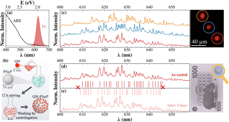 Figure 1
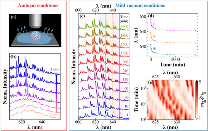 Figure 2
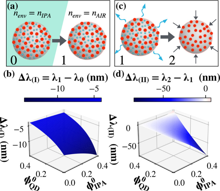 Figure 3
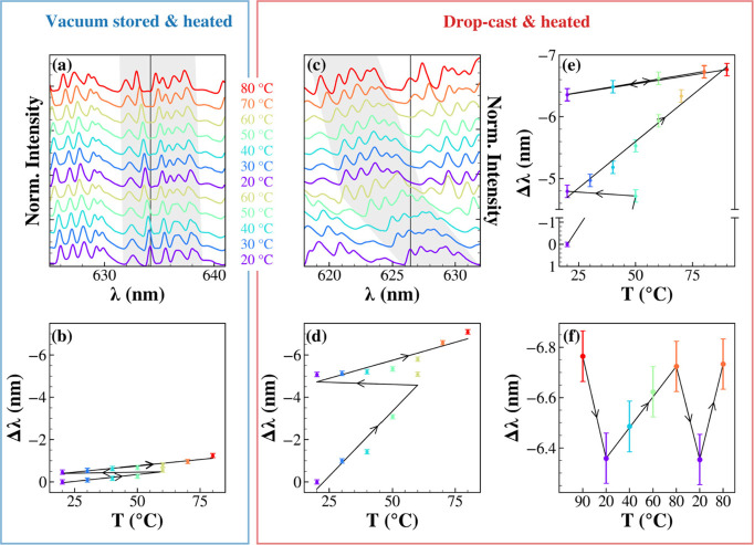 Figure 4
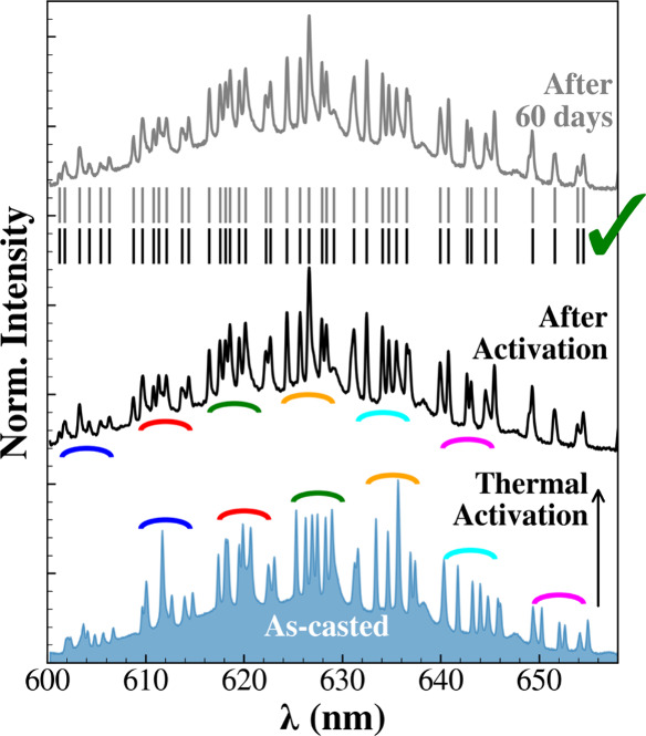 Figure 5
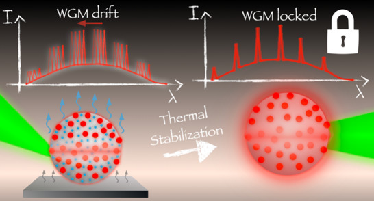 Figure 6
 Figure 7
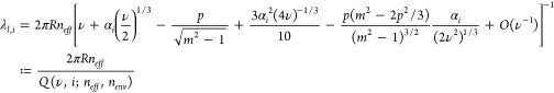 Figure 8
 Figure 9
 Figure 10
 Figure 11
- —H2020 European Research Council10.13039/100010663
- —Ministero dell’Istruzione, dell’Università e della Ricerca10.13039/501100003407
- —Ministero dell’Istruzione, dell’Università e della Ricerca10.13039/501100003407
Peer Reviews
No public reviews on file for this paper yet. If you reviewed it on a platform where reviews are public (OpenReview, ICLR, NeurIPS, ICML), you can paste yours below so the community can read it here.
Videos
No videos yet. Explain this paper in a talk, walkthrough, or lecture? Add one.
Taxonomy
TopicsPhotonic and Optical Devices · Mechanical and Optical Resonators · Advanced Fiber Laser Technologies
Optical microresonators exploit their symmetry to confine light near interfaces. As light circulates along a closed loop, constructive interference occurs when the optical path length matches an integer multiple of the circulating wavelength. When this condition is met, sharp resonances, known as Whispering Gallery Modes (WGMs), arise with ultrahigh quality factors Q = λ/Δλ and relatively small mode volumes. ?,? Together, these characteristics provide a platform of exceptional sensitivity for microscale optical sensing and spectroscopy. Minute variations in size or refractive index can produce measurable shifts, making WGMs ideal for applications ranging from biosensing ?,? and microlasing ?−? ? to optical labeling and anticounterfeiting technologies, ?−? ? where the dense resonance patterns serve as physically unclonable spectral fingerprint. Dielectric spheres, rings, toroids, and other more complex morphologies can serve as WGM microresonators. ?,? Despite their potential, practical use of WGM microresonators is often hindered by the complexity of light coupling into/from the resonator, requiring, for instance, carefully approaching a tapered fiber or prism to the surface of the microresonator. Active WGM microresonators have been developed to overcome these limitations by embedding a gain medium guest within the resonator host.? In such systems, spontaneous emission couples directly to the cavity modes, eliminating the need for external optics and enabling a straightforward optical readout while preserving the intrinsic sensitivity of WGMs.
Colloidal semiconductor quantum dots (QDs) are particularly attractive as a gain medium due to their high photoluminescence (PL) quantum yield, tunable emission, and photostability.? Polymeric microspheres, such as polystyrene microparticles (PSμPs), offer an ideal host, as they are optically transparent in the visible range, chemically inert, easy to functionalize, and can be prepared in various, well-controlled sizes. ?,? Coupling QDs to PSμPs via solvent-assisted loading is straightforward, providing a scalable strategy for the synthesis of active microresonators, ?−? ? ? ? with applications spanning microlasing, ?,? optical labeling,? enhanced electron or energy-transfers, ?,? and environmental sensing. ?,?,?
WGMs can act as ultrasensitive, noninvasive real-time probes of the resonator’s geometry or refractive index, thus enabling the monitoring of processes such as polymer relaxation,? internal refractive index changes,? and vapors or solvent uptake. ?−? ? However, the same exceptional sensitivity that makes WGMs such powerful probes also represents a well-recognized practical limitation: minute and often uncontrolled variations in the microresonator or its local environment can induce substantial spectral drifts, ultimately compromising specificity and long-term stability. ?−? ? Understanding the physicochemical origin of these drifts and how to suppress them is therefore essential for enabling reliable WGM-based technologies.
Here, we overcome this limitation by demonstrating an effective route to obtain drift-free active WGM microresonators. We use model hybrid microresonators in which colloidal QDs are coupled to PSμPs, providing a well-defined WGM fingerprint that allow us to identify slow solvent desorption as the dominant driver of irreversible blueshift of the resonances. Real-time spectral tracking under controlled environmental conditions directly links solvent loss to the optical response of the resonator. A thermal activation step fully suppresses these drifts, locking the WGM pattern into stable, low-thermosensitivity microresonators as robust and scalable platforms for secure photonic labeling and real-time monitoring of physicochemical changes on the nanoscale.
We synthesized CdSe/CdS core/shell QDs following the literature.? As reported in Figurea, the QDs exhibit an absorption spectrum dominated by the CdS shell edge below 550 nm, while the CdSe core excitonic feature remains discernible at around ∼610 nm (Figure S1). Figurea also shows the PL spectrum, centered at ∼625 nm (PL Quantum Yield 60%), which is compared in Figure S1 with the excitonic structure resolved in the absorption spectrum. We integrated these emitters into PSμPs (10 μm diameter) by using a solvent-assisted self-assembly strategy adapted from the literature and illustrated in Figureb.? Briefly, QDs dispersed in chloroform were added to a suspension of PSμPs in isopropanol while stirring. Chloroform is known to swell polystyrene? which may facilitate limited QD diffusion into the outer polymer layer, while its partial miscibility with isopropanol promotes mild colloidal destabilization that favors QD deposition onto the PSμPs. ?,? After 12 h, QD-PSμPS were separated from unbound QDs via centrifugation and redispersed in isopropanol. Further details can be found in the Experimental Methods section.
After dropcasting, we observe that individual PSμPs emit bright red light consistent with the photoluminescence of the QDs, indicating that QDs have been deposited onto the microparticles. Interestingly, the resulting microphotoluminescence (μ-PL) spectra of individual QD-PSμP (Figurec) show a dense array of narrow WGM peaks superimposed on the broader QD emission envelope. μ-PL spectra from distinct QD-PSμPs exhibit discrete WGM progressions, producing unique spectral patterns as exemplified in Figurec. This spectral diversity reflects the extreme sensitivity of WGM resonances to the geometry and refractive index of the microresonator. The resonance wavelength λ _ l _ , * i
- for a mode of angular mode number l, radial mode number i, and polarization p, which can be transverse electric (TE) or transverse magnetic (TM), is described by the asymptotic expansion derived from the Debye approximation of the Mie scattering solution: ?,?
where , R is the sphere radius, n _ eff _ is the effective refractive index of the sphere, m = n _ eff _/n _ env _ is the refractive index contrast between the sphere and the refractive index of the surrounding environment medium, α _ i _ the i-th zero of the Airy function, and p = m for TE or p = 1/m ^2^ for TM modes. As shown in the simulations reported in Figure S2, 0.1% variations in size or refractive-index produce nanometric shifts in the resonance positions. Consequently, each microparticle exhibits a unique and irreproducible spectral “fingerprint”. This inherent spectral individuality offers a compelling route toward several applications, such as photonic microtagging and anticounterfeting labels. Each QD-PSμP can act as an optical microlabel whose μ-PL spectrum encodes an unclonable optical signature, as conceptually illustrated in Figured-e. Indeed, the narrow WGM peaks function as a photonic barcode that cannot be replicated even under nominally identical fabrication conditions, thus enabling physically unclonable functions. ?,?,? Such optical microlabels could provide secure identifiers for anticounterfeiting or secure authentication technologies, where information is stored and verified through spectral readout. Potential applications extend to secure labeling of high-value goods, tracking of individual components in complex assemblies, or any scenario requiring the distinction of nominally identical micro-objects.
A crucial requirement for these applications is the temporal stability of the spectral barcode. To probe this stability, we tracked the μ-PL spectrum of individual QD-PSμPs over time. Surprisingly, the spectral fingerprints do not remain static. As shown in Figuree, the WGM pattern recorded 3 days after dropcasting under ambient laboratory conditions exhibits more than 1 nm blueshift compared to the as-cast spectrum (Figured). This spectral drift can stem from subtle morphological or environmental changessuch as polymer relaxation, QD redistribution, or adsorption/desorption of solvent moleculesthat alter the local refractive index and/or the effective cavity radius (see eq). ?,?,? Although this observation highlights the extraordinary sensitivity of WGMs to minute perturbations, it simultaneously challenges their use as reliable photonic labels, hinting that further control over the microresonator environment is needed.
We studied the spectral instability in different environments to identify its cause. In a first experiment, we monitored the evolution of μ-PL spectra of a QD-PSμP over time in the first few minutes after drop-casting, as schematically illustrated in Figurea. Immediately after drop-casting from isopropanol, the WGM structure is barely discernible on the PL envelope (Figureb), with broader peaks, which suggests a low Q-factor. This is likely due to the presence of solvent at the particle-environment interface, since the QD-PSμP is entirely exposed to liquid isopropanol as surrounding medium. Interestingly, the WGM spectrum displays several sets of two closely spaced resonances, each originating from a pair of a TE and a TM mode. The separation between two consecutive TE (or TM) modes is ∼7.5 nm, corresponding to the spacing between consecutive azimuthal mode numbers (Δl = 1). Using the standard expression for the free-spectral range? Δλ_Δ_ * l * =1 = λ^2^/2πRn _ eff _, and assuming a 10 μm PSμP with n _ eff _ = n _ PS _ = 1.58 at λ = 630 nm, we obtain Δλ ≈ 7.9 nm, in good agreement with the experimental value. This indicates that under these initial conditions where the refractive index contrast between the polystyrene sphere and the surrounding medium is reduced by residual isopropanol, the spectrum is dominated by fundamental radial modes (i = 1). After 2 min, the peaks dramatically narrow, additional modes appear, consistent with the emergence of higher-order radial modes,? and the progression of WGMs undergoes a blueshift. After five min, the spectrum appears to stabilize, corresponding to the evaporation of the isopropanol outside the QD-PSμP.
Then, we tested for the presence of residual solvent by placing a QD-PSμP under mild vacuum conditions (pressure 0.15 bar), measuring the spectrum at different intervals. Figure(c) shows a further progressive blue-shift of the WGM pattern. Interestingly, 10 min under reduced pressure induce a spectral shift exceeding the 1 nm shift observed after 3 days in air (compare with Figure), compatible with a sped-up isopropanol evaporation under reduced pressure. The temporal evolution of representative WGM peaks is plotted in Figured. A biexponential fit yields mean time constants of 25 and 364 min, both associated with shift amplitudes of 3.4 nm and likely reflecting internal solvent release from regions with different effective diffusion lengths within the polymer matrix. The spectral shift of a specific set of resonances is visually highlighted in Figuree, which presents a colormap of the total PL intensity (normalized to the broadband PL) to illustrate how WGM features shift during vacuum exposure. As more clearly evidenced in Figure S3, after 3600 min under low pressure, not only does the WGM spectrum exhibit a ∼7 nm blue shift but the substructures of certain modes become more distinctly resolved.
We propose that a two-step mechanism occurs (Figure) that rationalizes the spectral behavior observed during the evaporation of isopropanol. Additional details can be found in the Supporting Information.
We gain deeper insight into the solvent release process by monitoring the μ-PL spectrum in situ while gradually increasing the temperature of a QD-PSμP. For each temperature, a μ-PL spectrum was acquired after approximately 20 min of thermal equilibration. Temperatures above 90 °C were avoided since polystyrene exhibits a glass transition in the range 95–100 °C,? beyond which polymer morphology and optical properties could irreversibly change.
In a first experiment, we measured a QD-PSμP stored for 1 week under 0.15 bar (as in Figurec). Surprisingly, as the temperature ramp proceeds, an additional blueshift in the WGM pattern is produced, as shown in Figurea for a representative spectral region. Upon cooling back to 20 °C, the peaks did not return to their original positions, as evidenced in Figureb where the spectral shift of the mode highlighted in Figurea is followed as a function of temperature, revealing an irreversible shift. This behavior is consistent with the residual evaporation of isopropanol trapped within the PSμP, indicating that low-pressure storage at room temperature was insufficient to achieve complete solvent removal. A second heating ramp up to 80 °C caused a further blue-shift, with a total displacement of Δλ = −1.2 nm.
In a second experiment, we applied the same thermal protocol to freshly drop-cast QD-PSμP. The corresponding temperature evolution of the spectrum is shown in Figurec. In this case, the first heating ramp from 20 to 60 °C induced a more pronounced blueshift of Δλ ∼ −5 nm, again irreversible upon cooling (Figured). Heating to 80 °C resulted in a cumulative total blueshift of 7.2 nm relative to the initial spectrum, as better shown in Figuree for a representative peak. This larger displacement is consistent with a higher initial solvent content. Nevertheless, after cooling at 20 °C and repeating incremental heating steps of 10 °C, a progressive blueshift was still observed. We then performed a heating ramp to 90 °C to exceed the boiling point of isopropanol (82.6 °C at atmospheric pressure).? Interestingly, beyond this point, thermal cycling produced fully reversible spectral shifts, as shown in Figuree and highlighted in Figuref, indicating complete solvent removal.
Once all isopropanol is removed, irreversible shifts disappear, and the remaining small, reversible response can be attributed to the intrinsic thermoelastic properties of the polymeric microresonator. By differentiating eq and truncating at the first order of approximation, the temperature dependence of the WGM wavelength can be expressed as
where α and dn/dT are the thermal expansion and thermo-optic coefficients of the microresonator, respectively. When both have the same sign, their contributions increase, resulting in monotonic spectral shifts. For example, silica possesses α≈10^–6^ K^–1^ and dn/dT≈10^–6^ K^–1^,? leading to dominant redshifts upon heating. In contrast, polystyrene has a positive α ≈ 7 × 10^–5^ K^–1^ but a negative dn/dT ≈ −1.2 × 10^–4^ K^–1^,? so that the two contributions in eq nearly cancel each other, resulting in a very small net temperature dependence. This is consistent with the measured value of Δλ/ΔT ∼ −6 × 10^–3^nm/°C as calculated from data reported in Figuref, and confirms that once thermally stabilized, QD-PSμPs display relatively low thermal sensitivity. Therefore, QD-PSμP displays temporal (Figure) and thermal spectral drifts (Figure) which cannot be eliminated by a prolonged exposure to mild vacuum (Figure). However, the adopted thermal treatment does suggest an effective activation route for otherwise unstable photonic microlabels, yielding temperature- and time-invariant spectral fingerprints, appealing characteristics for robust photonic labeling applications.
Based on these results, we propose a thermal stabilization protocol where a freshly drop-cast QD-PSμP is heated at 85 °C for 1 h. Figure compares the μ-PL spectrum of the particle before treatment (blue-filled) and immediately after heating (black line), with colored arcs marking groups of peaks within the dense WGM pattern. Remarkably, when measuring the spectrum of the same particle after 60 days in ambient conditions (gray line), we observed outstanding spectral repeatability, maintaining an identical WGM progression, and preserved relative peak intensities. From a technological standpoint, this simple thermal treatment not only stabilizes the WGM spectral fingerprints, thus enabling reliable photonic labeling, but also establishes hybrid QD-PSμPS as versatile, ultrasensitive probes for monitoring solvent release and nanoscale environmental changes.
In summary, our study of WGMs in hybrid QD-PSμP microcavities shows that the unique spectral fingerprints of individual particles encode a highly sensitive record of their local environment. Temporal tracking of WGMs reveals that residual solvent drives irreversible blueshifts, while a simple thermal protocol suppresses these drifts, yielding long-term stable and reproducible WGM patterns. This thermal treatment is material-intrinsic and irreversible and requires no additional fabrication steps. After the stabilization protocol, the QD-PSμP resonators exhibit low thermal sensitivity due to near-compensation between the positive thermal expansion and the negative thermo-optic coefficient of polystyrene. Beyond secure photonic labeling, this approach establishes WGM spectroscopy as a noninvasive, real-time probe of structural and compositional dynamics in soft photonic architectures. More broadly, our method offers a general framework for designing responsive photonic materials, controlled-release platforms, and dynamically reconfigurable microresonators.
Experimental
Methods
QD Synthesis
CdSe/CdS QDs were synthesized by following the literature? with modifications reported in our previous publication.?
Assembly of
QD-PSμPs
Polystyrene microparticles (PSμPs, nominal diameter 10 μm) purchased from Sigma-Aldrich were loaded with synthesized QDs using a solvent-assisted procedure adapted from literature protocols.?
PSμPs were first dispersed in 450 μL of isopropanol (IPA) at a concentration of 9 × 10^6^ particles/mL in a glass vial and kept under continuous magnetic stirring. A total of 50 μL of a 1.6 μM(dot) solution of QDs in chloroform were added dropwise to PSμP suspension. Upon addition, chloroform is expected to partially penetrate the polystyrene matrix, inducing a transient swelling that generates nanoscale diffusion pathways within the polymer. These pathways facilitate the migration of QDs from the mixed solvent phase into the polymer network. Simultaneously, the limited miscibility of chloroform with isopropanol creates local composition gradients and modifies the solvation environment of the QD ligands. These effects contribute to a mild colloidal destabilization of the QDs, promoting their adsorption onto the PSμP surface and subsequent incorporation into the swollen polymer shell. The mixture was stirred for 12 h at room temperature. QD-PSμPs were then separated from unbound QDs by centrifugation at 6000 rpm for 5 min. The supernatant containing excess QDs was discarded, while the pellet was redispersed in fresh isopropanol and stored at 4 °C.
UV–Vis
Measurements
UV–vis absorption spectrum of diluted colloidal QD dispersion in chloroform was recorded using an Avantes fiber-optic spectrophotometer equipped with a multichannel CMOS detector, providing 1 nm spectral resolution. A combined deuterium/halogen lamp was used as a broadband illumination source.
Photoluminescence
Measurements
Photoluminescence (PL) spectrum of a diluted colloidal dispersion of QDs in chloroform was obtained by using a spectrophotometer (Acton SpectraPro 2300i monochromator coupled to a Princeton Instruments P400 CCD camera) under excitation from a 532 nm CW laser diode provided by Thorlabs. A 300 grooves/mm grating was used for the measurements, allowing for a spectral resolution of 1 nm.
Absolute quantum yield (QY) measurements of a diluted colloidal dispersion of QDs were obtained using an integration sphere (Labsphere) under 532 nm excitation provided by a diode laser (Thorlabs). The signal was detected on a fiber-optic spectrophotometer (Avantes) equipped with a multichannel CMOS detector. The QY value was estimated within a 5% uncertainty.
Microphotoluminescence Measurements
μPL measurements were carried out using a LabRam HR-Evolution Spectrometer system (HORIBA France SAS, Lyon, France) coupled to a confocal microscope. A pinhole aperture equal to 200 μm and a 50× LWD objective were used throughout the measurements. A laser excitation source (532 nm wavelength) was used to record all spectra. Laser power was reduced to 1 mW by using a ND filter to avoid damaging the samples with the intense focused light. The spectrometer was equipped with a 600 lines/mm grating, which provides a resolution better than 0.2 nm.
All temperature-dependent measurements were carried out by placing the samples into a THMS600-PS cell (Linkam Scientific Instruments, Salfords, United Kingdom). All temperature ramps were performed in an ambient atmosphere with an open cell lid. The set temperature was reached at a rate of 100 °C/min.
Supplementary Material
The reference list from the paper itself. Each links out to its DOI / PubMed record.
- 1Chiasera A.Dumeige Y.Féron P.Ferrari M.Jestin Y.Nunzi Conti G.Pelli S.Soria S.Righini G. C.Spherical Whispering-gallery-mode Microresonators Laser Photon Rev.20104345748210.1002/lpor.200910016 · doi ↗
- 2Yang S.Wang Y.Sun H.Advances and Prospects for Whispering Gallery Mode Microcavities Adv. Opt Mater.2015391136116210.1002/adom.201500232 · doi ↗
- 3Vollmer F.Arnold S.Whispering-Gallery-Mode Biosensing: Label-Free Detection down to Single Molecules Nat. Methods 20085759159610.1038/nmeth.122118587317 · doi ↗ · pubmed ↗
- 4Frustaci S.Vollmer F.Whispering-Gallery Mode (WGM) Sensors: Review of Established and WGM-Based Techniques to Study Protein Conformational Dynamics Curr. Opin Chem. Biol.201951667310.1016/j.cbpa.2019.05.00331202140 · doi ↗ · pubmed ↗
- 5Reale M.Castronovo P.Cannas M.Marino E.Sciortino A.Messina F.Excitation-Wavelength-Tunable Lasing in Individual Quantum Dot Superparticles Adv. Opt Mater.20251322250083810.1002/adom.202500838 · doi ↗
- 6Reynolds T.Riesen N.Meldrum A.Fan X.Hall J. M. M.Monro T. M.François A.Fluorescent and Lasing Whispering Gallery Mode Microresonators for Sensing Applications Laser Photon Rev.2017112160026510.1002/lpor.201600265 · doi ↗
- 7Kersuzan C.Celaj S.Ngangha A.Roux-Byl C.Xu X.Daney de Marcillac W.Maître A.Pons T.Monoexcitonic Lasing of Colloidal Semiconductor Nanocrystals in Polymeric Parabolic Microcavities ACS Nano 20251942374443745210.1021/acsnano.5c 1593841084908 · doi ↗ · pubmed ↗
- 8Okada D.Lin Z.-H.Huang J.-S.Oki O.Morimoto M.Liu X.Minari T.Ishii S.Nagao T.Irie M.Yamamoto Y.Optical Microresonator Arrays of Fluorescence-Switchable Diarylethenes with Unreplicable Spectral Fingerprints Mater. Horiz 2020771801180810.1039/D 0MH 00566 E · doi ↗