When Exophthalmos Reveals an Orbital Dermoid Cyst in a Child: A Case Report
Anas Douami, Daoud Bentaleb, Dalal Laoudiyi, Kamilia Chbani, Siham Salam

TL;DR
A 10-year-old child's eye pain and bulging led to the discovery of a rare benign orbital dermoid cyst through imaging techniques like CT and MRI.
Contribution
This case report highlights the importance of imaging in diagnosing rare orbital dermoid cysts in children.
Findings
Eye pain and unilateral exophthalmos in a child revealed an orbital cystic lesion.
CT and MRI imaging confirmed the lesion was consistent with an orbital dermoid cyst.
Abstract
Orbital dermoid cysts are benign tumors most commonly found in young children. Although rare, their intraorbital location should not be overlooked. Imaging enables diagnosis and guides the surgical approach. We report the observation of an orbital dermoid cyst revealed by eye pain and unilateral exophthalmos of the left eye, in which imaging revealed an orbital cystic lesion whose radiological characteristics mainly suggest a dermoid cyst on computed tomography (CT) scan and magnetic resonance imaging (MRI). We report a case of an orbital dermoid cyst in a 10-year-old child.
Genes, proteins, chemicals, diseases, species, mutations and cell lines named across the full text — each resolved to its canonical identifier and authoritative record.
Click any figure to enlarge with its caption.
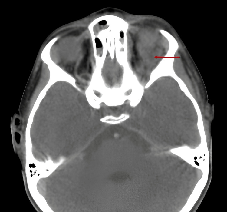 Figure 1
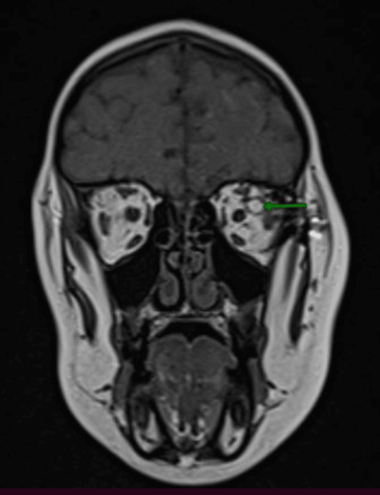 Figure 2
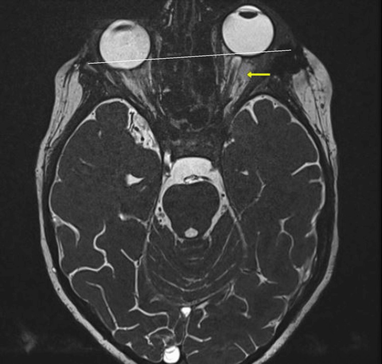 Figure 3
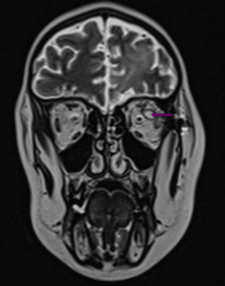 Figure 4
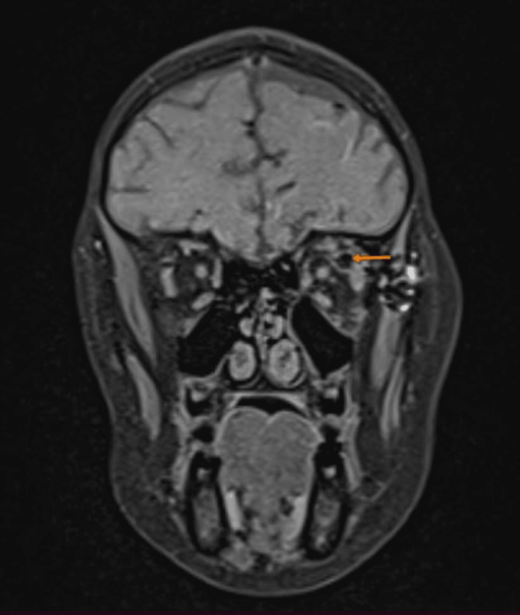 Figure 5
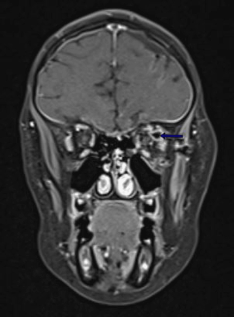 Figure 6
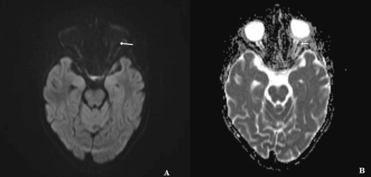 Figure 7
Peer Reviews
No public reviews on file for this paper yet. If you reviewed it on a platform where reviews are public (OpenReview, ICLR, NeurIPS, ICML), you can paste yours below so the community can read it here.
Videos
No videos yet. Explain this paper in a talk, walkthrough, or lecture? Add one.
Taxonomy
TopicsTeratomas and Epidermoid Cysts · Ocular Oncology and Treatments · Ocular Disorders and Treatments