A computer vision method to evaluate tumor-infiltrating lymphocytes and multiparametric modeling of neoadjuvant systemic therapy response in breast cancer
Mateusz Bielecki, Fang-I Lu, Angeline Vo, Eileen Rakovitch, Katarzyna J. Jerzak, Roberto Salgado, Raffi Karshafian, William T. Tran

TL;DR
This study uses computer vision and machine learning to predict breast cancer treatment response by analyzing tumor-infiltrating lymphocytes in pre-treatment biopsies.
Contribution
A novel computer vision method and multiparametric model for predicting neoadjuvant therapy response in breast cancer using TIL spatial features.
Findings
ML models using clinical and graph-based features achieved high predictive accuracy (AUC up to 0.955).
Ensemble models integrating clinical and graph features outperformed individual models in predicting pCR.
Significant differences in performance were observed for triple-negative breast cancer models.
Abstract
Neoadjuvant systemic therapy (NST) is often used to treat locally advanced breast cancer (BC) or patients with early-stage BC at high risk for micrometastatic spread. Pathological complete response (pCR) to NST in BC is associated with excellent prognostic outcomes; however, rates vary significantly. Tumor-infiltrating lymphocytes (TILs) are associated with NST response, suggesting potential as predictive biomarkers. To develop a computer vision approach to quantify spatial TIL parameters and a multiparametric machine learning (ML) model for predicting NST response. Retrospective, single institution study of 411 BC patients, combining clinical and graph-level pre-treatment histopathology data to predict response to NST using ML. Pre-treatment core needle biopsies were prepared, stained with hematoxylin and eosin, and digitized into whole slide images. Convolutional neural networks…
Genes, proteins, chemicals, diseases, species, mutations and cell lines named across the full text — each resolved to its canonical identifier and authoritative record.
Click any figure to enlarge with its caption.
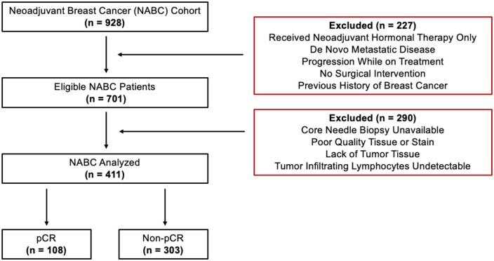 Figure 1
 Figure 2
 Figure 3
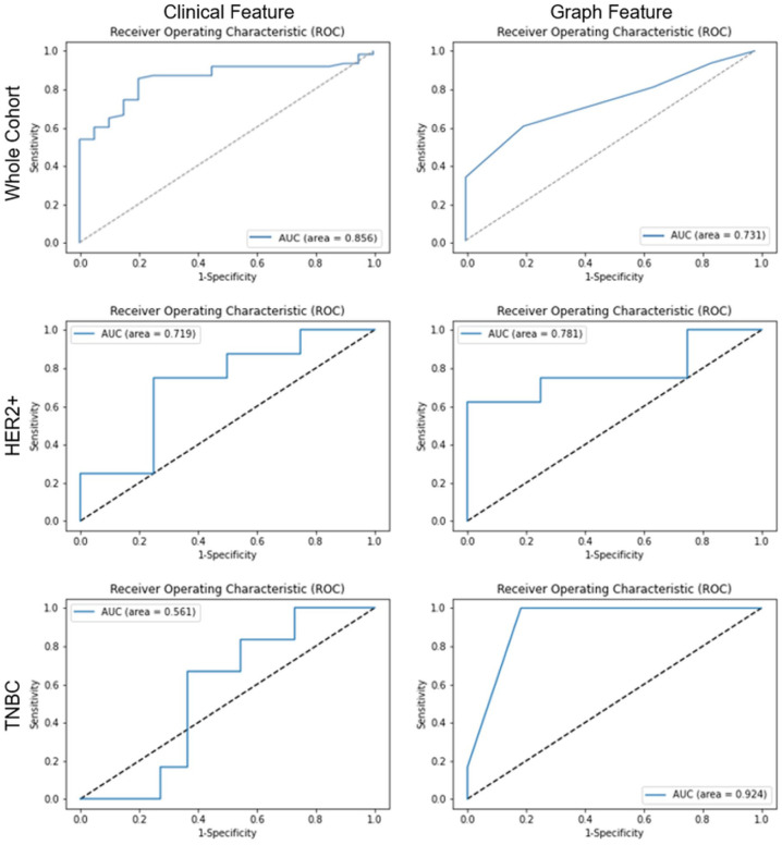 Figure 4
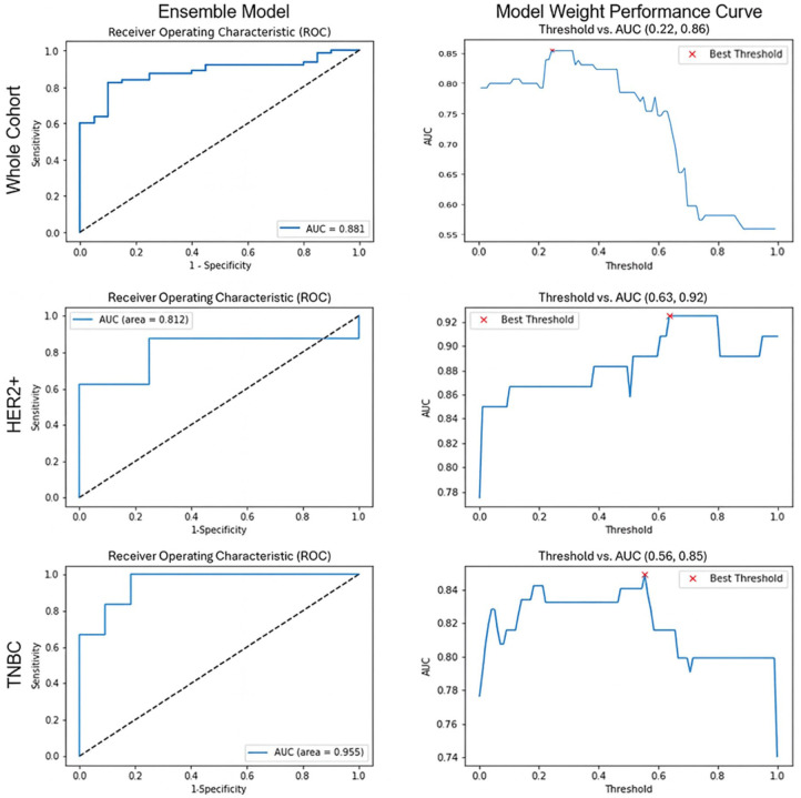 Figure 5
 Figure 6
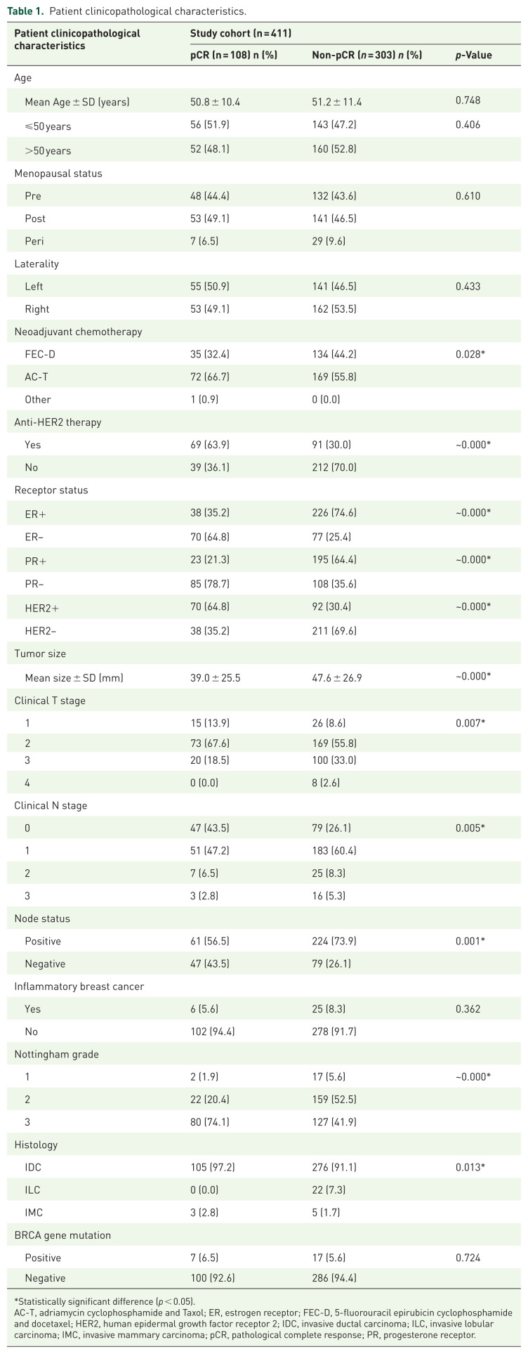 Figure 7
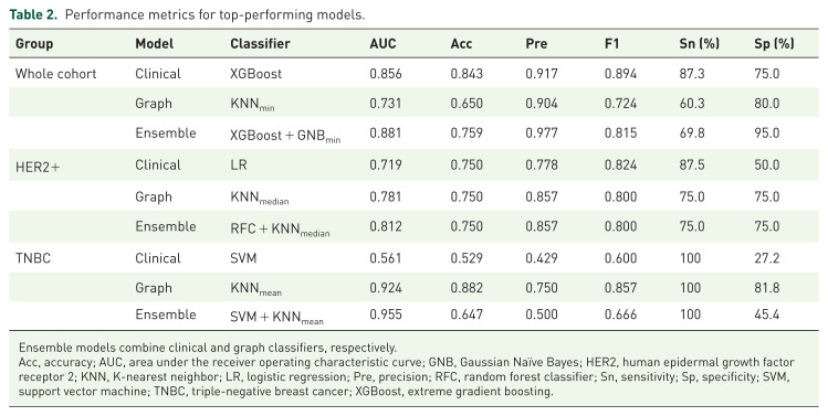 Figure 8
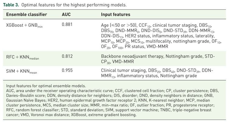 Figure 9
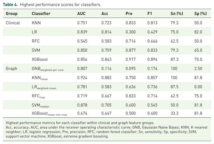 Figure 10
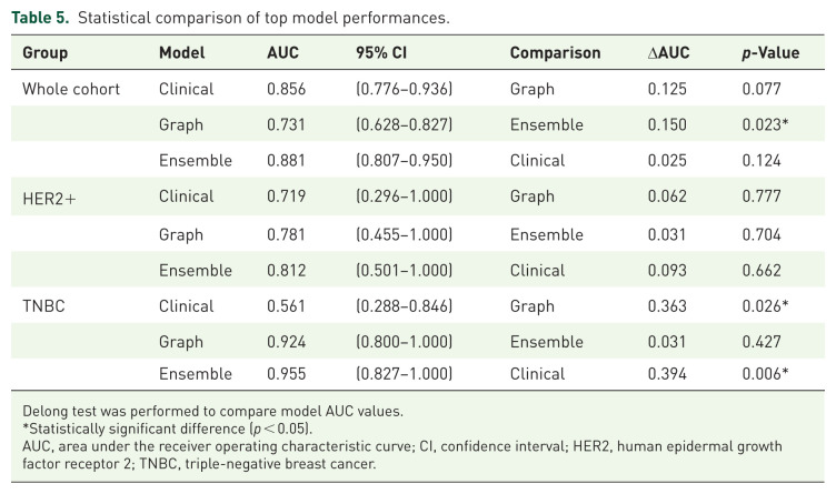 Figure 11
Peer Reviews
No public reviews on file for this paper yet. If you reviewed it on a platform where reviews are public (OpenReview, ICLR, NeurIPS, ICML), you can paste yours below so the community can read it here.
Videos
No videos yet. Explain this paper in a talk, walkthrough, or lecture? Add one.
Taxonomy
TopicsAI in cancer detection · Single-cell and spatial transcriptomics · Digital Imaging for Blood Diseases