Retrograde longitudinal imaging analyses of IDH-wildtype glioblastoma reveal its clinical timeline from radiological birth to death
Ryota Shigeeda, Ichiyo Shibahara, Yasushi Orihashi, Yoko Tanihata, Kazuko Fujitani, Mariko Toyoda, Yuri Hyakutake, Hajime Handa, Madoka Inukai, Sumito Sato, Mitsuhiro Shinoda, Hideto Komai, Kohei Uemasu, Takashi Kiga, Hiroyuki Koizumi, Daisuke Yamamoto, Kazuhiro Miyasaka

TL;DR
This study tracks the progression of a specific type of brain tumor from its earliest detectable stage to death, showing how long it takes to become visible on scans and how it evolves clinically.
Contribution
The study provides the first detailed longitudinal analysis of IDH-wildtype glioblastoma's clinical timeline from radiological birth to death.
Findings
Radiological birth of IDH-wildtype glioblastoma occurs about 0.83 years before diagnosis.
Tumors with certain genetic alterations progress more rapidly, while their absence delays progression.
The median time from radiological birth to death is 2.8 years.
Abstract
Glioblastoma (GB), IDH-wildtype, and low-grade glioma appear indistinguishable in their early pre-symptomatic phase, yet GB follows a far more aggressive clinical course. While genomic studies suggest a “biological birth” of GB years before diagnosis, when GB first becomes radiologically detectable (radiological birth) remains unknown. We analyzed longitudinal imaging data from 67 early-stage glioblastoma (earlyGB) cases, characterized by small, asymptomatic lesions that later progressed to classic magnetic resonance imaging appearance of GB (classicGB), comprising 44 institutional and 23 from published reports. A mathematical model integrating tumor volume, radius, imaging intervals, clinical data, and molecular features estimated radiological birth and its modifiers. The median interval from earlyGB to classicGB was 155 days (range: 35-1557) in our cohort and 113 days (range: 4-854)…
Genes, proteins, chemicals, diseases, species, mutations and cell lines named across the full text — each resolved to its canonical identifier and authoritative record.
Click any figure to enlarge with its caption.
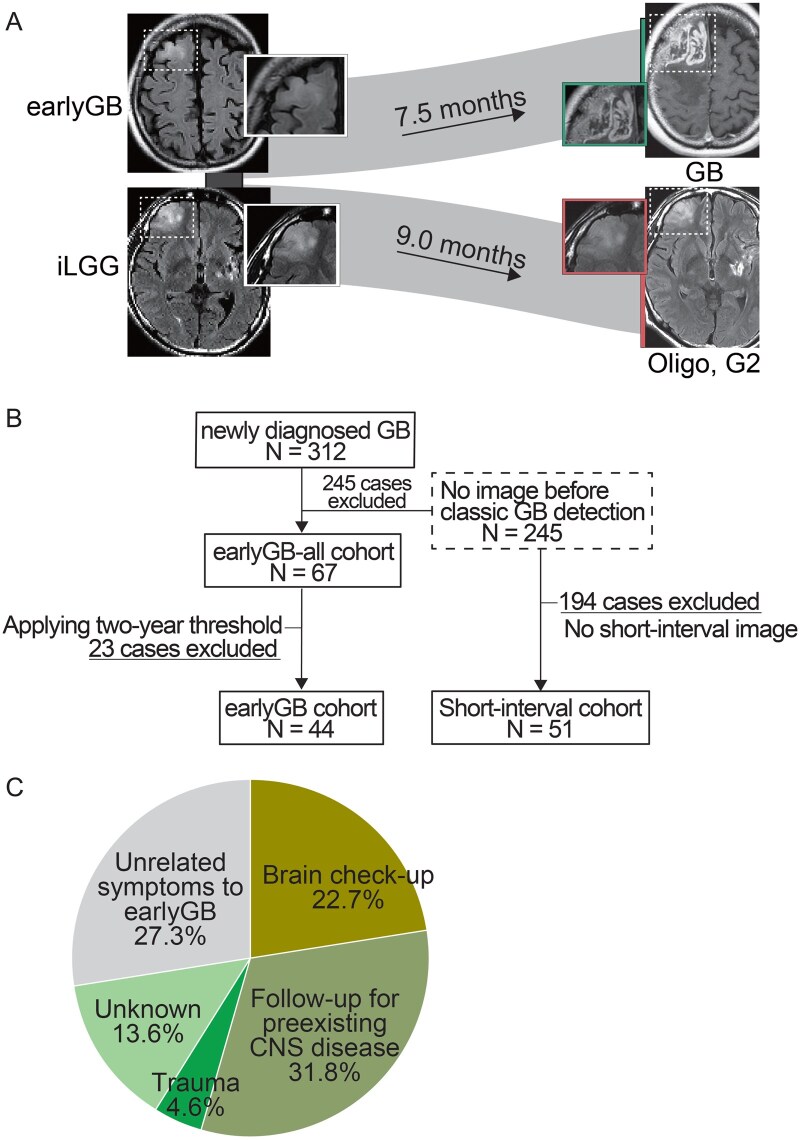 Figure 1
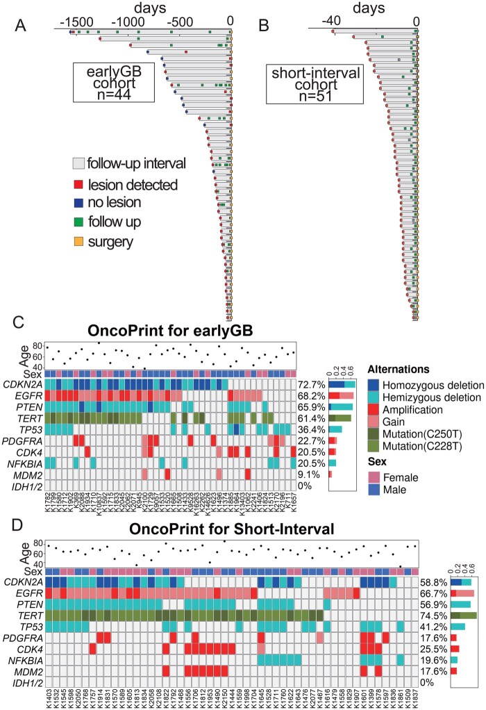 Figure 2
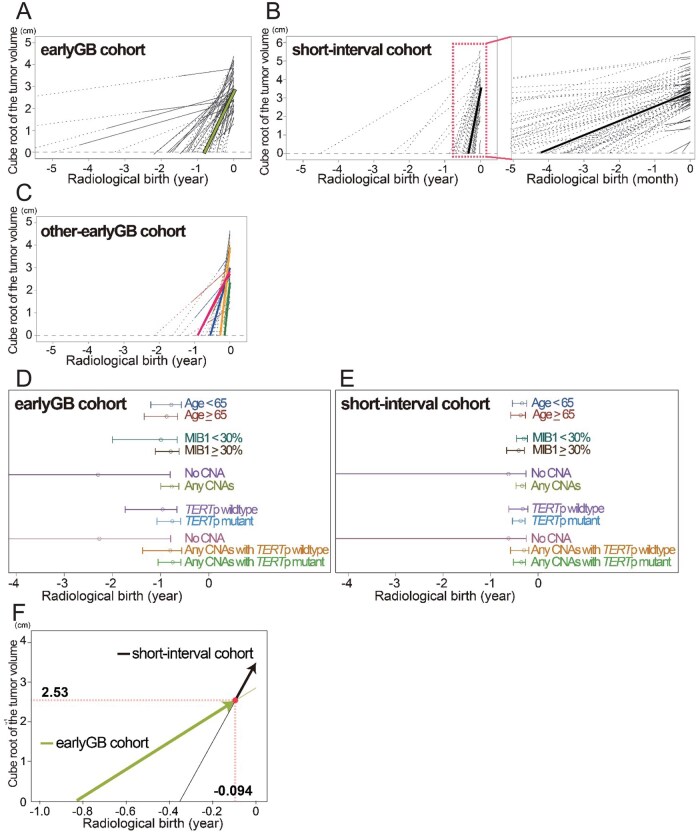 Figure 3
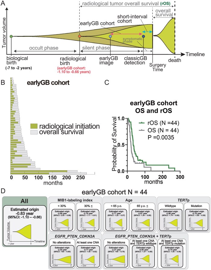 Figure 4
Peer Reviews
No public reviews on file for this paper yet. If you reviewed it on a platform where reviews are public (OpenReview, ICLR, NeurIPS, ICML), you can paste yours below so the community can read it here.
Videos
No videos yet. Explain this paper in a talk, walkthrough, or lecture? Add one.
Taxonomy
TopicsGlioma Diagnosis and Treatment · Brain Tumor Detection and Classification · Radiomics and Machine Learning in Medical Imaging