Avoiding splenectomy in splenic sclerosing angiomatoid nodular transformation through endoscopic ultrasound-guided tissue acquisition: a 36-month follow-up case report
Takaki Okuyama, Kazuyuki Matsumoto, Kosaku Morimoto, Shogo Kimura, Takayoshi Miyake, Takuya Satomi, Kensuke Takei, Shogo Inoue, Ryuta Takenaka

TL;DR
A patient with a splenic mass avoided surgery after a biopsy confirmed a rare condition, and the mass later regressed over 36 months.
Contribution
Demonstrates that endoscopic ultrasound-guided biopsy can confirm splenic sclerosing angiomatoid nodular transformation without splenectomy.
Findings
Endoscopic ultrasound-guided biopsy confirmed the diagnosis of splenic sclerosing angiomatoid nodular transformation.
The splenic lesion regressed in size after biopsy and remained stable for 36 months without splenectomy.
Spleen-preserving management is feasible for biopsy-confirmed cases of this condition.
Abstract
A 48-mm splenic mass was incidentally discovered in a 78-year-old man upon computed tomography. Follow-up imaging at 12 months revealed enlargement to 60 mm, prompting endoscopic ultrasound-guided tissue acquisition with a 22-gauge needle. Histopathological analysis confirmed that it was a sclerosing angiomatoid nodular transformation. The patient was asymptomatic and had no hematologic abnormalities; therefore, splenectomy was not performed. After biopsy, the lesion regressed from 60 mm to 46 mm, possibly owing to hematoma formation or vascular disruption, and remained stable during 36 months of follow-up. Although splenectomy has been performed in most reported cases of sclerosing angiomatoid nodular transformation because of diagnostic uncertainty, a few recent reports have demonstrated that sclerosing angiomatoid nodular transformation can be diagnosed by endoscopic…
Genes, proteins, chemicals, diseases, species, mutations and cell lines named across the full text — each resolved to its canonical identifier and authoritative record.
Click any figure to enlarge with its caption.
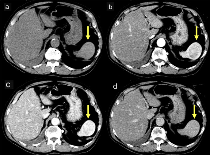 Figure 1
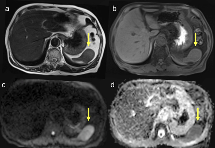 Figure 2
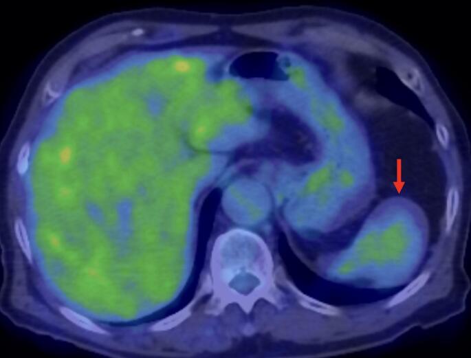 Figure 3
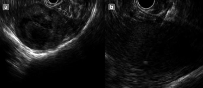 Figure 4
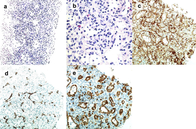 Figure 5
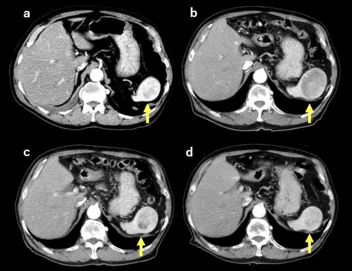 Figure 6
Peer Reviews
No public reviews on file for this paper yet. If you reviewed it on a platform where reviews are public (OpenReview, ICLR, NeurIPS, ICML), you can paste yours below so the community can read it here.
Videos
No videos yet. Explain this paper in a talk, walkthrough, or lecture? Add one.
Taxonomy
TopicsAbdominal Trauma and Injuries · Hepatocellular Carcinoma Treatment and Prognosis · Abdominal vascular conditions and treatments