Brain texture alterations predict subtle visual perceptual dysfunctions in recent onset psychosis and clinical high-risk state
Rebekka Lencer, Andreas Sprenger, Inga Meyhöfer, Udo Dannlowski, Georg Romer, Lana Kambeitz-Ilankovic, Joseph Kambeitz, Theresa Lichtenstein, Marlene Rosen, Stephan Ruhrmann, Shalaila S. Haas, Raimo K. R. Salokangas, Christos Pantelis, Carolina Bonivento, Frauke Schultze-Lutter

TL;DR
This study shows that brain texture changes can predict subtle visual perceptual issues in early psychosis and high-risk individuals.
Contribution
The study introduces brain texture features as a novel predictor of visual perceptual dysfunction in early psychosis and clinical high-risk states.
Findings
Brain texture features predicted visual perceptual dysfunction with over 77% accuracy in recent onset psychosis.
The prediction models also showed 64% accuracy in clinical high-risk samples and 50% in recent onset depression.
Brain features were significantly linked to symptom severity and psychosocial functioning scores.
Abstract
Deeper understanding of Subtle Visual Dysfunctions (VisDys) in the early stage of mental illness and their neurobiological underpinnings, as reflected by microstructural brain texture features, could advance our understanding of the underlying disease perceptual mechanisms that mediate susceptibility to psychosis. In this study, we aim a) to investigate the utility of brain texture features for the prediction of VisDys in recent onset psychosis (ROP) and clinical high-risk syndromes for psychosis (CHR-P), respectively, b) to test prediction models established in ROP and CHR-P in an independent validation sample with recent onset depression (ROD) diagnoses and c) to test for symptom expression related brain features associated with VisDys. sMRI were acquired in a training sample including 128 ROP (67 patients with VisDys), 134 CHR-P (71 patients with VisDys). Independent validation sets…
Genes, proteins, chemicals, diseases, species, mutations and cell lines named across the full text — each resolved to its canonical identifier and authoritative record.
Click any figure to enlarge with its caption.
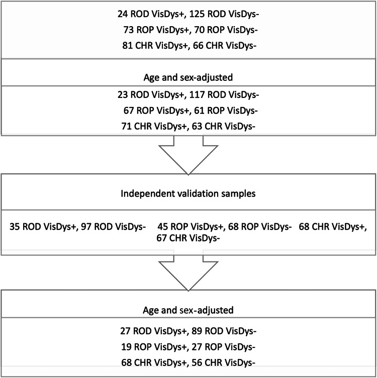 Figure 1
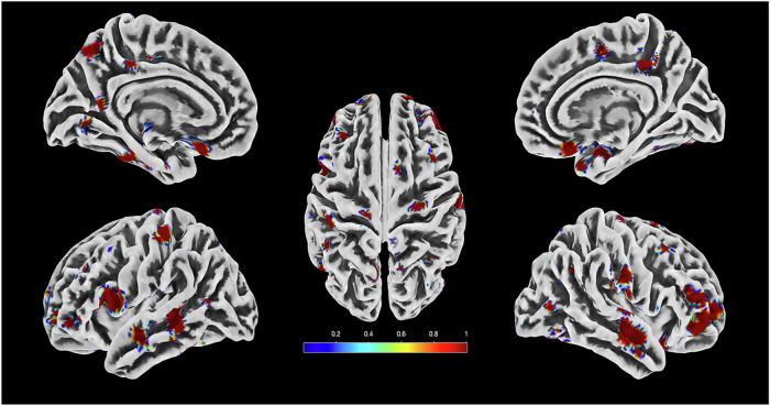 Figure 2
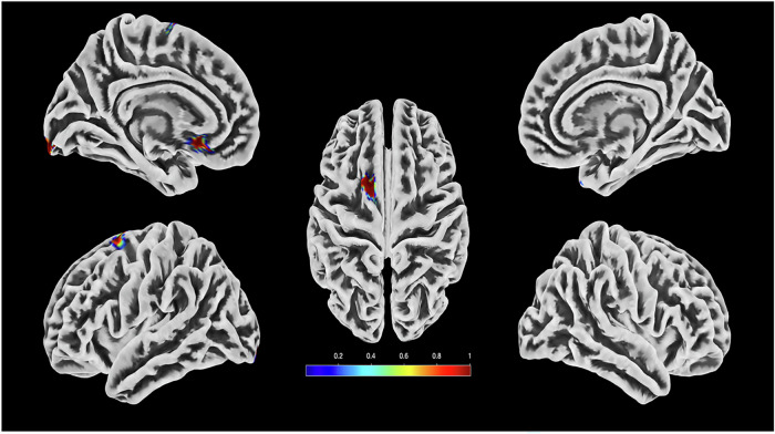 Figure 3
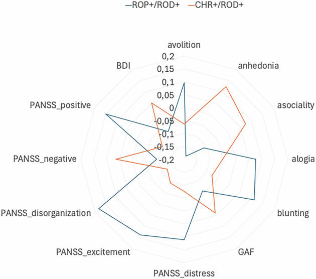 Figure 4
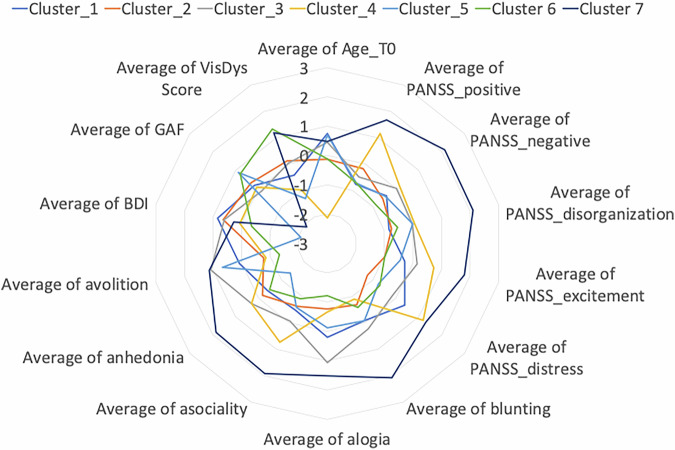 Figure 5
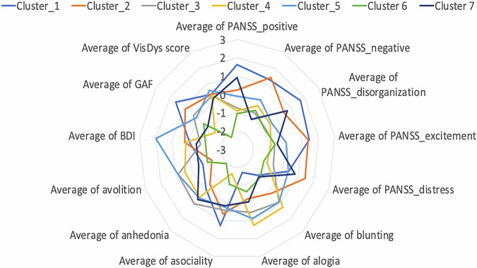 Figure 6
Peer Reviews
No public reviews on file for this paper yet. If you reviewed it on a platform where reviews are public (OpenReview, ICLR, NeurIPS, ICML), you can paste yours below so the community can read it here.
Videos
No videos yet. Explain this paper in a talk, walkthrough, or lecture? Add one.
Taxonomy
TopicsSchizophrenia research and treatment · Functional Brain Connectivity Studies · Advanced Neuroimaging Techniques and Applications