Structural Fiber Tract Alterations in Relation to Surgery in Children With a Posterior Fossa Tumor
Pien E. J. Jellema, Jannie P. Wijnen, Karina J. Kersbergen, Martijn Froeling, Maarten H. Lequin, Wouter P. Nieuwenhuis, Alberto De Luca, Eelco W. Hoving

TL;DR
This study examines how surgery for posterior fossa tumors in children affects brain fiber tracts, particularly the dentato-rubro-thalamic tract, which may be linked to post-surgery speech issues.
Contribution
The study introduces a method to assess macro- and microstructural changes in the DRTT during surgery using intraoperative diffusion MRI tractography.
Findings
Intraoperative reductions in DRTT and AF tract volumes and diameters were observed during posterior fossa tumor surgery.
Patients with postoperative speech disturbances showed higher variability in microstructural measures along the DRTT.
Microstructural changes in the DRTT near the resection cavity were more pronounced compared to the arcuate fasciculus.
Abstract
Cerebellar mutism syndrome (CMS) is a potential complication of pediatric posterior fossa tumor (pPFT) resection and may be related to disruption of the dentato‐rubro‐thalamic tract (DRTT). Intraoperative tractography allows assessment of changes to the DRTT during surgery. We evaluated macro‐ and microstructural changes of the DRTT by comparing pre‐ and intraoperative diffusion MRI (dMRI) tractography in patients with pPFT. Pre‐ and intraoperative T1‐weighted and dMRI data were acquired to reconstruct the DRTT, while the arcuate fasciculus (AF) was reconstructed as a control tract. To evaluate macrostructural intraoperative alterations, tract volume and diameter were calculated. Microstructural changes were assessed using fractional anisotropy (FA) and mean diffusivity (MD), both globally and along the tract. Speech disturbances, as a characteristic symptom of CMS, were scored…
Genes, proteins, chemicals, diseases, species, mutations and cell lines named across the full text — each resolved to its canonical identifier and authoritative record.
Click any figure to enlarge with its caption.
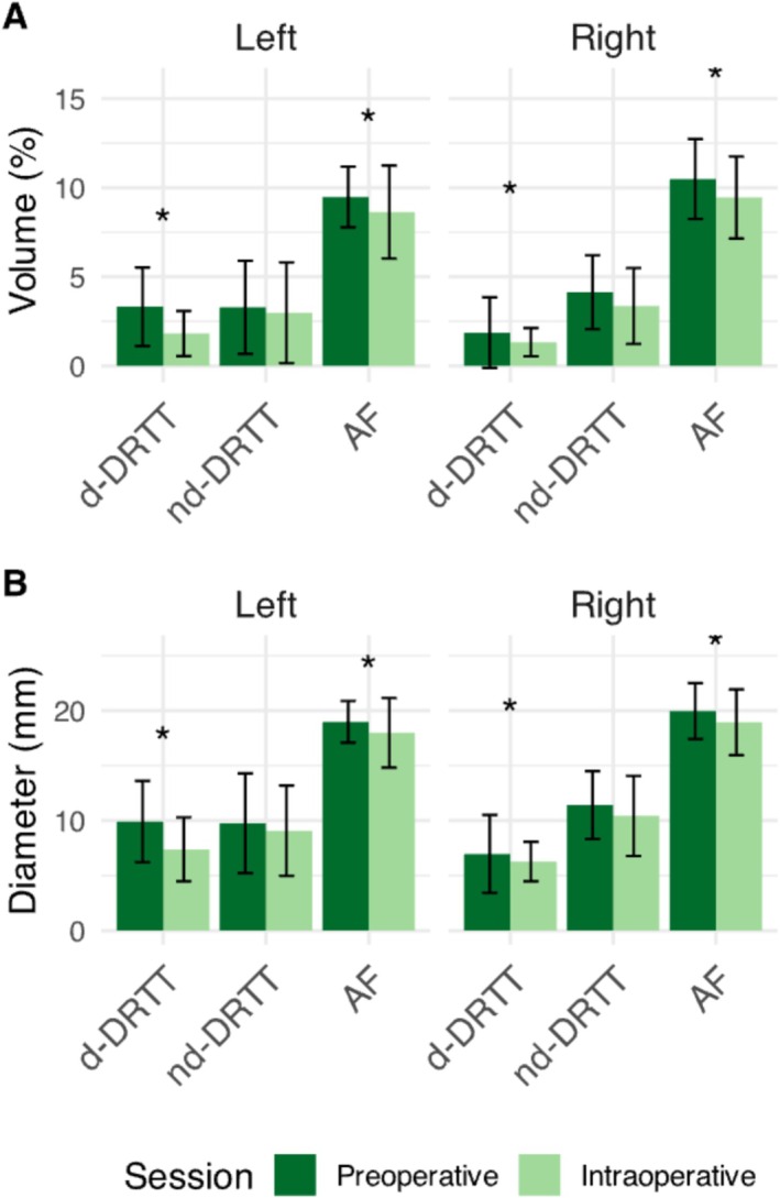 Figure 1
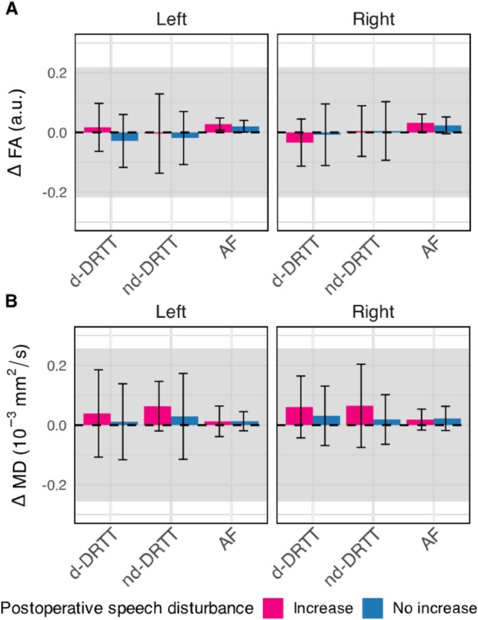 Figure 2
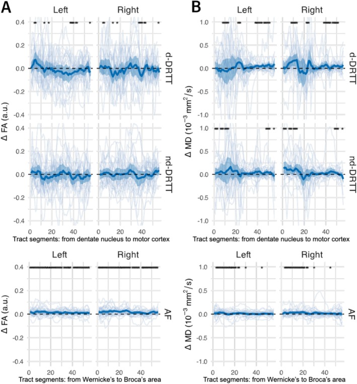 Figure 3
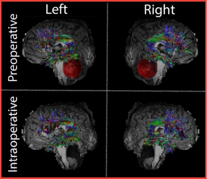 Figure 4
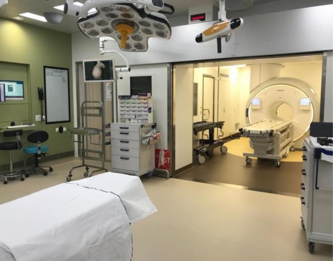 Figure 5
 Figure 6
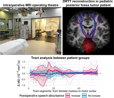 Figure 7
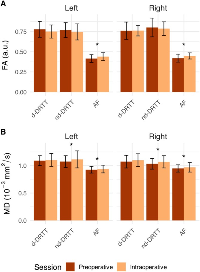 Figure 8
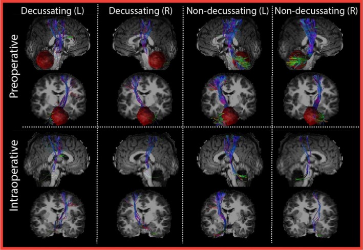 Figure 9
Peer Reviews
No public reviews on file for this paper yet. If you reviewed it on a platform where reviews are public (OpenReview, ICLR, NeurIPS, ICML), you can paste yours below so the community can read it here.
Videos
No videos yet. Explain this paper in a talk, walkthrough, or lecture? Add one.
Taxonomy
TopicsAdvanced Neuroimaging Techniques and Applications · Epilepsy research and treatment · Trigeminal Neuralgia and Treatments