3D heterotypic models of glioblastoma reveal the impact of microglia on cellular organization and the production of a distinct secretome
Clara García-Sáez, Josune Alonso-Marañón, Mikel García-Puga, Ane Rubio-Zulaika, Irati de Goñi-Garcia, Lorea Blázquez, Sandra Camarero-Espinosa

TL;DR
This study shows that microglia in glioblastoma tumors help cancer cells grow more and resist drugs, by forming protective structures and promoting immune evasion.
Contribution
The study introduces a 3D heterotypic model showing how microglia influence glioblastoma growth and drug resistance through structural and secretory changes.
Findings
Heterotypic spheroids with microglia showed increased proliferation and drug resistance compared to homotypic spheroids.
A core-shell structure formed in heterotypic spheroids, with microglia creating a protective shell around glioma cells.
Heterotypic spheroids induced monocyte migration and M2 polarization, enhancing immune evasion.
Abstract
Glioblastoma (GBM) is a deadly brain tumor with a very poor prognosis. Development of new therapeutics is hindered by the lack of appropriate preclinical models that reflect the complexity of the tumor microenvironment, especially the crucial role of microglia. In this study, we investigated the impact of microglia on GBM models using humanized 3D spheroids. Homotypic and heterotypic spheroids were created out of a GBM-derived cell line (DKMG) or patient-derived glioma stem cells (GB22-13), along with a microglia cell line (HMC3). Heterotypic glioma-HMC3 spheroids exhibited increased proliferation and greater drug resistance to chemotherapy drug Temozolomide compared with homotypic spheroids. Heterotypic spheroids also grew larger, developed multinucleated structures within 7 days, and had a greater invasive potential. Additionally, a distinct core-shell structure emerged in the…
Genes, proteins, chemicals, diseases, species, mutations and cell lines named across the full text — each resolved to its canonical identifier and authoritative record.
Click any figure to enlarge with its caption.
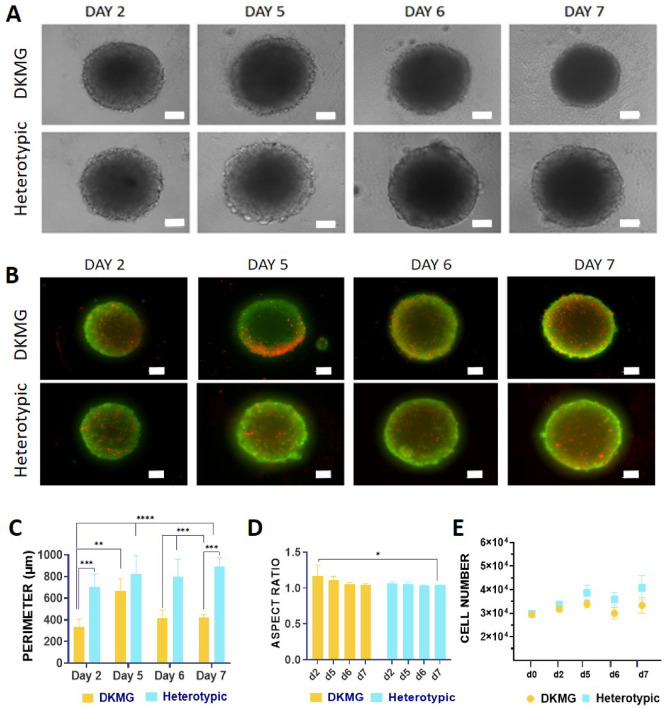 Figure 1
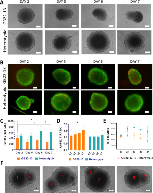 Figure 2
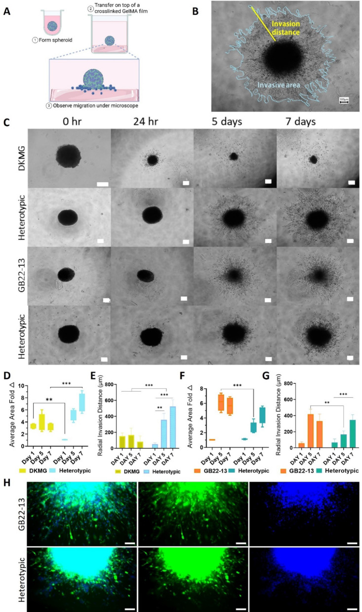 Figure 3
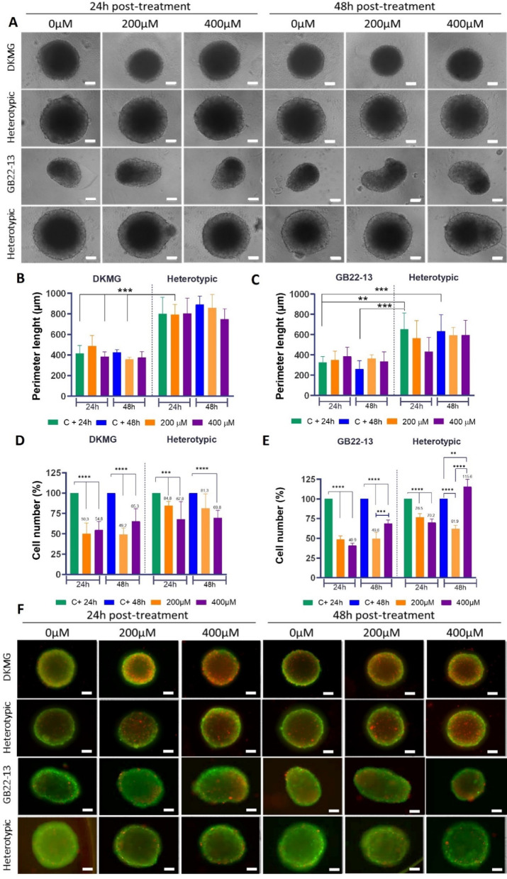 Figure 4
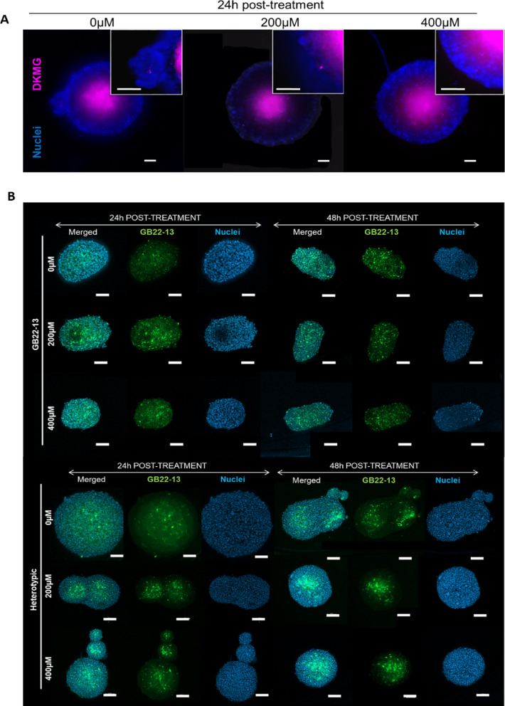 Figure 5
 Figure 6
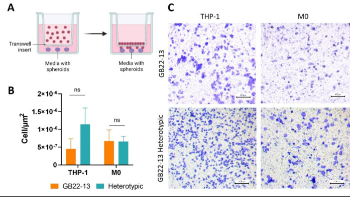 Figure 7
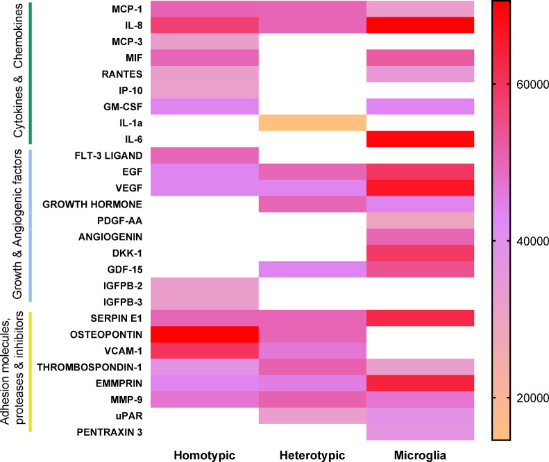 Figure 8
Peer Reviews
No public reviews on file for this paper yet. If you reviewed it on a platform where reviews are public (OpenReview, ICLR, NeurIPS, ICML), you can paste yours below so the community can read it here.
Videos
No videos yet. Explain this paper in a talk, walkthrough, or lecture? Add one.
Taxonomy
TopicsGlioma Diagnosis and Treatment · Immune cells in cancer · Neuroinflammation and Neurodegeneration Mechanisms