Research progress in electrochemical determination of Janus kinase inhibitors and proposals for amplifying clinical applicability
Peter Mikus, Michal Hanko, Zuzana Zelinkova, Jan Labuda

TL;DR
This paper reviews recent advances in electrochemical methods for detecting Janus kinase inhibitors, comparing them to traditional techniques and suggesting ways to improve their clinical use.
Contribution
The paper introduces electrochemical detection of JAK inhibitors as a novel, sensitive method with potential for real-time point-of-care therapeutic drug monitoring.
Findings
Electrochemical methods achieve detection limits as low as 10−9 M to 10−12 M for JAK inhibitors.
Modified electrodes with nanomaterials and imprinted polymers enhance sensitivity and specificity.
Electrochemical approaches show promise for real-time, personalized drug monitoring in clinical settings.
Abstract
Janus kinase inhibitors (JAKis) represent a class of drugs that treat inflammatory and autoimmune diseases by blocking Janus kinase (JAK) enzymes. Monitoring of precise drug concentration ensures therapeutic effect while minimizing the risk of toxicity. In parallel to conventional methods based on high performance liquid chromatography, liquid chromatography − mass spectrometric or spectrometric methods, sensitive electrochemical methods for the detection of JAKis have been developed in very recent years. The procedures utilize conventional bare glassy carbon electrode or boron-doped diamond electrode and, particularly, chemically modified electrodes incorporating nanomaterials and their composites as powerful catalysts as well as imprinted polymers. The linear concentration ranges and limits of detection achieve very low 10− 9 M to 10− 12 M (µg to ng/mL) values, matching clinically…
Genes, proteins, chemicals, diseases, species, mutations and cell lines named across the full text — each resolved to its canonical identifier and authoritative record.
Click any figure to enlarge with its caption.
 Figure 1
 Figure 2
 Figure 3
 Figure 4
 Figure 5
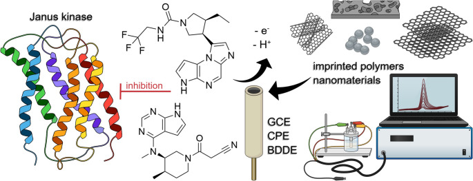 Figure 6
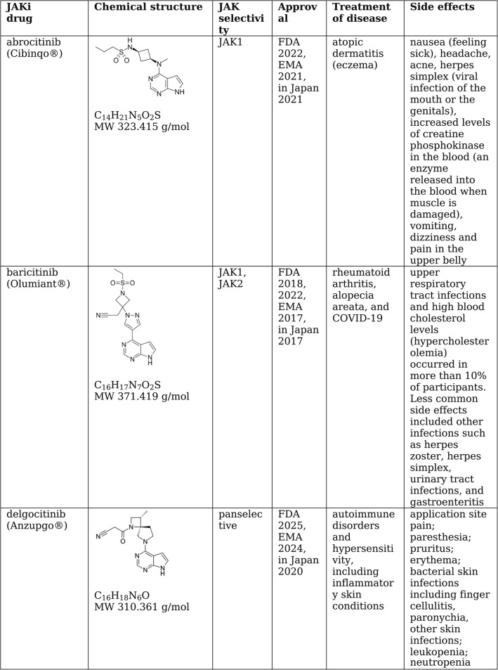 Figure 7
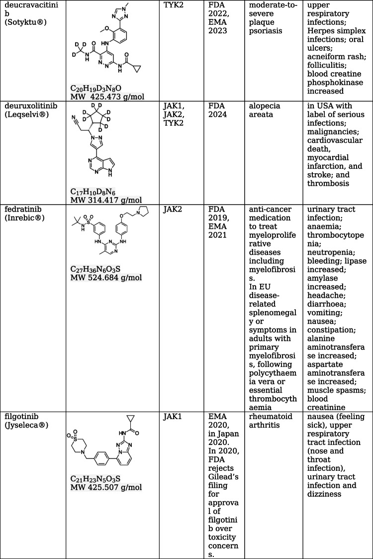 Figure 8
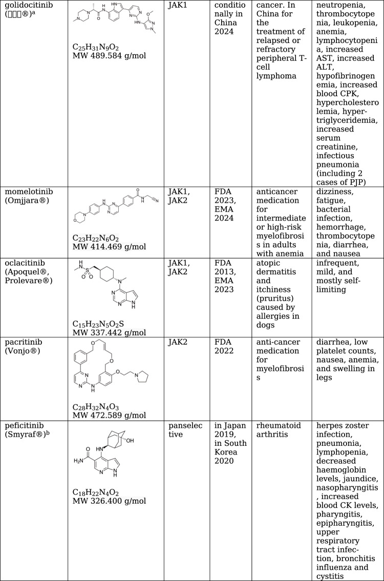 Figure 9
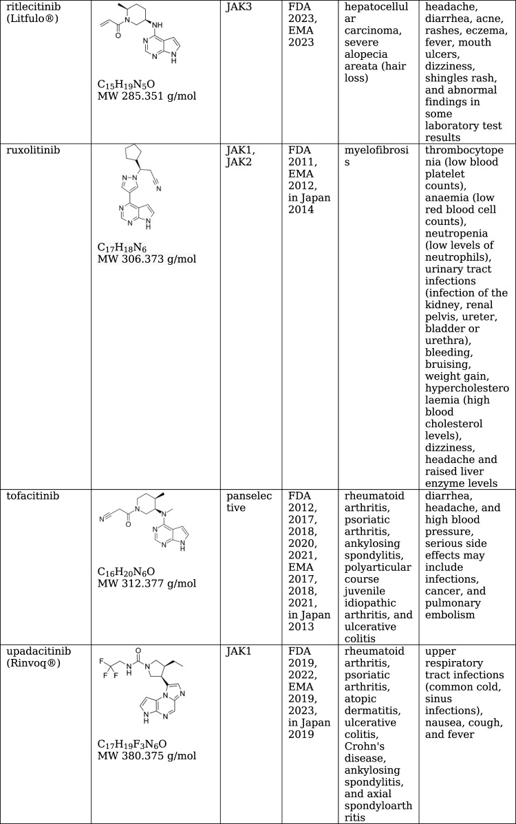 Figure 10
Peer Reviews
No public reviews on file for this paper yet. If you reviewed it on a platform where reviews are public (OpenReview, ICLR, NeurIPS, ICML), you can paste yours below so the community can read it here.
Videos
No videos yet. Explain this paper in a talk, walkthrough, or lecture? Add one.
Taxonomy
TopicsCytokine Signaling Pathways and Interactions · Ocular Diseases and Behçet’s Syndrome · interferon and immune responses