Quantification of Kidney Inflammation Using Nanobubble-mediated Contrast Enhanced Ultrasound
Niloufar Rostam Shirazi, Xiaolin He, Dana Dranka, Omar Falou, Eno Hysi, Agata A. Exner, Darren Yuen, Michael C. Kolios

TL;DR
This study introduces a non-invasive ultrasound method using nanobubbles to measure kidney inflammation, offering a potential alternative to biopsies.
Contribution
The novel use of Cy5-labeled nanobubbles with contrast-enhanced ultrasound to quantify kidney inflammation in a preclinical model.
Findings
CEUS imaging with nanobubbles revealed impaired microvascular perfusion and increased retention in inflamed kidneys.
Decorrelation time mapping showed prolonged nanobubble retention, indicating increased capillary permeability in injured kidneys.
Imaging metrics correlated with histological injury scores, validating the non-invasive method's accuracy.
Abstract
Kidney inflammation is a central driver of acute kidney injury (AKI) and its progression to chronic kidney disease (CKD). While several imaging and biomarker-based approaches are under development, clinically validated non-invasive methods to directly quantify renal inflammation remain limited. This study introduces a novel approach using contrast-enhanced ultrasound (CEUS) with Cy5-labeled nanobubbles (NBs) to address this critical knowledge gap. Using a murine ischemia-reperfusion injury (IRI) model, CEUS imaging enabled real-time visualization of inflammation-induced changes in kidney perfusion and vascular integrity. Parametric analyses of non-linear imaging revealed delayed time-to-peak (TTP) and increased area under the falling curve (AUfC) in IRI kidneys, suggesting impaired microvascular perfusion and NB retention. Decorrelation time (DT) mapping further identified prolonged NB…
Genes, proteins, chemicals, diseases, species, mutations and cell lines named across the full text — each resolved to its canonical identifier and authoritative record.
Click any figure to enlarge with its caption.
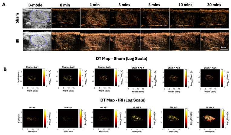 Figure 1
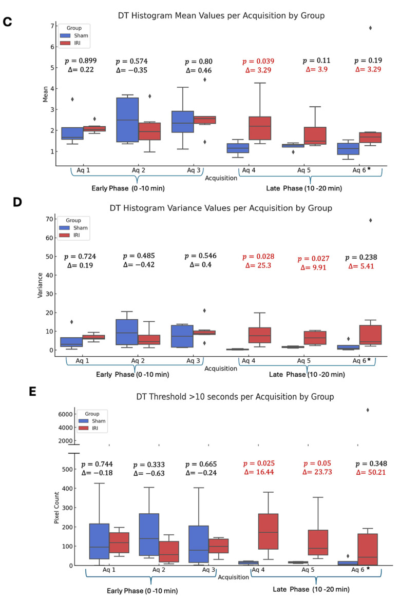 Figure 2
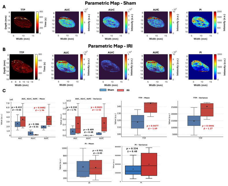 Figure 3
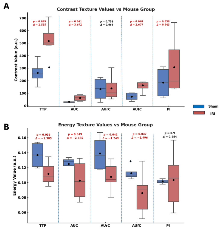 Figure 4
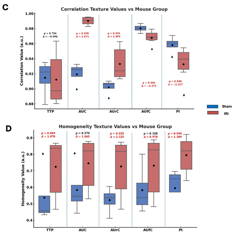 Figure 5
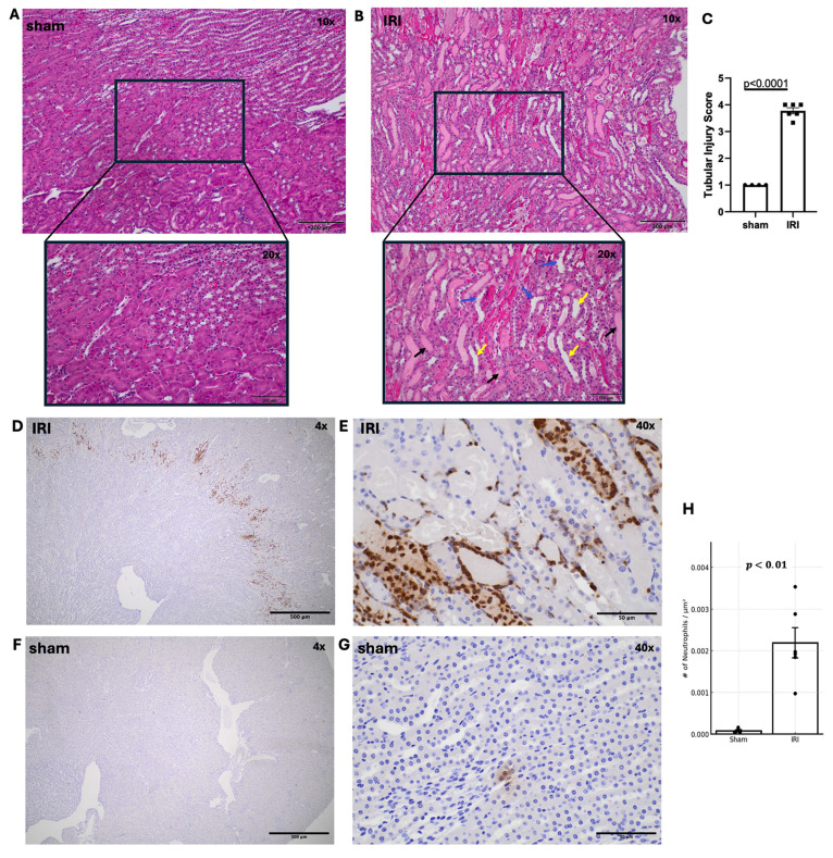 Figure 6
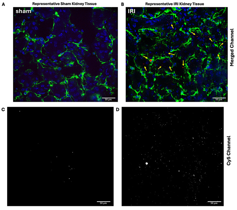 Figure 7
 Figure 8
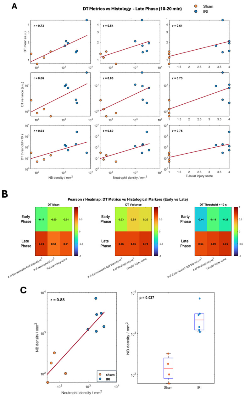 Figure 9
 Figure 10
 Figure 11
 Figure 12
Peer Reviews
No public reviews on file for this paper yet. If you reviewed it on a platform where reviews are public (OpenReview, ICLR, NeurIPS, ICML), you can paste yours below so the community can read it here.
Videos
No videos yet. Explain this paper in a talk, walkthrough, or lecture? Add one.
Taxonomy
TopicsUltrasound and Hyperthermia Applications · Photoacoustic and Ultrasonic Imaging · Kidney Stones and Urolithiasis Treatments