Subungual Glomus Tumor With Normal Findings on Imaging: A Case Report
Youssef A Abdelmegeid, Ahmed Yousry Saber, Victoria Deans

TL;DR
A case report describes a subungual glomus tumor diagnosed clinically despite normal imaging results.
Contribution
Highlights the importance of clinical diagnosis for glomus tumors when imaging results are normal.
Findings
Glomus tumor was diagnosed based on clinical findings despite normal ultrasound and MRI.
Surgical removal resolved symptoms and histology confirmed the diagnosis.
Normal imaging results should not rule out glomus tumors in clinical evaluation.
Abstract
Glomus tumors are frequently associated with cold sensitivity, pain, and tenderness. They are benign tumors arising from the glomus body and typically present with subungual pinpoint pain. Diagnosis is primarily based on clinical findings, with ultrasound or MRI often used to confirm the diagnosis. We report the diagnosis and surgical management of a 49-year-old female with a classic glomus tumor. Despite normal ultrasound and MRI findings, she was diagnosed based on history and physical examination. Surgical removal of the lesion resulted in complete resolution of her symptoms, and histology subsequently confirmed the diagnosis of a glomus tumor. Clinicians should suspect a glomus tumor based on clinical findings, and a normal ultrasound or MRI should not exclude its presence.
Genes, proteins, chemicals, diseases, species, mutations and cell lines named across the full text — each resolved to its canonical identifier and authoritative record.
Click any figure to enlarge with its caption.
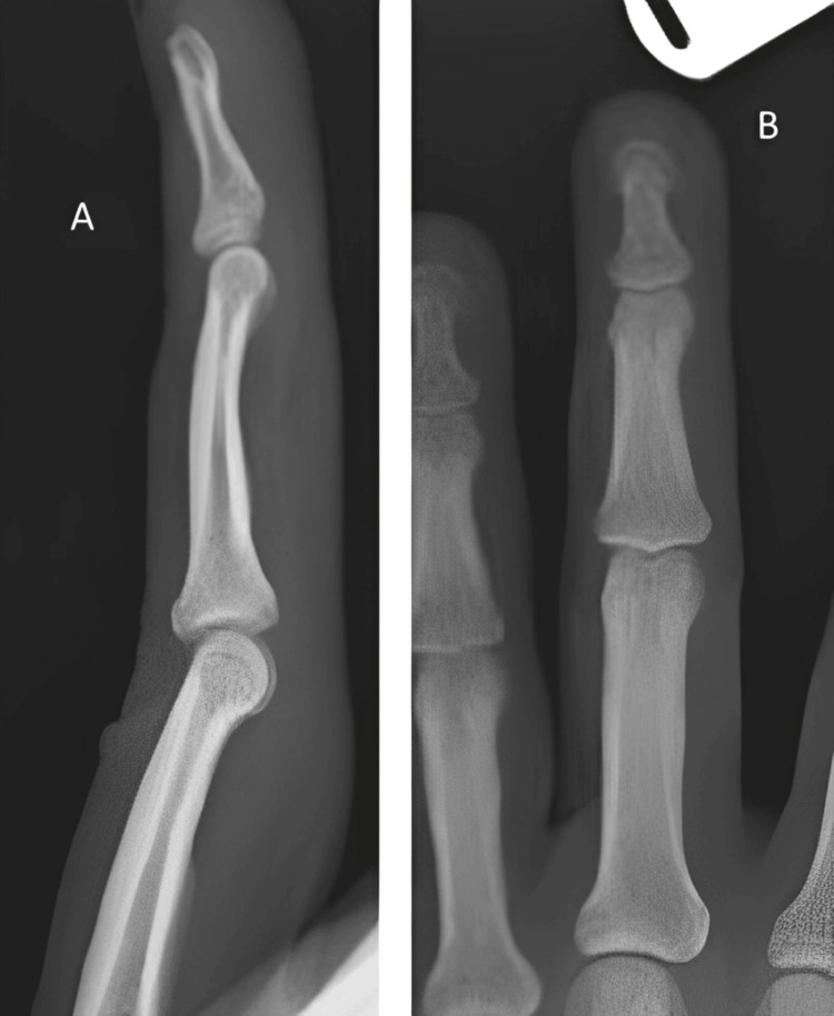 Figure 1
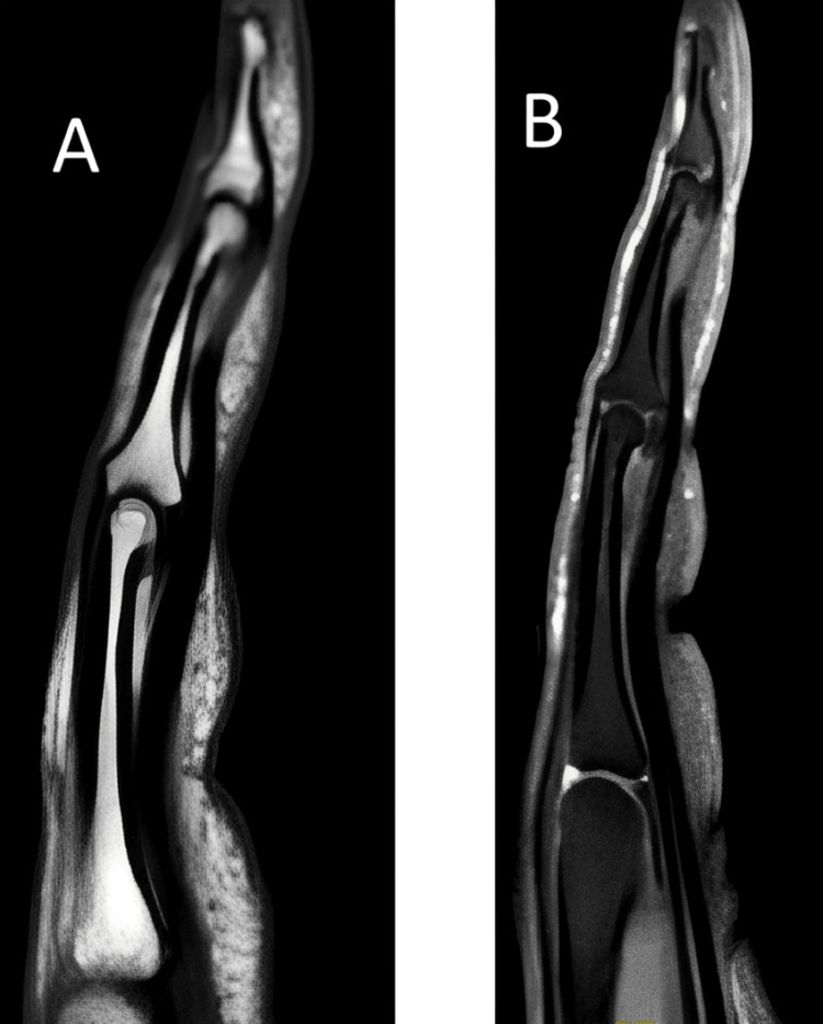 Figure 2
Peer Reviews
No public reviews on file for this paper yet. If you reviewed it on a platform where reviews are public (OpenReview, ICLR, NeurIPS, ICML), you can paste yours below so the community can read it here.
Videos
No videos yet. Explain this paper in a talk, walkthrough, or lecture? Add one.
Taxonomy
TopicsSoft tissue tumors and treatment · Head and Neck Anomalies · Teratomas and Epidermoid Cysts