Structure and dynamics of a multidomain nitric oxide synthase regulated by a C2 domain
Dhruva Nair, Brian R. Crane

TL;DR
This paper reveals the structure and dynamic regulation of a multidomain nitric oxide synthase enzyme using cryo-electron microscopy.
Contribution
The study provides the first detailed structural view of a full-length Synechococcus nitric oxide synthase with a regulatory C2 domain.
Findings
The full-length Synechococcus NOS enzyme contains a regulatory C2 domain, a NOD module, and a pseudoglobin.
Pterin binding is coupled to conformational changes in the oxygenase domain.
The C2 domain moves over 85 angstroms to regulate either the NOS or NOD heme center.
Abstract
Nitric oxide synthase (NOS) is a widely studied multidomain redox enzyme that produces the key signaling molecule and cytotoxic agent nitric oxide (NO) for functions that range from mammalian vasodilation to prokaryotic antibiotic resistance. NOS enzymes from metazoans and cyanobacteria rely on dynamic associations of their oxygenase and coupled diflavin reductase domains that have largely evaded detailed structural characterization. Cryo–electron microscopy studies of a representative dimeric six-domain Synechococcus NOS reveal the architecture of the full-length enzyme, which contains an unusual regulatory C2 domain, and additional nitric oxide dioxygenase (NOD) and pseudoglobin modules. Five distinct structural states depict how pterin binding couples to tight and loose oxygenase conformations and how the Ca2+-sensitive C2 domain moves over 85 angstroms to alternatively regulate…
Genes, proteins, chemicals, diseases, species, mutations and cell lines named across the full text — each resolved to its canonical identifier and authoritative record.
Click any figure to enlarge with its caption.
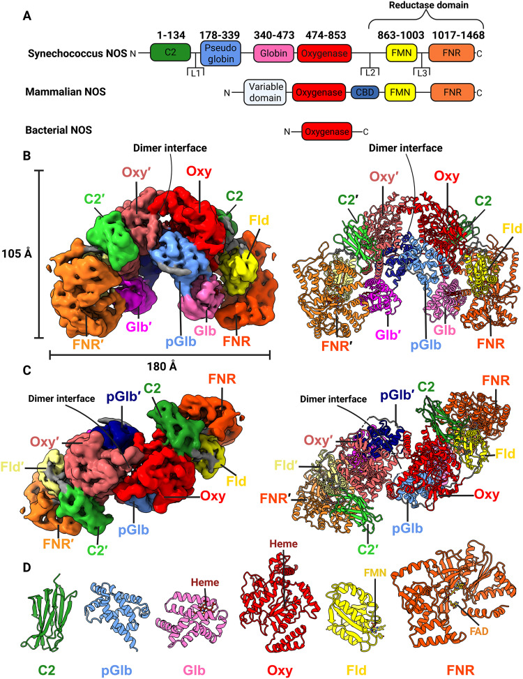 Fig. 1
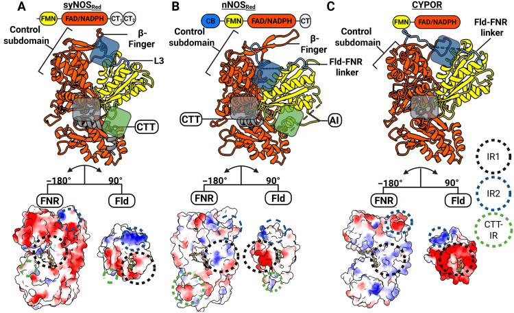 Fig. 2
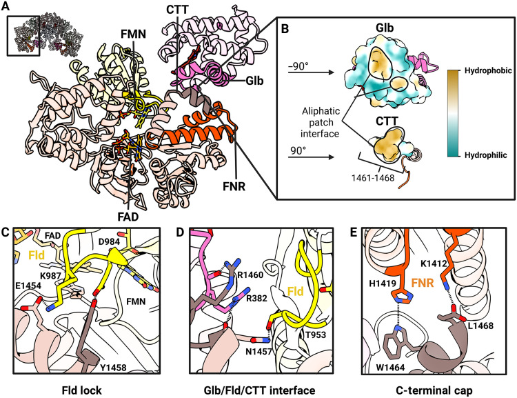 Fig. 3
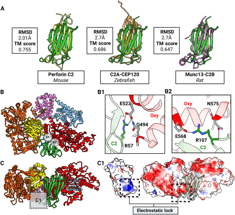 Fig. 4
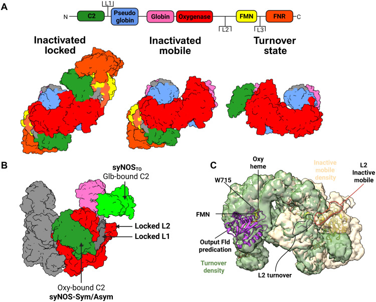 Fig. 5
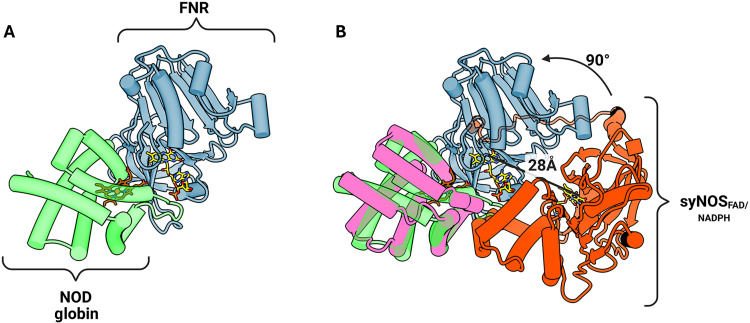 Fig. 6
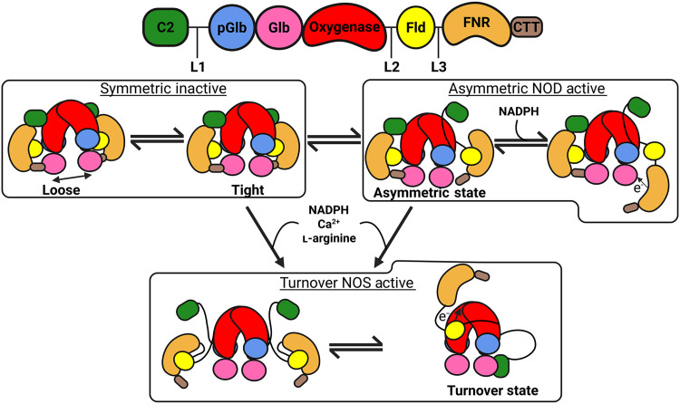 Fig. 7
- —National Science Foundation MCB
Peer Reviews
No public reviews on file for this paper yet. If you reviewed it on a platform where reviews are public (OpenReview, ICLR, NeurIPS, ICML), you can paste yours below so the community can read it here.
Videos
No videos yet. Explain this paper in a talk, walkthrough, or lecture? Add one.
Taxonomy
TopicsPhotosynthetic Processes and Mechanisms · Metal-Catalyzed Oxygenation Mechanisms · Nitric Oxide and Endothelin Effects
INTRODUCTION
Nitric oxide (NO) is a potent free-radical signal and cytotoxic agent found throughout biology (1–8). Nitric oxide synthases (NOS) are homodimeric P450-like enzymes that produce NO through the five-electron oxidation of l-arginine (l-Arg) to l-citrulline (9–12). NOSs comprise a heme-containing oxygenase domain (NOS_Oxy_) that is reductively activated by either an appended reductase domain (NOS_Red_; characteristic of metazoans) or an auxiliary reductase partner (characteristic of prokaryotes) (Fig. 1A) (10–12).
Ca2+-free syNOS has a locked architecture.(A) Gene architecture of Synechococcus NOS, mNOS, and a typical bacterial NOS. Note that some noncyanobacterial prokaryotic NOSs have auxiliary domains. (B) Side view of the electron density (left; threshold = 0.0298, C2 symmetry applied) and corresponding ribbon representation (right) for the locked syNOS homodimer (NOS-Sym). Subunit 1 (right) is colored as designated by the gene architecture, and subunit 2 (left) is colored as follows: C2′, lime green; pGlb′, navy; Glb′, magenta; Oxy′, Indian red; Fld′, pale yellow; FNR′, bright orange . (C) Top view of (B). (D) Individual syNOS domains with cofactors indicated. Created in BioRender. Crane, B. (2026) https://BioRender.com/o28o838.
Mammalian NOSs (mNOSs) are multidomain enzymes composed of an N-terminal NOS_Oxy_ and a C-terminal nicotinamide adenine dinucleotide phosphate [NADP(^+^/H)]–dependent NOS_Red_, bridged by a calmodulin (CAM)–binding domain (NOS_CBD_; Fig. 1A). NOS_Red_ belongs to the cytochrome P450 reductase (CYPOR) family and, as such, is further divided into a NOS_Oxy_-proximal flavin mononucleotide (FMN)–binding flavodoxin domain (NOS_Fld_) and a flavin adenine dinucleotide (FAD) ferredoxin–nicotinamide adenine dinucleotide phosphate (NADP^+^) reductase module (NOS_FNR_) (10–13). NOS_Oxy_ uses tetrahydrobiopterin (BH_4_) or tetrahydrofolate for rapid heme reduction during oxygen activation (14). To reduce heme for oxygen binding, electrons flow from NADPH → FAD → FMN(H)• → Heme-Fe^3+^. Ca^2+^-CAM enhances heme reduction by facilitating the traverse of NOS_Fld_ ~70 Å from a shielded position in a conformationally locked state of NOS_Red_ to a productive binding interaction with NOS_Oxy_ (15–18).
Given their medical importance (19), NOSs have been extensively studied in mammals, which have three isoforms: (i) endothelial NOS (eNOS; regulation of the vasculature), (ii) neuronal NOS (nNOS; regulation of neuronal function), and (iii) inducible NOS (iNOS; immune responses to pathogens and cancer cells) (11, 20–22). Most prokaryotic NOSs have fewer domains than their metazoan counterparts and have diverse functions that include participation in stress responses, antibiotic resistance, and biosynthesis of secondary metabolites (23–27).
To mitigate NO cytotoxicity, prokaryotes oxidize NO to NO_3_^−^ with flavohemoprotein (Hmp), which combines a heme-reducing flavin reductase with an oxygen-activating globin domain. In contrast, mammals oxidize NO with hemoglobin, cytoglobin, and neuroglobin, which obtain reduced equivalents from various environmental factors (28–30).
Cyanobacterial NOSs represent a bridge between metazoan and prokaryotic NOSs (26, 31–34). They comprise an mNOS architecture fused to an N-terminal globin domain, as exemplified by the 1468-residue NOS from Synechococcus sp. PCC 7335 (i.e., syNOS; Fig. 1A). Like mNOS, Ca^2+^ activates syNOS, despite syNOS lacking a CBD and Synechococcus lacking an obvious CAM homolog (34). The syNOS globin domain (NOS_Glb_) oxidizes NO to NO_3_^−^ as a nitric oxide dioxygenase (NOD) and, when considered with syNOS_FNR_, resembles Hmp (34).
Ca^2+^-CAM regulates mNOS activity by controlling the motions and associations of NOS_Oxy_ and NOS_Red_ that allow electron transfer (ET) between them. Owing to the dynamic nature of turnover and inactivated states of full-length NOSs, they have evaded characterization by direct structural methods. Single-particle electron microscopy (EM) in conjunction with chemical cross-linking and hydrogen-deuterium exchange mass spectrometry (HDX-MS) have yielded informative, if incomplete, structural information (35–39). These studies, along with biochemical and spectroscopic characterization (12, 40–44) have motivated a model of mNOS conformational dynamics that involves three states: (i) input, wherein NOS_Fld_ closely associates with NOS_FNR_ so that the NOS_Red_ FAD can be reduced by NADPH; (ii) intermediate, wherein NOS_Fld_ transitions between its closed position next to NOS_FNR_ and an interaction with NOS_Oxy_; and (iii) output, wherein Ca^2+^-CAM promotes docking of NOS_Fld_ onto the NOS_Oxy_ of the adjacent subunit (44–47). Although cryo–electron microscopy (cryo-EM) has provided residue-level information on NOS_Oxy_, NOS_Red_ positioning has been challenging to discern owing to its conformational heterogeneity (35, 46).
Herein, we resolve domain configurations of the multidomain syNOS by cryo-EM in conjunction with further characterizing ET properties associated with its NOS and NOD activities. We capture resting and dynamic states of the enzyme and identify an atypical C2-like domain that acts as a reversible lynchpin to mediate Ca^2+^ sensitivity. An elaborated C-terminal tail (CTT) (22) of NOS_Red_, key for regulation of mNOS, maintains a conformationally locked dimer and represents an expansion of the regulatory mechanisms seen within the diflavin reductase family.
RESULTS AND DISCUSSION
SyNOS expression, purification, and cross-linking mass spectrometry
Full-length syNOS (syNOS_FL_) was fused to an N-terminal Twin-Strep affinity tag and coexpressed along with Escherichia coli HSP90 and glyceraldehyde-3-phosphate dehydrogenase (GAPDH) in BL21(DE3) and purified through a series of affinity and size exclusion chromatography (SEC) steps (see Methods and fig. S1) (46, 48, 49). As found previously (34), syNOS purified as a mixture of monomers and dimers. SEC fractions were tested for NO production with l-Arg and NADPH, and active fractions were applied to glow-discharged Cu-Quantifoil grids, vitrified, and imaged on either a Talos Arctica or Titan Krios. Before freezing, samples were incubated with or without Ca^2+^/l-Arg/NADPH and excess BH_4_ (table S1).
To complement cryo-EM and gain further insight into domain dynamics, we subjected purified syNOS dimer fractions to disuccinimidyl dibutyric urea (DSBU) cross-linking mass spectrometry (XL-MS), under inactive (Ca^2+^-depleted) and turnover conditions (see Methods for assay conditions). Peptide coverage was relatively high for both (inactive state, 91%; turnover state, 93%), but only a limited number of intra/intersubunit Lys-to-Lys cross-links were identified (table S2).
Structure determination of inactivated syNOSFL
SyNOS datasets and the states within are summarized in table S1 and described below. Two datasets of syNOS samples lacking Ca^2+^, l-Arg, and NADPH were collected on a Talos Arctica equipped with a K3 detector. Dataset 1 (DS1) (1215 micrographs) and DS2 (4055 micrographs) were initially preprocessed using CryoSPARC (figs. S2 and S3) and then subjected to particle picking using CRYOLO and lastly refined in CryoSPARC (50, 51). During two-dimensional (2D) classification multiple classes emerged resembling a symmetric full-length dimer (syNOS-Sym). DS1 produced a syNOS-Sym overall resolution of 3.8 Å (47K particles), and DS2 produced a NOS-Sym map of 4.7-Å resolution (84K particles).
In addition, an asymmetric class (syNOS-Asym) became apparent in both DS1 and DS2 that lacked density for one syNOS_Red_ and one syNOS_C2_. DS1 produced a syNOS-Asym map with the nominal resolution of 3.67 Å (67K particles), whereas DS2 yielded a 4.00-Å resolution map (265K particles). In syNOS-Asym, uninterpretable electron density surrounds the position of the vacated syNOS_Red_ indicative of conformational disorder for this subunit.
3D refinement of syNOS-Sym gave clear electron density for secondary structure, cofactors, and, in many cases, residue side chains. Portions of each subunit were less well defined yet still allowed unambiguous placement of the constituent domains. Inspection of the electron density indicated that the homodimer interface was structurally heterogeneous. To better reconstruct the full-length dimer, we combined the syNOS-Sym particle stacks from DS1 (47K particles) and DS2 (84K particles) and rerefined the density alignment for 3D variability analysis (3DVA) and 3D flexible refinement (3DFlex) analysis. We explored C1, C2, and C2 (relaxed) symmetries, which yielded 3.99-, 3.95-, and 3.95-Å nominal resolutions, respectively, with the C2(relaxed) refinement chosen for further flexibility analysis (fig. S4 and discussed below). The C2 symmetry–imposed electron density was sufficient for model building and refinement (table S3).
Structure determination of turnover syNOS with Ca2+ and l-Arg
DS3 (fig. S5) (5019 micrographs) represented syNOS incubated with Ca^2+^, additional BH_4_, and l-Arg and was collected on a Titan Krios equipped with a K3 detector. Micrographs were initially preprocessed using CryoSPARC (fig. S5) and then subjected to particle picking with CRYOLO and lastly refined in CryoSPARC. During 2D classification, multiple classes emerged, resembling not only NOS-Asym but also a state that lacked density for syNOS_Red_ on both subunits. The latter state resembles previously determined nNOS cryo-EM structures that resolved a NOS_Oxy_ homodimer but lacked density for NOS_Red_ owing to conformational disorder (36). We refer to this class as the a “loose” turnover state (NOS-TO_L_) because the syNOS_Oxy_ interface is expanded relative to a typical “tight” mNOS_Oxy_ dimer. 2D classes of NOS-TO_L_ gave density plumes at the interface characteristic of poor signal alignment, indicative of a conformationally heterogeneous particle, hence a composite map from two local refinements was created [Protein Data Bank (PDB): 9Q15]. syNOS-Asym (PDB: 9Q0Y) was refined to a nominal resolution of 3.19 Å (71K particles), whereas syNOS-TO_L_ (PDB: 9Q0X) was resolved to a 3.6-Å resolution (61K particles; table S3).
DS4 was collected from the same conditions on the 200-kV Talos Arctica equipped with a K3 detector. These grids, which had thicker cubic ice, produced an intermediate resolution turnover state with a tight syNOS_Oxy_ dimer interface (syNOS-TO_T_) that more closely resembles crystal structures of BH_4_-bound mNOS_Oxy_ (fig. S6). Single particle analysis (SPA) is possible from cubic ice conditions, which is known to favor contraction (shrinkage) of protein structure, although the mechanisms for this behavior are largely unknown (52).
Structure determination of monomeric syNOS under turnover conditions
DS5 (9036 micrographs) of syNOS incubated with additional BH_4_, Ca^2+^, l-Arg, and NADPH was collected on a Titan Krios equipped with a Falcon 3 detector (fig. S7). For this dataset, the homodimers appeared primarily dissociated into monomers (syNOS-Mon). Unlike syNOS-Asym, in syNOS-Mon, even syNOS_Oxy_ appeared as a monomer. The micrographs were processed through CryoSPARC with multiple rounds of 3D particle stack cleaning to remove damaged particles (fig. S7). The syNOS-Mon (PDB: 9Q05) density was refined to a 3.09-Å resolution (45K particles), with discernible side chains and cofactors, although neither Ca^2+^ nor l-Arg could be identified in the map (table S3).
syNOSFL molecular architecture
In the absence of Ca^2+^, the syNOS-Sym (PDB: 9Q06) state of the full-length enzyme (syNOS_FL_) forms a compact C2-symmetric homodimer composed of six domains per subunit (Fig. 1, B to D); the domain arrangements divide the protein into two redox modules: (i) an Fld-FNR module with an N-terminal C2 domain (residues 1 to 134) bound at the Oxy-FNR interface and (ii) a Glb-Oxy module, which also contains an eight-helical pseudoglobin domain that does not bind heme (syNOS_pGlb_; residues 179 to 339). Three extended linkers connect the most mobile domains, with L1 (44 residues) connecting syNOS_C2_ to syNOS_pGlb_, L2 (10 residues) connecting syNOS_Oxy_ to syNOS_Fld_, and L3 (13 residues) connecting syNOS_Fld_ to syNOS_FNR_ (fig. S8).
In this symmetric “locked” state, syNOS dimerizes through interactions of syNOS_Oxy_ (residues 474 to 852), and syNOS_pGlb_, which bridges the gap between syNOS_Glb_ (residues 340 to 473) and syNOS_Oxy_ (Fig. 1, B and C). SyNOS does not contain a β-hairpin region and tetrahedral zinc-binding site formed at the mNOS_Oxy_ dimer interface; instead, syNOS_pGlb_ occupies this position, although an insertion between αD/αE (residues 245 to 263), that would superimpose on the mNOS zinc-binding site is not well defined by the electron density (fig. S9, A to C). The syNOS_Oxy_ C-terminal helical hairpin acts as an interaction hub at the center of the subunit by contacting syNOS_Fld_, syNOS_FNR_, syNOS_Glb_, and the syNOS_C2_ domain.
3DVA and 3DFlex conducted on syNOS-Sym (PDB: 9Q06) revealed that syNOS subunits “breathe” between tight, ordered associations that bind BH_4_ within the helical lariats and more separated looser associations with lower density for these regions (movie S1), not unlike the “tight” and “loose” dimer states previously observed for Bacillus subtilis NOS (53–55). Most structures, except for NOS-TO_T_, lacked density for the helical lariats and BH_4_, which compose the center of the syNOS_Oxy_ dimer interface (fig. S9). XL-MS indicated that syNOS under turnover conditions produced three additional interfacial cross-links between syNOS_pGlb_ and syNOS_Oxy_ compared to the inactive protein (fig. S10, A and B). Thus, the active state favors a tighter dimer interface, consistent with BH_4_ binding. Samples collected in thicker ice favored this tight association (syNOS-TO_T_). In addition to an ordered NOS_Oxy_ interface and density for BH_4_, NOS-TO_T_ displayed new contacts between L1 and αA (syNOS_pGlb_) on one subunit and α9 (syNOS_Oxy_) on the opposing subunit. Hence, L1 may influence syNOS_pGlb_ positioning, which, in turn, propagates structural stability to the dimer interface (fig. S9B).
The previously unannotated and uncharacterized N-terminal domain (residues 1 to 134) takes the form of a tightly packed eight-stranded β-sandwich, capped by peripheral loops at each end (Fig. 1, C and D). This structure strongly resembles a C2 domain (syNOS_C2_), which is usually associated with Ca^2+^-dependent membrane targeting in eukaryotes and typically not found in prokaryotes (56, 57). SyNOS_C2_ brackets the backside of the syNOS_Oxy_ heme pocket and syNOS_FNR_ (residues 1017 to 1468; Fig. 1C). In NOS-Sym, syNOS_FNR_ latches onto syNOS_Glb_ through the ordered CTT, which also binds to syNOS_Fld_ (residues 863 to 1003; Fig. 1, B and C).
SyNOS_Fld_ and syNOS_FNR_ comprise a canonical NOS reductase unit in a closed conformation with the FMN-FAD isoalloxazine cofactors aligned in a planar end-to-end configuration (Fig. 2). To facilitate ET between subunits, mNOSs rearrange to position the NOS_Fld_ domain <15 Å from the NOS_Oxy_ heme on the opposing subunit (58). Residue conservation, cross-linking, and HDX-MS indicate that NOS_Fld_ interacts dynamically on the backside of the heme pocket at a conserved Trp residue (356 in human eNOS) (36–38, 55). SyNOS_C2_ blocks this position and would have to move to allow productive ET from syNOS_Fld_, which also must displace from its shielded interaction with syNOS_FNR_ (38). Notably, syNOS lacks the mNOS CBD (~30 residues) that, upon CAM binding, favors the ET-competent “output state” (Fig. 2, A to C) (17, 38, 59, 60). However, the syNOS interdomain linkers also likely mediate domain motions. These linkers generally have extended conformations, with the longest, L1, traversing ~60 Å. The threading of L1 beneath L2 implies that any C2 movement will displace L2 and thereby cause syNOS_Fld_ to move “up-and-over” toward the adjacent syNOS_Oxy_ subunit (fig. S8). In comparison to the interdomain interactions implied by the 25-Å resolution nNOS + CAM structure by negative-stain EM (37), syNOS_Fld_ likely undergoes a different trajectory to interact with syNOS_Oxy_ in part because syNOS_Glb_ will block the CAM-directed conformational change proposed for nNOS.
Structural contributions to the closed conformations of syNOSRed, nNOSRed, and CYPOR reveal their evolutionary relationships.(A to C) The diflavin closed conformation is defined by the L3 loop [situated between the Fld and the FNR domain; blue in (A) and (B)], the Fld domain, and what we define as IR1, IR2 and the CTT-IR. Domain architectures (top), the corresponding ribbon diagrams (middle), and key interfaces (bottom) mediated by electrostatic interactions for syNOSRed (A), nNOSRed (PDB: 1TLL; B), and CYPOR (PDB: 1AMO; C) Surfaces are colored by coulombic electrostatic potential (blue, positive; white, neutral; red, negative) with the Fld and FNR domains separated and rotated to view the interface. IR1 (basic-to-acidic patch interactions between the FAD and FMN binding pockets), IR2 (Fld interaction–to–FNR control region), and CTT-IR (CTT interaction with Fld) are denoted by colored circles in the electrostatic surface representation and semitransparent rectangles in ribbons. Created in BioRender. Crane, B. (2026) https://BioRender.com/go6wpy9.
SyNOSRed is distantly related, but structurally similar, to CYPOR
SyNOS_Red,_ mNOS_Red_, sulfite reductase diflavin reductase (SIRFP), and methionine synthase reductase (MTRR) belong to the CYPOR family of diflavin reductases (fig. S11) (61–64). Diflavin reductases perform three-separate ET reactions: (i) hydride transfer from NADPH/NADH to the FNR FAD cofactor, (ii) sequential ET from the FNR to the Fld, and (iii) ET from the Fld to an electron acceptor. Activities (i) and (ii) occur when the diflavin reductase is trapped in a “closed” conformation, permitting first hydride and then ET along the sequence NADPH → FAD → FMN(H)• with activity (iii) requiring an “open” conformation that frees the Fld to reduce the acceptor [e.g., NOS_Oxy_ or cytochrome c (Cc)].
Analysis by sequence clustering, phylogenetic estimation using maximum likelihood (PhyML), and multiple structure alignment (MSTA) indicated that syNOS_Red_ and mNOS_Red_ are outgroups relative to CYPORs (fig. S11); yet, syNOS_Red_ is more closely related in structure to CYPOR than to nNOS_Red_ or other diflavin reductases (Fig. 2 and fig. S11) (22). Nevertheless, key differences arise in the structural and electrostatic features that stabilize the reductase closed conformation in syNOS (NOS-Sym) compared to CYPOR (Fig. 2, A to C). CYPORs and mNOS_Red_ stabilize the closed conformation by way of an interfacing region 1 (IR1) between an acidic patch surrounding the CYPOR_Fld_ FMN cofactor and a complementary basic patch surrounding the CYPOR_FNR_ FAD cofactor (Fig. 2, B and C). An additional IR2 associates the Fld internal helices to the FNR control region. SyNOS_Red_ lacks electrostatic complementarity within IR1 and residue complementary within IR2. Instead, regions of the CTT (CTT-IR) and the C2 domain mediate interactions between the Fld and FNR in the syNOS_Red_ closed conformation (Fig. 3A).
CTT interactions that stabilize the NOSRed-NOSGlb locked state.(A) Ribbon diagram of syNOSRed and syNOSGlb from the boxed region of inactivated syNOSFL (upper left inset) showing key stabilizing interfaces. (B) Aliphatic surface patch between syNOSCTT and syNOSGlb shown by hydrophobicity (yellow) after separating and rotating each domain to view the interface. (C) Fld-to-CTT lock interface mediated by Lys987(Fld)/Glu1454(CTT) salt bridge and Asp984(Fld)/Tyr1458(FNR) and Thr953/Asn1457 hydrogen bonds. (D) Fld-to-Glb interface mediated by Asn1457(CTT)/Thr953(Fld) hydrogen bond, Arg1460(CTT)/Arg382-carbonyl(Glb) hydrogen bond, and Asn1457-carbonyl(CTT)/Arg382(Glb) hydrogen bond. (E) C-terminal cap interface mediated by Trp1419(CTT)/His1419(FNR) hydrogen bond and a Leu1468-carboxyl(CTT)/Lys1412 salt bridge. Created in BioRender. Crane, B. (2026) https://BioRender.com/e3wdvr8.
The CTT, shared by syNOS_Red_ and mNOS_Red_, are important for redox flux in NOS but are not found in canonical CYPORs (Fig. 2, A to C) (22, 46). These enzymes also share a β-finger (syNOS residues 1100 to 1132) that resides closer to the Fld domain in syNOS_Red_ (~7.5 Å) compared to mNOS_Red_ (~17 Å), thereby suggesting either a differing structural roles for this element or revealing a conformational state that nNOS_Red_ can also assume (Fig. 2C). syNOS_Red_ also lacks the mNOS_Red_ Fld autoinhibitory regulatory loop (AI; nNOS residues 836 to 849) that destabilizes CAM binding at low levels of Ca^2+^ (Fig. 2B) (61, 65). Although syNOS, nNOS_Red_, and CYPOR stabilize their closed conformations through different interactions, they all produce a coplanar orientation and similar separation between the FAD and FMN cofactors (~13 Å from flavin N_5_-to-N_5_).
NOS CTTs are enzyme-specific conformational regulators of ET
CTTs, thought to be an mNOS addition within the conserved CYPOR family (66), are 20- to 30-residue helical extensions that autoinhibit reduction of NOS_Fld_ by NOS_FNR_ (Fig. 3). Sequence alignments show no discernable residue similarity between the syNOS CTT and CTTs of the three mNOS isoforms or that of the most similar eukaryotic NOS sequence from the diatom Tribonema minus (fig. S12). The lack of sequence similarity between syNOS_CTT_ and mNOS_CTT_, despite substantial similarity in the preceding syNOS_FNR_ domains, implies convergent evolution of these CTTs, likely in concert with the addition of syNOS_Glb_.
Unlike previous NOS_Red_ structures that capture only a portion of the CTT, the syNOS structure resolves the entire CTT and its interactions with syNOS_Fld_ and syNOS_Glb_ (Fig. 3 and fig. S13A). Compared to other NOSs, the 27-residue syNOS CTT divides into two perpendicular helices (CTT_1_ and CTT_2_; residues 1440 to 1468; Fig. 3A). CTT_1_ locks the position of syNOS_Fld_ in the canonical Fld-FNR closed conformation. CTT_1_ (residues 1446 to 1458) interacts with the FMN ligating loops of NOS_Fld_ (residues 984 to 989 and 951 to 960). In this interface, key residue contacts likely influence ET-competent conformational states of syNOS_Fld_ (Fig. 3, C and D). CTT_1_ Lys^987^ likely modulates Fld dynamics by ordering residues that coordinate the FMN pyrophosphate groups. The Lys^987^Ala variant, which disrupts the Lys^987^/Glu^1454^ salt bridge, has a much more extended, flexible structure by small-angle x-ray scattering (SAXS) than does the wild type (WT) (fig. S14). The second CTT helix (CTT_2_; residues 1460 to 1468) is absent in other NOSs and CYPORs, likely because it binds to the atypical syNOS_Glb_ (Fig. 3A). An aliphatic patch on CTT_2_ interacts with the hydrophobic crevice between residues 358 and 392 on syNOS_Glb_ (Fig. 3, A and B). Specific salt bridges and hydrogen bonds among CTT_2_, syNOS_Glb_, and syNOS_Fld_ help to stabilize the Ca^2+^-free closed conformation of syNOS (Fig. 3, D and E). Under turnover conditions, XL-MS revealed a loss of cross-links between the CTT and FNR, which underscores the role of the CTT in mediating dynamics of syNOS_Red_ (fig. S10C and table S2).
SyNOS_Red_ has distinct reactivity toward the surrogate electron acceptor Cc, compared to mNOS_Red_ and other CYPOR family members. The CTT lowers Cc reduction by NOS_Fld_ in both syNOS and nNOS, but the Ca^2+^ dependence is opposite. In the absence of Ca^2+^, removal of the CTT does not affect Cc reduction by syNOS (fig. S15). Ca^2+^ decreases Cc reduction by almost fourfold, but removal of the CTT reactivates Cc reduction (fig. S15). In mNOS, Ca^2+^-CAM increases, rather than decreases, Cc reduction (67, 68) and CTT removal increases activity further. Furthermore, replacing the mNOS_FNR_ domain with CYPOR_FNR_, which lacks a CTT, switches the Ca^2+^-CAM dependence of mNOS such that Cc reduction now decreases ~4-fold with Ca^2+^-CAM (67, 68), much like for syNOS + Ca^2+^. These Cc reduction behaviors among NOS proteins likely reflect differences in the relative strength of interactions among NOS_Fld_, NOS_FNR_, the CTT, NOS_Oxy_ and Cc itself. syNOS structures in the Ca^2+^-depleted states (NOS-Sym, NOS-Asym, and NOS-Mon) revealed a stable, closed Fld, whereas the Ca^2+^ turnover states (NOS-TO_1,2_) had a mobile undiscerned Fld domain, seemingly more conducive to Cc reduction, not less. However, in the Ca^2+^-state, the Fld domain may partition more frequently toward NOS_Oxy_, which would compete with Cc and thereby reduce Cc reduction rates. In mNOS, once Ca^2+^-CAM relieves a strong Fld-to-CTT contact, the Fld may favor interactions with Cc over the Oxy domain.
The CTT-stabilized closed conformation of syNOSRed limits flavin reduction
SyNOS_FL_ and syNOS_Red_ rates of flavin reduction by NADPH, measured through anaerobic stopped-flow experiments, were biphasic and resembled the flavin reduction kinetics of CYPOR and nNOS_Red_ bound to Ca^2+^-CAM (fig. S16A) (69). However, NADPH reduces syNOS_FL_ at an ~74-fold slower rate than syNOS_Red_, implying that the closed conformation of syNOS_FL_ restricts hydride transfer from NADPH to FAD (fig. S16, B to D). The lower FAD reduction rate in syNOS_FL_ is unexpected because syNOS_Red_ lacks an NADP(H)-ligating Arg (1400 in rat nNOS) known to inhibit electron flow in CAM-free nNOS/eNOS (70). In nNOS, the Arg^1400^Ser substitution increases flavin reduction while decreasing NO synthesis (70). Because syNOS contains an Asn residue at the 1400 position, it would be predicted to have a faster flavin reduction rate than nNOS_Red_ (70). However, the syNOS_Glb_-to-CTT interaction, unique to syNOS, may especially stabilize conserved CTT residue Trp^1440^ (71), which blocks the nicotinamide group of NADPH from approaching the FAD isoalloxazine ring for hydride transfer (fig. S16B). The CTT enforced rigidity in syNOS (also reflected in the XL-MS data; fig. S10C) may compensate for the modified NADP^+^-binding pocket that lacks an autoinhibitory Arg^1400^ equivalent. Notably, compared to nNOS and CYPOR the nicotinamide moiety in syNOS-Mon adopts an atypical conformation that is intermediate to the reduction incapable NADP^+^ conformation seen in nNOS (PDB: 1TLL) and the NADP^+^/FAD stacked conformation seen in the CYPOR W677X variants (PDB: 1AJO; fig. S16D).
Ca2+ regulates syNOS activity through a C2-like domain
The lack of a CAM-CBD interaction in syNOS renders its Ca^2+^sensitivity enigmatic. However, syNOS_C2_ structurally resembles C2 domains that mediate membrane recruitment by binding Ca^2+^ (Fig. 4), particularly mouse perforin C2 (Fig. 4A) (56, 57, 72).
syNOSC2 mediates interactions with NOSFNR and NOSOxy.(A) Superimposition of syNOS (C2, green) with perforin C2 (PDB: 4Y1S, yellow), C2A-CEP120 (PDB: 6EWL, orange), and Munc13-C2B (PDB: 6NYT, magenta) as labeled, with root mean square deviation (RMSD) of Cα positions (RMSD) and template modeling (TM) scores boxed. (B) Ribbon diagram of a syNOS subunit, colored by domain with key C2-Oxy interactions shown in insets B1 and B2. (B1) Salt bridge between Arg57(C2)/Glu523(Oxy) with Glu523 stabilized by Gln494. (B2) Salt bridge between Arg107(C2)/Glu568(Oxy) with the Arg107 amide stabilized by Asn575. (C) Ribbon diagram of a syNOS subunit showing the electrostatic lock interface between syNOSC2 and syNOSOxy in box inset C1. (C1) C2 rotated 90° (left) and FNR [right; orientation as in (C)], colored by electrostatic surfaces (blue, positive; white, intermediate; red, negative). C2 (green, ribbons) is also shown in its original position for reference. Created in BioRender. Crane, B. (2026) https://BioRender.com/iifeel9.
In the closed conformation, syNOS_C2_ forms a tripartite interface with the syNOS_Oxy_ domain and the FNR control region (Fig. 4B). Two regions of salt bridges mediate the C2-Oxy interface (Fig. 4B). In particular, the interaction between syNOS_C2_ Arg^107^ and NOS_Oxy_ Glu^568^, positions the C2 domain beside Trp^715^, the proposed site of mNOS_Fld_ binding onto mNOS_Oxy_ (38). A basic patch on the C2 domain and a complementary acidic patch on α helix 1162 to 1173 of the FNR control domain wedges the C2 between syNOS_Oxy_ and syNOS_Fld_ (Fig. 4C). Because syNOS_C2_ blocks the putative Fld-Oxy contact, displacement of syNOS_C2_ would decrease conformational restraints on syNOS_Red_ (particularly the L2 linker) and relieve steric hindrance on the backside of the heme pocket.
A variant devoid of the C2 domain (syNOS_ΔC2_; residues 179 to 1468) has no NOS activity but retains Cc reduction activity (fig. S14). Notably, syNOS_∆C2_ has a high WT-like Cc reduction rate that is uninhibited by Ca^2+^. In keeping with an enzyme unconstrained by inhibitory interactions. SAXS data indicate that the global flexibility of syNOS_ΔC2_ exceeds that of the WT and the more flexible K987A variant (fig. S14). Thus, the C2 domain serves to stabilize NOS_Oxy_ and influences redox domain mobility of syNOS in response to Ca^2+^, akin to the role that Ca^2+^-CAM plays in the regulation of mNOSs. In other systems, Ca^2+^ binding to C2 domains neutralizes acidic regions to allow association with negatively charged phospholipid head groups (73). Although syNOS_C2_ lacks the typically conserved Asp residues that coordinate Ca^2+^, isothermal calorimetry (fig. S17) indicated that the syNOS_C2_ binds Ca^2+^ with a 1.6 μM Kd (dissociation constant), which compares to the Ca^2+^ affinity of PLDβ C2 domains (0.8 μM) (74).
Various stimuli produce surges in cyanobacterial intracellular Ca^2+^ concentrations from 10 to 200 nM and even the micromolar range (75, 76). In vitro, syNOS activity responds to Ca^2+^ with a Michaelis constant (Km) of ~200 μM (34). This larger value than the measured C2 K_d_ may reflect the conformational coupling of the C2 domain movement to Ca^2+^ binding and possibly cellular context. Some types of cyanobacteria uptake Ca^2+^ at very high concentrations to mineralize CaCO_3_ (77).
Ca2+ causes a large movement of the C2 domain
In the presence of Ca^2+^ and l-Arg, syNOS adopts a more flexible state, wherein most of syNOS_Red_ is undiscerned by cryo-EM (NOS-TO_L,T_). In this turnover state, the C2 domain has moved away from the Oxy/FNR interface and now binds at the entrance of the syNOS_Glb_ heme pocket, nearly 85 Å from its original position (Fig. 5A). The new C2 position blocks the syNOS_Glb_ heme-binding center and substantially shifts the αB-D helices and constituent residues that compose the distal heme pocket where NO is oxidized (Fig. 5B). The highly conserved C2 basic patch (Lys^35^, Lys^37^, and Arg^81^) that formally bound syNOS_FNR_ in the locked dimer interacts with the heme-propionic acid groups of syNOS_Glb_ and the 81 to 89 loop, also conserved among syNOS_C2_ domains, reaches over to block the distal heme pocket. Furthermore, the movement of syNOS_C2_ displaces the L2 linker, which swivels by ~135° and now directs syNOS_Fld_ toward the backside of the heme pocket on the opposing subunit (Fig. 5C), consistent with the trans ET mechanism characteristic of mNOSs (58). As syNOS_C2_ now resides where the electron-donating FNR modules of Hmps interact with their cognate globin domains, we tested whether removal of the C2 domain (syNOS_ΔC2_) affected syNOS_Glb_ reduction in syNOS_FL_. Unexpectedly, we observed little change in syNOS_Glb_ reduction rates in syNOS_ΔC2,_ nor did Ca^2+^ have a substantial effect on syNOS_Glb_ reduction rates in syNOS_FL_ (fig. S18).
Ca2+ activates C2 mobility.(A) syNOS gene architecture colored by domain, with linkers labeled L1 to L3 (top). Surface representation of three separate syNOS states with relative orientations of the C2 domain. (B) Superimposed surface representations of the turnover state (C2, lime green) and inactivated states (C2, forest green; Oxy, red; Glb, hot pink) reveal markedly different C2 positions. (C) L2 in the inactivated state (tan electron density) connects to the Fld (yellow ribbons) but then swivels 135° in the turnover state (green density) to direct the NOSFld to the adjacent NOSOxy subunit (purple ribbons of AlphaFold prediction). Created in BioRender. Crane, B. (2026) https://BioRender.com/oyeigux.
syNOSGlb reduction requires an altered configuration of syNOSFNR
Flavohemoglobins reductively activate dioxygen with an FNR domain to oxidize NO to NO_3_^−^; however, in principle, either syNOS_Fld_ or syNOS_FNR_ could reduce syNOS_Glb_. In the locked state, both the syNOS_Fld_ FMN and the syNOS_FNR_ FAD are too far from the syNOS_Glb_ Fe^3+^ to yield reasonably fast ET rates (27 and 31 Å, respectively). When considering that syNOS_Glb_ has a lower reduction potential (E^0^′^^ = −250 mV) than syNOS_Fld_ (E^0^′^^m = −170 mV), but not syNOS_FNR_ (E^0^′^^m = −290 mV), syNOS_FNR_ is likely the better electron donor. However, the relatively low expected driving force for reduction between syNOS_FNR_ and syNOS_Glb_ (table S5) and a distance of separation of ~31 Å suggest that syNOS_FNR_ displaces from its position in NOS-Sym to reduce syNOS_Glb_ at the observed rate (Fig. 6) (78). The isolated syNOS_FNR_ domain reduces the syNOS_Glb_ domain with saturation behavior (Km = 11.5 μM) at nearly the same rate (kcat = 0.25 s^−1^) as syNOS_Glb_ is reduced in full-length Ca^2+^-free syNOS (kobs = 0.22 s^−1^; fig. S18). Furthermore, if syNOS_Fld_ in the locked conformation can directly reduce syNOS_Glb_, we would expect the conformationally destabilized syNOS_ΔC2_ to show slower syNOS_Glb_ reduction, but this is not the case (fig. S16). A modest decrease in syNOS NOD activity with Ca^2+^ suggests an inhibitory effect of syNOS_C2_ (fig. S18), which would be expected from its relocation to the site where FNR domains typically bind globin domains in Hmps.
syNOSFNR likely requires a large conformational change to reduce syNOSGlb.(A) Cartoon representation of E. coli Hmp (PDB: 1GVH) with the NOD domain (lime green) and FNR (metallic blue) domains orientated to show the heme-to-FAD separation of 17.8 Å. (B) syNOSGlb (purple) and associated syNOSFNR domain (red; control region residues 1084 to 1224 not shown) superimposed on the Hmp Glb (green) and associated FNR (blue). syNOSFNR would need to rotate 90° and translate 28 Å to adopt an equivalent Hmp conformation. Created in BioRender. Crane, B. (2026) https://BioRender.com/cyrqlf8.
The asymmetric state may act as a conformational intermediate between NOS and NOD activities
The mNOS input, intermediate, and output states are not discrete but sampled within a dynamic continuum (18, 35). In the output state, Fld-CAM moves 70 Å to dock onto an opposing NOS_Oxy_ domain at the backside of the heme pocket. Similarly, syNOS-Asym (PDB: 9Q0Y and 9Q15) represents an intermediate structural state between the locked dimer and the turnover state that is likely in dynamic equilibrium with NOS-Sym and NOS-TO. An AlphaFold3 prediction of the Oxy dimer unit with one Fld domain places syNOS_Fld_ at the experimentally validated position on syNOS_Oxy_ found by HDX-MS and cross-linking studies of mNOS (38). Upon superimposing this prediction onto either NOS-Asym or NOS-Sym, the Fld binding surface on syNOS_Oxy_ is blocked by the syNOS_C2_ (Fig. 5C and fig. S19). Furthermore, compared to Hmp, syNOS_FNR_ would have to rotate 90° and move 28 Å to reduce syNOS_Glb_ (Fig. 6). This configuration is likely accessible in the dynamic asymmetric subunit but would be prevented in syNOS_TO_ (PDB: 9Q0X) by the rearranged syNOS_C2_. Hence, it is likely that syNOS-Asym is capable of syNOS_Glb_ reduction but not syNOS_Oxy_ reduction.
The intermediate conformation of syNOS-Asym (PDB: 9Q0Y and 9Q15) may be important for the switch between NOS and NOD activities and explain the inhibitory effect of Ca^2+^ on Cc reduction. In the absence of Ca^2+^, syNOS-Sym locks down and inhibits NOS activity, but NOS-Asym allows syNOS_FNR_ to reduce syNOS_Glb_, and syNOS_Fld_ to reduce Cc. In syNOS-Asym (PDB: 9Q0Y and 9Q15), syNOS_Fld_ cannot reduce syNOS_Oxy_ because syNOS_C2_ blocks syNOS_Fld_ from the syNOS_Oxy_ interaction site on the opposing subunit. Ca^2+^ promotes the relocation of the C2 domain from syNOS_Oxy_ to syNOS_Glb_; however, some C2 displacement from NOS_Oxy_ may occur in the absence of Ca^2+^ because the C2 domain is not well defined in the more mobile subunit of syNOS-Asym. In addition to preventing syNOS_FNR_ from reducing the syNOS_Glb_, C2 movement disrupts L2, destabilizing the interaction of syNOS_Fld_ with syNOS_FNR_ and favors partitioning of syNOS_Fld_ toward the unblocked syNOS_Oxy_ and away from Cc (Fig. 7).
Model for NOS to NOD activity switching in syNOS.In the inactivated locked state, syNOSOxy equilibrates between a loose and tight dimer, the latter stabilized by BH4. This state can transition into the asymmetric NOD active state, which involves a release of a C2 domain from one subunit and increased mobility of NOSFld and NOSFNR, such that NOSFNR can reduce the syNOSGlb to activate NOD activity. syNOSGlb reduction coincides with a loosening of the CTT to allow hydride transfer from NADPH to FAD and movement of syNOSFNR. In the presence of Ca2+/NADPH/l-Arg, syNOS rearranges to facilitate NOS activity. The displacement of syNOSC2 from NOSOxy and its binding to NOSGlb relieve constraints on L2 and exposes the Trp715 site such that NOSFNR can relocate to NOSOxy on the opposing subunit and reduce the NOSOxy heme for NO production. Created in BioRender. Crane, B. (2026) https://BioRender.com/dbzlggg.
Similar to mNOS, syNOS assumes a continuum of conformations that circumscribe defined enzymatic states. Hence, to generate changes in physiologically relevant activities, the relative weighting of states within the conformational ensemble must be regulated and responsive to cellular context. It is notable that the syNOS_C2_ moves between a location that blocks syNOS_Oxy_ reduction to one that blocks syNOS_Glb_ reduction, thereby suggesting counterregulation of NO synthesis and NO oxidation. Such a mechanism may allow the enzyme to tune the balance of NO production versus NO detoxification, which would be of benefit for signal transduction, nitrogen metabolism, and cellular homeostasis. Overall, these syNOS structural studies demonstrate how a multidomain reductase and its associated regulatory components can undergo large scale rearrangements to modulate reactivity in response to environmental conditions.
MATERIALS AND METHODS
Molecular cloning
The native coding sequence for syNOS_FL_ (UniProt ID: B4WU43) was polymerase chain reaction (PCR) amplified and cloned into a modified pet28-a construct between restriction sites NdeI and XhoI using the NEBuilder HiFi DNA Assembly master mix (E2621L). The construct contained sequences for an N-terminal Twin-Strep Tag with a Tobacco Etch Virus protease (TEV) cleavage site. To produce the hptG and gapA construct, htpG and gapA were amplified from K-12 E. coli and placed into the MCS-1 and the MCS2 of a tagless pAYC-Duet vector using the NEBuilder HiFi DNA Assembly master mix (E2621L). Individual point mutants were constructed using Kinase-Ligase-DpnI (KLD) method using primers in table S4 (New England Biosciences). All plasmid molecular cloning and plasmid material generation were carried out with NEB 5-alpha competent E. coli (New England Biosciences). Successful transformants were validated through Sanger sequencing or Nanopore sequencing (Eurofins Genomics).
Protein expression and purification
SyNOS and/or variants were expressed in BL21(DE3) as previously described (34) with the modification of coexpression with hptG/gapA, which improved yield and reproducibility. Cell pellets were stored at −80°C before purification. The pellet was thawed and resuspended in lysis buffer [50 mM Hepes (pH 7.5), 150 mM NaCl, 1 mM Tris(2-carboxyethyl)phosphine (TCEP), 10% glycerol, and 10 μM BH_4_)], followed by high-speed centrifugation at 100,000g and application on Strep-Tactin XT 4-Flow resin (IBA Life Sciences). Elution of the affinity-bound protein fraction was carried out in batch with 50 mM biotin-supplemented lysis buffer. Postelution samples were concentrated using Viva-Spin columns (100-kDa cutoff), before application onto 10/300 Superose 6 Increase Column (Cytiva) using Gel Filtration Buffer 1 [GFB1l 25 mM Hepes (pH 7.5), 150 mM NaCl, 1 mM TCEP, 10% glycerol, and 10 μM BH_4_)].
Size exclusion chromatography–small-angle x-ray scattering
SEC-SAXS was performed at the ID7A1 beamline at the Cornell High Energy Synchrotron Source (CHESS). Photon flux was 2.3 × 10^12^ ph/s, and scattering data were collected 0.005 Å^−1^ ≤ q ≤ 0.7 Å^−1^ on an EIGER 4M detector (Dectris). Samples were injected into an inline 3.2/150 Superdex 200 column in gel filtration buffer. The entire SEC-SAXS trace was captured using a 2-s exposure throughout the elution. The flow-cell format of the experiment should limit x-ray photoreduction of the sample, but it is possible that the NOS cofactors undergo some photoreduction during the experiment (79). All SEC-SAXS data processing was performed through RAW (80).
Single turnover anaerobic globin reduction
Anaerobic syNOS_Glb_ heme reduction assays were carried out using an Agilent 8453 ultraviolet-visible (UV-vis) spectrometer with an attached Hewlett Packard 89090A Peltier temperature controller for maintaining constant temperature at 20°C and constant stirring at 400 rpm (Agilent & Hewlett Packard). Before reaction initiation with 1 mM NADPH, syNOS or variants (1 μM) were incubated anaerobically in the presence of the glucose oxidase/catalase (GOD/CAT) oxygen scavenging system, as previously described (81). Samples were run in either in the presence of 1 mM Ca^2+^ or 1 mM EGTA, as noted. In the case of syNOS_FNR_ (residues 1027 to 1468)/syNOS_Glb_ (residues 337 to 469) reduction trials, syNOS_Glb_ was fixed at 2.2 μM and syNOS_FNR_ was varied between 1 and 21 μM with no EGTA/Ca^2+^ present. All data were processed using homemade scripts in R Studio (Posit PBC) with rates of syNOS_Glb_ reduction with to a single exponential to determine kobs. For the truncated domains, kcat and Km were calculated by plotting kobs for each syNOS_FNR_/syNOS_Glb_ pair against syNOS_FNR_ concentration using Prism (GraphPad).
Stopped-flow pre-steady-state flavin reduction kinetics
Anaerobic stopped-flow mixing of NADPH with syNOS_Red_ or syNOS_Fl_ in the absence of Ca^2+^ was used to characterize flavin reduction rates. After purification, syNOS_Red_ contains oxidized FAD and FMN. Attempts to prereduce the enzyme to FMNH• with sodium dithionate, dithiothreitol (DTT), and ascorbate caused precipitation without an observable spectral shift to FMNH•. Thus, flavin reduction rates reflect conversion from fully oxidized syNOS_Red_. All stopped flow experiments were carried out anaerobically using a Stopped Flow SX20 instrument (Applied Photophysics). Samples were preincubated anaerobically in the presence of the GOD/CAT oxygen scavenging system (see above). Changes in absorbance were detected using a single-wavelength Photomultiplier Tube detector. syNOS_FL_ (10 μM)/syNOS_Red_ (40 μM) were mixed with 4x excess NADPH. All data were processed using homemade scripts in R Studio (Posit PBC).
Cryo-EM grid preparation and data collection—syNOS ligand free (dataset 1 and 2)
A 4 μl-volume of syNOS solution (1.0 mg ml^−1^), in GFB1, was applied to glow-discharged grids (200-mesh Quantifoil Cu, R1.2/1.3; Electron Microscopy Sciences). After 10 s of incubation, the excess solution was blotted for 3.5 s with filter paper by a Vitrobot Mark IV and plunged into liquid ethane for vitrification (Thermo Fisher Scientific). Cryo-EM images of syNOS were collected on a Talos Arctica (Thermo Fisher Scientific) operated at 200 keV at a nominal magnification of 63,000× with a Gatan GIF Quantum LS Imaging energy filter, using a Gatan K3 direct electron camera in super-resolution counting mode, corresponding to a pixel size of 1.31 Å. A total of 1214 images stacks were obtained with a defocus range of −0.8 to −2.0 μm by Serial EM (University of Colorado-Boulder). Each stack video was recorded for a total dose of 50 electrons/Å^2^.
Cryo-EM grid preparation and data collection—Ca2+ and l-Arg conditions (dataset 3)
A 4-μl volume of syNOS (1.0 mg ml^−1^) in the presence of 1 mM Ca^2+^ and 1 mM l-Arg solution was applied to glow-discharged grids (300-mesh Quantifoil Au, R1.2/1.3). After 10 s of incubation, the excess solution was blotted for 2.5 s with filter paper by a Vitrobot Mark IV and plunged into liquid ethane for vitrification (Thermo Fisher Scientific). Cryo-EM images of syNOS were collected on a Talos Krios (Thermo Fisher Scientific) operated at 300 keV at a nominal magnification of 165,000× with a Gatan GIF Quantum LS Imaging energy filter, using a Gatan K3 direct electron camera in super-resolution counting mode, corresponding to a pixel size of 0.422 Å. A total of 5019 images stacks were obtained with a defocus range of −0.6 to −2.0 μm by Leginon (82). Each stack video was recorded for a total dose of 50.5 electrons/Å^2^.
Cryo-EM grid preparation and data collection—Turnover conditions (dataset 5)
A 4-μl volume of syNOS-ligand free solution (1.0 mg ml^−1^) in GFB1 was applied to glow-discharged grids (200-mesh Quantifoil Cu, R1.2/1.3). After 10 s of incubation, the excess solution was blotted for 3.5 s with filter paper by a Vitrobot Mark IV and plunged into liquid ethane for vitrification (Thermo Fisher Scientific). Cryo-EM images of syNOS were collected on a Talos Krios (Thermo Fisher Scientific) operated at 300 keV at a nominal magnification of 165,000× with a Gatan GIF Quantum LS Imaging energy filter, using a Gatan K3 direct electron camera in super-resolution counting mode, corresponding to a pixel size of 0.730 Å. A total of 5001 images stacks were obtained with a defocus rage of −0.6 to −2.0 μm by Leginon (82). Each stack video was recorded for a total dose of 44.9 electrons/Å^2^.
Cryo-EM image processing and structural reconstruction
All cryo-EM density refinement was performed using CryoSPARC (50). In brief, micrographs were motion corrected using patch motion followed by contrast transfer function (CTF) estimation using patch CTF. Particles were picked using CRYOLO and run through 2D classification and initial density reconstructions. Candidate densities were then selected for further refinement, including junk classes to eliminate contaminating/damaged particles. For dataset-dependent details, please refer to figs. S2 to S6, as illustrated by the procedures illustrated in figs. S2 to S6.
For syNOS-Asym (DS4; EMD-72110), nonuniform refinement obscured the smaller subunit. To improve the density quality we locally refined each subunit. ChimeraX was used to create the two masks, centered at each subunit, for the local refinement (fig. S5). Gaussian noise with an SD of 3 was applied to EMD 72110, producing a new density that obscured secondary structure features. The Segger tool (83) was used to segment the map into each subunit of the composite map (EMD-72116) produced from DS4. The electron density was resampled into the appropriate box size and imported into CryoSPARC where masks were created using the 3D Volume tool and subsequently used for local refinement. The refined maps were aligned and merged using the vop maximum command in ChimeraX to produce the composite map.
An AlphaFold3 prediction of a syNOS homodimer (residues 1 to 1468) was used as the input model and docked into the appropriate electron density using ChimeraX (50, 84, 85). Ligand and poor fitting regions were built and real-space refined using with Coot (85). The resulting models were passed through ISOLDE (86) to approach thermodynamically rational rotamers/conformations. The outputs from these initial models were subjected to a series of real-space refinement with PHENIX (87), and conformational outlier residues were then adjusted manually using Coot (85).
Cc reduction assay
Cc was used as an electron acceptor to measure the efficiency of electron flow from NOS_Red_. Cc accepts an electron from the fully reduced FMNH_2_ and not from FADH_2_/FADH^-•^/FAD^•^/FMNH^•^. Cc reduction assays were carried out as previously described (88) using an Agilent 8453 UV-vis spectrometer with an attached Hewlett Packard 89090A Peltier temperature controller for constant temperature at 20°C and constant stirring at 400 rpm (Agilent & Hewlett Packard). All reported reactions were carried out in Cc buffer [100 mM Tris (pH 7.5) and 200 mM NaCl]. Cc was preoxidized with 1 mM potassium ferrocyanide and then buffer exchanged into Cc buffer. Reactions were conducted with 10 μM Cc and the sample of interest (3 nM). Reactions were initiated with 1 mM NADPH and were run either in the presence of 1 mM Ca^2+^ or 1 mM EGTA as noted. All data were processed using homemade scripts in R Studio (Posit PBC).
NOS activity assay
All activity assays were carried out for 30 min in 100 mM Tris (pH 7.5), 200 mM NaCl, 1 mM DTT, and 1 mM l-Arg and initiated with 1 mM NADPH (25°C). Each sample was evaluated in the presence of 1 mM Ca^2+^ or 1 mM EGTA. Postreaction, samples were boiled at 70°C to halt NOS activity. NADPH was then precipitated using the ZnCl_2_/NaCO_3_ method and subsequently centrifuged at 15,000g at room temperature. The supernatant was then quantified for total NO_2_^−^ and NO_3_^−^ content through the Griess assay as implemented in the Nitrate/Nitrite Colorimetric Assay Kit (Cayman Chemicals).
Oxygen consumption assay to probe NOD activity
The Oxygraph+ system (Hansatech Instruments) was used to measure oxygen consumption of syNOS in the presence of the NO donor, 3-(2-hydroxy-1-methyl-2-nitrosohydrazino)-N-methyl-1-propanamine (NOC-7). A 1 μM solution of an enzyme (syNOS_Fl_ or syNOS_∆C2_) was incubated in buffer containing 50 mM Tris and 150 mM NaCl at pH 7.5, with 10 μM NOC-7 for 5 min. The reaction was initiated with 1 mM NADPH. Each sample was evaluated in the presence of 1 mM Ca^2+^ or 1 mM EGTA. Three separate controls were performed to rule out superoxide or nonenzymatic NO oxidation: NOC-7 + NADPH, NOC-7 + syNOS_FL_, and syNOS + NADPH.
Isothermal calorimetry
Thermodynamic binding properties between Ca^2+^ and syNOS_C2_ (1 to 138) were measured on a Nano ITC Low Volume isothermal titration calorimeter (TA Instruments). SyNOS_C2_ was prepared in a 50 mM N-cyclohexyl-2-aminoethanesulfonic acid) (CHES) and 150 mM NaCl buffer at pH 9.0 at 0.072 mM with 200 μl in the sample cell. Lower pH values also gave indication of Ca^2+^ binding but led to aggregation of the C2 domain that prevented quantitative assessment. CaCl_2_ was prepared in the same buffer at a concentration of 0.250 mM; 2.5 μl was titrated every 250 s for a total of 30 injections. Thermal data were analyzed using an independent binding model with the NanoAnalyze software.
Reduction potential determination
Measurements were performed using the isolated domains syNOS_Glb_ (337 to 469), syNOS_FMN_ (856 to 1027), and syNOS_FNR_ (1027 to 1468), which were expressed and purified separately. Reduction potential measurements were performed using a dye calibration method (89, 90), wherein reduction was driven by a deazaflavin/EDTA photochemical system in 50 mM NaPO_3_ and 10% glycerol at pH 7.0, with the following modifications: (i) 9 μM 5-deazariobflavin and 5 mM EDTA were used as the chemical reductant and (ii) reactions were equilibrated anaerobically in a Coy chamber while being supplemented with the GOD/CAT oxygen scavenging system. All measurements were carried out using an Agilent 8453 UV-vis spectrometer with an attached Hewlett Packard 89090A Peltier temperature controller for maintaining constant temperature at 20°C and constant stirring at 400 rpm (Agilent & Hewlett Packard), with the reaction being photoinitiated with a 15-mW, 300-nm laser (Thorlabs) placed above the clear septum capping the cuvette. Calibrating dyes per domain were selected on the basis of literature values of the published redox potentials (table S5). Note that the quoted reduction potentials for syNOS_Fld_ and syNOS_FNR_ will be the average of the single electron couples between semiquinone and oxidized and hydroquinone and semiquinone.
DSBU cross-linking, in-gel protein digestion, and extraction
Purified syNOS that correspond to the dimer fraction from SEC was split into two conditions: (i) inactive: GFB1 and (ii) turnover: GFB1 + 1 mM Ca^2+^ + 1 mM NADPH +100 μM BH_4_. Both conditions were then incubated with 1 mM (100 molar excess) DSBU (Thermo Fisher Scientific) for 30 min at room temperature before quenching with 20 mM Tris (pH 8.0). Samples were then centrifuged and run on an SDS–polyacrylamide gel electrophoresis (PAGE) gel using Laemmli sample buffer.
Bands corresponding to dimers were excised, digested, and extracted as previously described (91), apart from the proteases of choice being Lys-C/Trypsin, before liquid chromatography–tandem mass spectrometry (LC-MS/MS) analysis.
Cross-link identification by nanoLC-MS/MS
The digested product was characterized by nanoLC-MS/MS analysis at the Cornell Biotechnology Resource Center. The analysis was carried out using an Orbitrap Fusion Tribrid (Thermo Fisher Scientific, San Jose, CA) mass spectrometer equipped with a nanospray Flex Ion Source and coupled with a Dionex UltiMate 3000 RSLCnano system (Thermo Fisher Scientific, Sunnyvale, CA). Each sample was loaded onto a nano Viper PepMap C18 trapping column (5 μm, 100 μm by 20 mm, 100 Å; Thermo Fisher Scientific) at a flow rate of 20 μl/min for rapid sample loading. After 3 min, the valve switched to allow peptides to be separated on an Acclaim PepMap C18 nano column (2 μm, 75 μm by 25 cm; Thermo Fisher Scientific) at 35°C in a 93-min gradient of 5 to 40% and then 93 to 98 min of 40 to 60% buffer B (95% acetonitrile with 0.1% formic acid) at 300 nl/min. The Orbitrap Fusion was operated in positive ion mode with the nanospray voltage set at 1.5 kV and source temperature at 275°C. External calibrations for Fourier transform, ion trap, and quadrupole mass analyzers were performed before the analysis. Samples were analyzed using the Higher-Energy Collisional Dissociation (HCD)-MS2 workflow, in which the MS scan range was set to 300 to 1800 mass/charge ratio (m/z) and the resolution was set to 60,000. Precursor ions with charge states 2 to 8 were selected for HCD-MS2 acquisitions in an Orbitrap analyzer with a resolution of 30,000 with a collision energy of 25 and 30%, normalized Automatic Gain Control (AGC) of 200% (92). The precursor isolation width was 1.6 m/z, and the maximum injection time was 70 ms. All data were acquired under Xcalibur 4.4 operation software (Thermo Fisher Scientific).
Cross-link identification and visualization
Raw spectra were searched using Proteome Discoverer 2.4 (Thermo Fisher Scientific, San Jose, CA) with the XlinkX v2.0 algorithm for identification of cross-linked peptides. The search parameters were as follows: four missed cleavages for either double digestion or triple digestion with fixed carbamidomethyl modification of cysteine, variable modifications of methionine oxidation, asparagine/glutamine deamidation, protein N-terminal acetylation, serine/threonine/tyrosine phosphorylation, and lysine diglycine tag. Only lysine-to-lysine cross-links were considered. The peptide mass tolerance was 10 ppm (parts per million), and the MS2 fragment mass tolerance was 0.6 Da. A custom database (246 sequences) containing syNOS, common E. coli chaperones, and common contaminates was used for the search, with >1% FDR (false discovery rate) for reporting cross-link results. Identified cross-linked peptides were filtered for Max. XlinkX Score > 40, a confidence score that reflects the quality of peptide spectrum matching, peptide fragmentation, and cross-link doublet identification. Spectra were manually analyzed to validate potential cross-links. The cross-linked peptides were analyzed using XMAS (93).
The reference list from the paper itself. Each links out to its DOI / PubMed record.
- 1R. M. Palmer, S. Moncada, A novel citrulline-forming enzyme implicated in the formation of nitric oxide by vascular endothelial cells. Biochem. Biophys. Res. Commun. 158, 348–352 (1989).2912454 10.1016/s 0006-291x(89)80219-0 · doi ↗ · pubmed ↗
- 2B. R. Crane, J. Sudhamsu, B. A. Patel, Bacterial nitric oxide synthases. Annu. Rev. Biochem. 79, 445–470 (2010).20370423 10.1146/annurev-biochem-062608-103436 · doi ↗ · pubmed ↗
- 3J. A. Mc Cleverty, Chemistry of nitric oxide relevant to biology. Chem. Rev. 104, 403–418 (2004).14871130 10.1021/cr 020623 q · doi ↗ · pubmed ↗
- 4M. Cossenza, R. Socodato, C. C. Portugal, I. C. L. Domith, L. F. H. Gladulich, T. G. Encarnaçao, K. C. Calaza, H. R. Mendonça, P. Campello-Costa, R. Paes-de-Carvalho, “Nitric oxide in the nervous system: Biochemical, Developmental, and Neurobiological Aspects” in Nitric Oxide, G. Litwack, Ed. vol. 96 of Vitamins and Hormones (Elsevier, 2014), pp. 79–125.
- 5C. Farah, L. Y. M. Michel, J. L. Balligand, Nitric oxide signalling in cardiovascular health and disease. Nat. Rev. Cardiol. 15, 292–316 (2018).29388567 10.1038/nrcardio.2017.224 · doi ↗ · pubmed ↗
- 6J. O. Lundberg, E. Weitzberg, Nitric oxide signaling in health and disease. Cell 185, 2853–2878 (2022).35931019 10.1016/j.cell.2022.06.010 · doi ↗ · pubmed ↗
- 7M. A. Marletta, Revisiting nitric oxide signaling: Where was it, and where is it going? Biochemistry 60, 3491–3496 (2021).34096266 10.1021/acs.biochem.1c 00276 PMC 8714492 · doi ↗ · pubmed ↗
- 8M. Shepherd, D. Giordano, C. Verde, R. K. Poole, The evolution of nitric oxide function: From reactivity in the prebiotic Earth to examples of biological roles and therapeutic applications. Antioxidants 11, 1222 (2022).35883712 10.3390/antiox 11071222 PMC 9311577 · doi ↗ · pubmed ↗