Typical Imaging Features of Perianal Mucinous Adenocarcinoma
Hyeji Kim, Seung Soo Kim, Dong Hyun Kang

TL;DR
This paper describes the typical imaging features of perianal mucinous adenocarcinoma, helping in its accurate diagnosis.
Contribution
The study identifies specific imaging characteristics unique to perianal mucinous adenocarcinoma.
Findings
The tumor appears as a multilocular cystic mass around the anus.
It shows peripheral irregular enhancement and calcification on imaging.
It has slightly higher signal intensity than muscle on T1-weighted imaging.
Abstract
Teaching point: Typical imaging features of perianal mucinous adenocarcinoma are multilocular cystic mass around the anus, with peripheral irregular enhancement, calcification, and slightly higher signal intensity than muscle on T1-weighted imaging.
Genes, proteins, chemicals, diseases, species, mutations and cell lines named across the full text — each resolved to its canonical identifier and authoritative record.
Click any figure to enlarge with its caption.
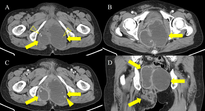 Figure 1
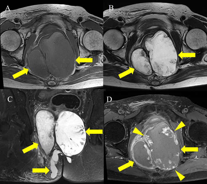 Figure 2
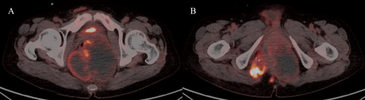 Figure 3
Peer Reviews
No public reviews on file for this paper yet. If you reviewed it on a platform where reviews are public (OpenReview, ICLR, NeurIPS, ICML), you can paste yours below so the community can read it here.
Videos
No videos yet. Explain this paper in a talk, walkthrough, or lecture? Add one.
Taxonomy
TopicsColorectal and Anal Carcinomas · Urinary and Genital Oncology Studies · Cancer and Skin Lesions
Case History
A 65-year-old woman was referred to the hospital for evaluation of a painful perianal mass. Her past medical history was unremarkable. The serum white blood cell count (10,610/mm^3^) and C-reactive protein level (77.08 mg/L) were slightly elevated, but tumor markers were within the normal range, including carbohydrate antigen (CA 19-9), 27.7 U/ml, and carcinoembryonic antigen (CEA), 4.85 ng/ml. Contrast-enhanced computed tomography (CT) was performed for evaluation of the perianal mass. An axial and coronal reformatted contrast-enhanced CT image (Figure 1) demonstrated a 13-cm multilocular cystic mass (arrows) around the rectum and anus, with calcifications (open arrowheads) and an enhancing solid portion (arrowhead). On magnetic resonance imaging (MRI), the multilocular mass (arrows) showed slightly high signal intensity (SI) compared to muscle on axial T1-weighted imaging (T1WI) (Figure 2A) and marked high SI on axial T2-weighted imaging (T2WI) and coronal fat-suppressed T2WI (Figure 2B and 2C). Axial gadolinium-enhanced fat-suppressed T1WI (Figure 2D) revealed peripheral irregular enhancement (arrowheads) of the mass. Positron emission tomography-CT (PET-CT) (Figure 3) showed fluorodeoxyglucose uptake (maxSUV, 11.3) in the mass periphery. The patient underwent mass excision, and the final diagnosis was perianal mucinous adenocarcinoma (PMAC).
CT images demonstrating multilocular cystic mass around the rectum and anus.
MRI images showing multilocular cystic mass with T1 hyperintensity and peripheral irregular enhancement.
Fluorodeoxyglucose uptake in the cystic mass.
Comment
PMAC is an extremely rare malignant neoplasm that represents approximately 2–19% of all anal carcinomas, which represent 2% of all neoplasms of the gastrointestinal tract [1]. Although the exact histogenesis of PMAC is unclear, the tumor may arise from an anal gland or a chronic fistulous tract. The average age of patients with this condition is 55 years, and it is slightly more common in men [1].
Mucinous adenocarcinoma contains abundant extra-cellular mucin and demonstrates typical CT and MRI features. PMAC typically shows multiple conglomerated cystic masses around the anus, with calcifications on CT. Locules filled with extracellular mucin show higher SI than muscle on T1WI, and SI can differ among them. PMAC usually reveals marked high SI on T2WI. PET-CT may have limitations in evaluating mucinous adenocarcinoma, but fluorodeoxyglucose uptake can be observed [1]. Differential diagnosis from perianal abscess, tail gut cyst, sacrococcygeal teratoma, and pseudomyxoma involving the pelvic retroperitoneum can be difficult. However, a high SI with various levels among locules on T1WI and peripheral irregular enhancement on contrast-enhanced imaging can be helpful in differentiating PMAC from other tumors [1]. Patients with PMAC larger than 5 cm have a poor prognosis [1].
The reference list from the paper itself. Each links out to its DOI / PubMed record.
- 1Ho CM, Tan CH, Ho BC. Clinics in diagnostic imaging (143). Perianal mucinous adenocarcinoma arising from chronic fistula-in-ano. Singap Med J. December 2012; 53(12):843–848; quiz p. 9. Pub Med PMID: . Epub 2012/12/27. eng.23268160 · pubmed ↗