Improvement of Occlusal Function After Clear Aligner Orthodontics Verified by T-Scan Novus Digital Analysis
Tanya Bozhkova, Nina Musurlieva, Velina Stoeva

TL;DR
This study shows that clear aligners can improve bite balance, as measured by a digital tool called T-Scan Novus, in a single patient over nine months.
Contribution
The study demonstrates the use of T-Scan Novus to objectively measure occlusal changes after clear aligner therapy in a clinical case.
Findings
Occlusal force distribution became more balanced after treatment, shifting from 67.9% left to 52.4% left.
Occlusion and disocclusion times improved to within physiological ranges (0.25 s and 0.08 s, respectively).
Abstract
Background and Clinical Significance: Clear aligner therapy has become a widely used orthodontic treatment, particularly among adults seeking esthetic and comfortable alternatives to fixed appliances. Achieving a stable and functional occlusion remains one of the primary objectives in orthodontics. The T-Scan digital occlusal analysis system offers an innovative and objective method for quantifying occlusal contact distribution and timing, thereby improving diagnostic accuracy and follow-up. This report aims to present a clinical case demonstrating the use of the T-Scan Novus system for evaluating occlusal balance before and after clear aligner therapy, highlighting its role in documenting short-term functional occlusal changes. Case presentation: A 42-year-old female patient with Class II malocclusion, deep bite, and anterior crowding was treated with Smilers® clear aligners over nine…
Genes, proteins, chemicals, diseases, species, mutations and cell lines named across the full text — each resolved to its canonical identifier and authoritative record.
Click any figure to enlarge with its caption.
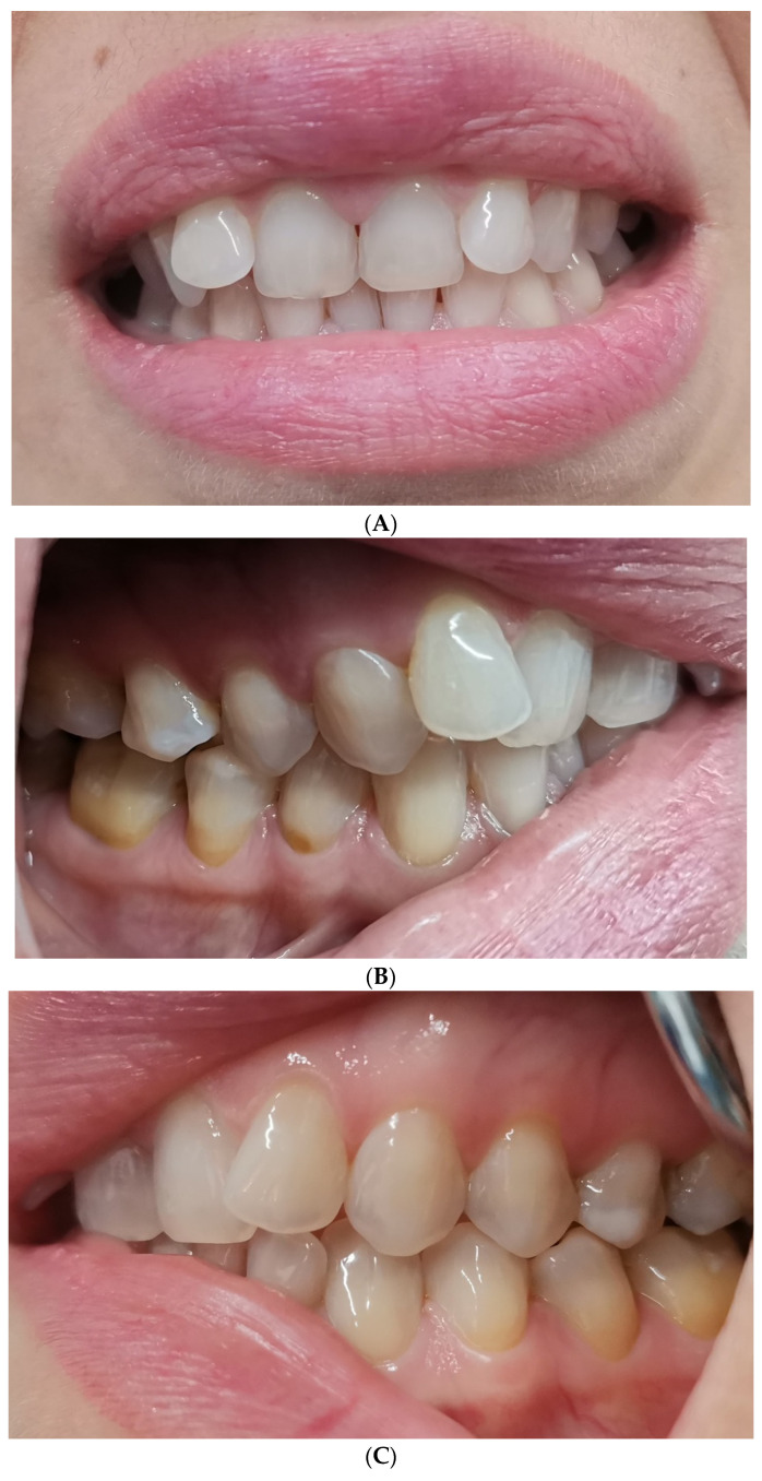 Figure 1
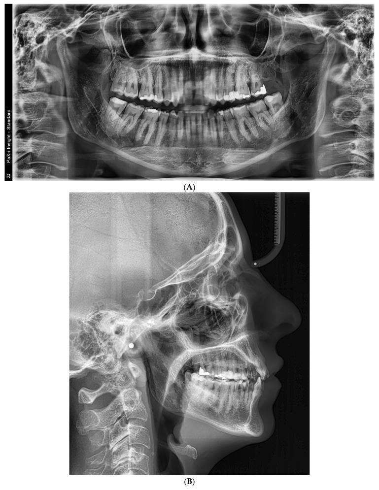 Figure 2
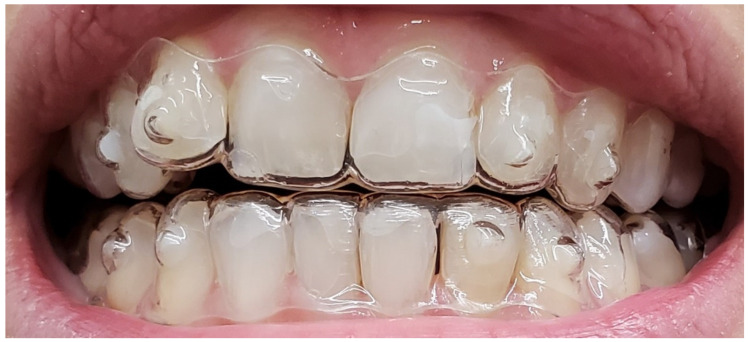 Figure 3
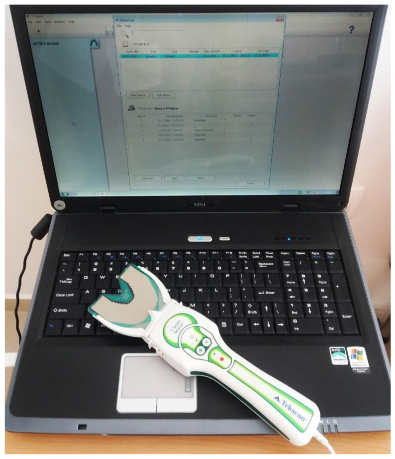 Figure 4
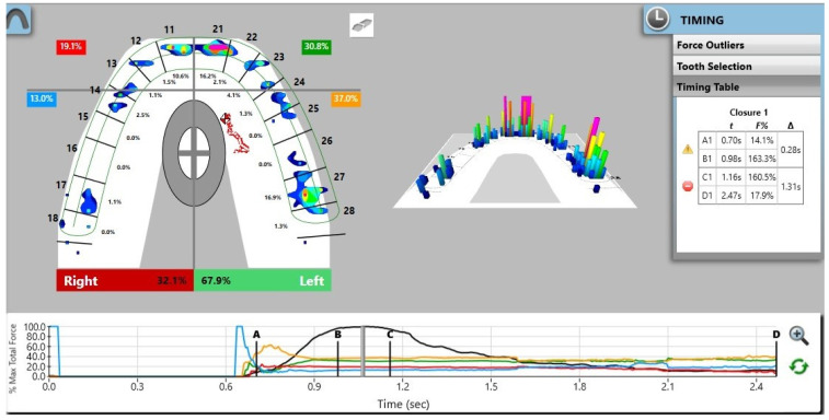 Figure 5
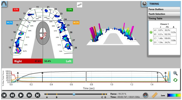 Figure 6
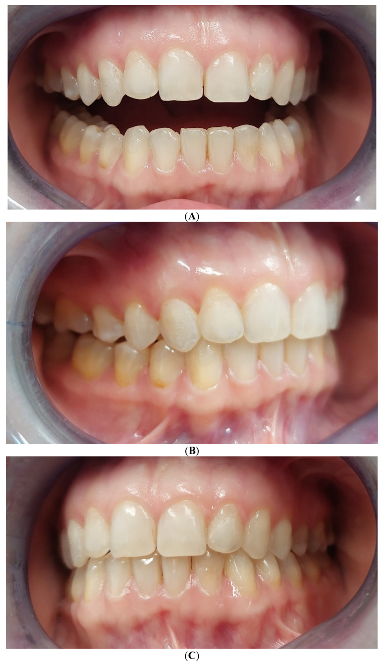 Figure 7
Peer Reviews
No public reviews on file for this paper yet. If you reviewed it on a platform where reviews are public (OpenReview, ICLR, NeurIPS, ICML), you can paste yours below so the community can read it here.
Videos
No videos yet. Explain this paper in a talk, walkthrough, or lecture? Add one.
Taxonomy
TopicsTemporomandibular Joint Disorders · Orthodontics and Dentofacial Orthopedics · Musicians’ Health and Performance
1. Introduction and Clinical Significance
The demand for adult orthodontic treatment has steadily increased, driven by a preference for esthetic, comfortable, and minimally invasive treatment modalities [1,2]. Clear aligner therapy (CAT) has therefore become a widely accepted alternative to fixed appliances, offering advantages such as improved oral hygiene, enhanced patient comfort, and reduced visibility [3,4].
An additional benefit of aligner therapy is the avoidance of metallic components, which may be particularly relevant for patients with known or suspected metal hypersensitivity [5]. Thermoplastic aligner materials are generally considered biocompatible and well tolerated intraorally [6]. Nevertheless, the increasing global use of aligners has raised concerns regarding environmental sustainability, as the sequential manufacturing and disposal of multiple aligners per patient contribute to plastic waste [7].
Beyond esthetic alignment, the primary objective of orthodontic treatment is the establishment of a stable and functional occlusion [8]. Asymmetric occlusal force distribution and prolonged contact timing may compromise masticatory efficiency and long-term occlusal stability, even in patients without overt temporomandibular symptoms [9]. Conventional occlusal indicators, such as articulating paper, provide primarily qualitative information and lack objective quantification of force magnitude and timing [10].
Digital occlusal analysis using the T-Scan Novus system enables dynamic and reproducible assessment of occlusal force distribution and contact timing [11,12]. Although previous studies have investigated occlusal parameters following orthodontic treatment at a population level [13,14], clinical illustrations focusing on digital occlusal assessment after clear aligner therapy remain limited.
The novelty of this case report lies in the clinical illustration of digital occlusal analysis using the T-Scan Novus system to objectively document short-term changes in occlusal force distribution and contact timing following clear aligner therapy.
Aim of the Study
This report aims to present a clinical case demonstrating the use of the T-Scan Novus system for evaluating occlusal balance before and after clear aligner therapy, highlighting its contribution to documenting short-term functional occlusal changes.
2. Case Presentation
2.1. Diagnosis and Etiology
A 42-year-old female patient presented with esthetic concerns related to anterior dental misalignment. Clinical and radiographic examination revealed a Class II malocclusion with retroclination of the maxillary central incisors, increased overbite (deep bite), and mild anterior crowding. No clinically relevant transverse discrepancies or midline deviations were observed. Periodontal tissues were clinically healthy at baseline, and the patient reported no temporomandibular joint pain or masticatory discomfort.
Baseline intraoral photographs and radiographic findings are presented in Figure 1 and Figure 2.
2.2. Treatment Progress
The patient declined fixed orthodontic treatment and opted for clear aligner therapy. Intraoral scans were obtained using a Medit i500 scanner and submitted to Smilers^®^ (Biotech Dental, Salon-de-Provence, FranceB, France) for digital treatment planning.
The treatment plan included limited interproximal enamel reduction in the anterior region to relieve crowding and facilitate alignment. A total of 18 sequential aligners were prescribed and changed every two weeks, resulting in a total treatment duration of nine months. Attachments were placed according to the digital treatment plan. No intermaxillary elastics or post-treatment occlusal adjustments were performed prior to the post-treatment occlusal analysis. Figure 3 illustrates the aligners following completion of the initially planned interproximal reduction.
2.3. Digital Occlusal Analysis
Digital occlusal analysis was performed using the T-Scan Novus System (Tekscan Inc., South Boston, MA, USA) (Figure 4) with a Novus HD sensor. Prior to each recording session, the system was calibrated according to the manufacturer’s instructions.
All recordings were performed by the same experienced operator. The patient was seated upright with standardized instructions to close into maximum intercuspation with comfortable force. Three consecutive recordings were obtained at each time point (pre-treatment and one-month post-treatment), demonstrating consistent patterns; representative values are reported descriptively. A new sensor was used for each recording session.
The system automatically quantified occlusion time (OT), disocclusion time (DT), force distribution per quadrant, and center of force (COF) trajectory.
Measurements were performed before and one month after orthodontic treatment using T-Scan Novus v.9.
2.4. Results
Digital occlusal analysis was performed using the T-Scan Novus system before orthodontic treatment with clear aligners and one month after completion of active therapy. At each point, occlusion time (OT), disocclusion time (DT), and center of force (COF) trajectory were recorded and analyzed.
2.4.1. Occlusal Force Distribution
Pre-treatment recordings demonstrated a marked asymmetry in occlusal force distribution, with a predominance on the left side (67.9%) compared with the right side (32.1%) (Figure 5). Post-treatment analysis revealed a more balanced bilateral force distribution, with approximately 52.4% of the total occlusal force on the left side and 47.6% on the right side (Figure 6). These findings indicate a reduction in left–right force imbalance compared with baseline. The percentage distribution of occlusal forces before and after treatment is summarized in Table 1.
2.4.2. Occlusion and Disocclusion Timing
Before treatment, prolonged occlusal contact timing was observed, as reflected by increased occlusion time (OT) and disocclusion time (DT) values. Following aligner therapy, a reduction in both parameters was recorded (OT = 0.25 s; DT = 0.08 s) (Table 2). Although these values suggest an improvement in contact timing relative to the pre-treatment condition, they represent measurements from a single patient at a single post-treatment time point and should therefore be interpreted descriptively rather than as evidence of definitive functional normalization.
2.4.3. Center of Force (COF) Trajectory
Analysis of the center of force trajectory demonstrated a more centralized and stable path during maximum intercuspation in the post-treatment recordings compared with the pre-treatment condition. The COF trajectory remained within the physiologic target zone throughout the closing phase, indicating a more symmetrical distribution of occlusal contacts at peak intercuspation.
2.4.4. Qualitative Interpretation
No visually apparent premature contacts were identified in the post-treatment T-Scan recordings. This observation is based on digital occlusal mapping and should be considered a supportive finding rather than a standalone diagnostic criterion.
Figure 7 illustrates the occlusal condition recorded one month after completion of active aligner therapy. In addition, follow-up clinical examinations and digital occlusal assessments were scheduled and performed at three and six months after treatment to monitor occlusal adaptation and short-term stability.
3. Discussion
This clinical case illustrates the application of digital occlusal analysis for the descriptive assessment of occlusal force distribution and contact timing following orthodontic treatment with clear aligners. The T-Scan Novus system allows real-time visualization of occlusal contact sequences and relative force distribution, providing objective, dynamic information that complements conventional qualitative occlusal indicators such as articulating paper [1,2].
Before treatment, the patient demonstrated pronounced asymmetry in occlusal force distribution, with a clear predominance on one side. Post-treatment recordings showed a shift toward a more balanced bilateral force distribution. Similar trends have been described in cross-sectional orthodontic studies using T-Scan III and Novus systems; however, such population-level observations cannot be directly extrapolated to individual clinical outcomes [5].
Occlusion time (OT) reflects the interval between initial tooth contact and maximum intercuspation, whereas disocclusion time (DT) represents the transition from full occlusal contact to anterior guidance. Although reference values for these parameters have been proposed in the literature, reported thresholds vary depending on methodology, sensor calibration, and patient-specific factors [4,6]. In the present case, a reduction in DT was observed after treatment, while OT increased at the one-month follow-up. These findings likely reflect short-term occlusal adaptation rather than definitive functional stabilization, particularly given the limited follow-up period.
The visualization of force distribution patterns and timing parameters may facilitate the identification of occlusal imbalances that are not readily detectable with static clinical methods alone. Previous studies suggested that digital occlusal analysis may be useful during splint therapy, prosthetic rehabilitation, orthodontic finishing, and retention phases as a supportive tool for monitoring occlusal changes over time [1,8]. Nevertheless, its output remains technique-sensitive and should be interpreted in conjunction with clinical findings rather than as isolated diagnostic endpoints.
Taken together, this case demonstrates the feasibility of using the T-Scan Novus system to document short-term changes in occlusal parameters following clear aligner therapy. However, given the single-patient design, absence of repeated measurements, and short follow-up, the findings should be regarded as illustrative. Larger prospective studies with standardized recording protocols and longer observation periods are required to clarify the clinical relevance and reproducibility of digital occlusal metrics in orthodontic outcome assessment [2,4].
3.1. Limitations of the Study
The primary limitation of this report is its single-case design, which precludes generalization of the findings. Additionally, the short post-treatment follow-up period does not allow conclusions to be made regarding long-term occlusal stability. Measurement variability inherent to digital occlusal analysis and the absence of patient-reported functional outcomes should also be acknowledged.
3.2. Recommendations for Future Research
Future studies should include larger patient cohorts, longer follow-up periods, and repeated measurements to assess occlusal stability over time. The integration of patient-reported outcomes and complementary functional assessment tools may further enhance the clinical relevance of digital occlusal analysis in orthodontic and prosthetic workflows.
4. Conclusions
In the present clinical case, digital occlusal analysis using the T-Scan Novus system enabled an objective assessment of occlusal force distribution and contact timing before and after clear aligner therapy. The results indicated an improvement toward more balanced occlusal loading and shorter contact timing, supporting short-term functional occlusal stability. Although these findings underscore the clinical usefulness of T-Scan Novus analysis, further studies involving larger patient samples and longer follow-up are necessary to support its routine integration into orthodontic and prosthetic treatment protocols.
The reference list from the paper itself. Each links out to its DOI / PubMed record.
- 1Wang M. Liu L. Ma X. Jin X. Zhang Z. Jia X. Fan J. Tang H. Li Y. Computerized dynamic occlusal analysis and its correlation with static characters in post-orthodontic patients using the T-Scan system and the ABO objective grading system BMC Oral Health 20232331210.1186/s 12903-023-02868-537217888 PMC 10204290 · doi ↗ · pubmed ↗
- 2Zhe Z. Wang Q. Li J. Zhou M. Tang K. Chen J. Wang F. Construction of a Novel Digital Method for Quantitative Analysis of Occlusal Contact and Force BMC Oral Health 2023231904353700564310.1186/s 12903-023-02899-y PMC 10067253 · doi ↗ · pubmed ↗
- 3Filippon I. Tanner C. von Jackowski J. Schulz G. Töpper T. Müller B. Aligner-induced tooth movements in three dimensions using clinical data of two patientsar Xiv 20242405.1043310.48550/ar Xiv.2405.10433 · doi ↗
- 4Abutayyem H. Annamma L. Desai V. Alam M. Evaluation of occlusal bite force distribution by T-Scan in orthodontic patients with different occlusal characteristics: A cross sectional-observational study BMC Oral Health 20232388810.1186/s 12903-023-03544-437986159 PMC 10662916 · doi ↗ · pubmed ↗
- 5Thumati P. Digital analysis of occlusion using T-Scan III in orthodontics J. Indian Orthod. Soc.20165019620110.1177/0974909820160313 · doi ↗
- 6Reslan M. Rayyan M. Ballouli D. Said M. Sayed M. Mekkawi W. Fouad M. Digital Correction of Occlusal Force Imbalance: T-scan III Assessment before and after Guided Equilibration in Postorthodontic Patients J. Contemp. Dent. Pract.2026261197120510.5005/jp-journals-10024-4001 · doi ↗
- 7Chen Y.J. Yao C.C. Chang Z.C. Lai H.H. Hsu L.F. Hsu T.H. Kok S.H. Occlusal function and electromyographic activity of masticatory muscles in skeletal Class III patients with different patterns of mandibular asymmetry J. Oral Rehabil.20235027628510.1111/joor.1341236597189 · doi ↗ · pubmed ↗
- 8Qadeer S. Yang L. Sarinnaphakorn L. Kerstein R.B. Comparison of closure occlusal force parameters in post-orthodontic and non-orthodontic subjects using T-Scan® III DMD occlusal analysis Cranio 20163439540110.1080/08869634.2015.112227726917279 · doi ↗ · pubmed ↗