Contrastive learning enables epitope overlap predictions for targeted antibody discovery
Clinton M. Holt, Alexis K. Janke, Parastoo Amlashi, Parker J. Jamieson, Toma M. Marinov, Ivelin S. Georgiev

TL;DR
The paper introduces machine learning methods to predict antibody binding sites, enabling faster discovery of therapeutic antibodies.
Contribution
A contrastive learning framework for antibody language models that accurately predicts epitope overlap across diverse antibodies.
Findings
Contrastive fine-tuning achieves 97% accuracy in predicting epitope overlap for SARS-CoV-2 antibodies.
AbLang-PDB outperforms sequence-based methods with a 5-fold improvement in average precision.
70% of selected HIV-1 antibody candidates demonstrated specificity and 50% competed for binding.
Abstract
Computational epitope prediction remains an unmet need for therapeutic antibody development. We present three complementary approaches for predicting epitope relationships from antibody sequences. First, by analyzing approximately 18 million antibody pairs targeting around 250 protein families, we establish that over 70% of heavy-chain complementarity-determining region 3 (CDRH3) sequence identity among antibodies sharing both V genes reliably predicts overlapping epitopes. Second, we develop a supervised contrastive fine-tuning framework for antibody large language models that enriches embeddings with epitope information. Applied to SARS-CoV-2 receptor-binding-domain antibodies, this approach achieves 97% total accuracy in predicting high levels of structural overlap. Third, we create AbLang-PDB, a generalized model achieving 5-fold improvement in average precision over sequence-based…
Genes, proteins, chemicals, diseases, species, mutations and cell lines named across the full text — each resolved to its canonical identifier and authoritative record.
Click any figure to enlarge with its caption.
 Figure 1
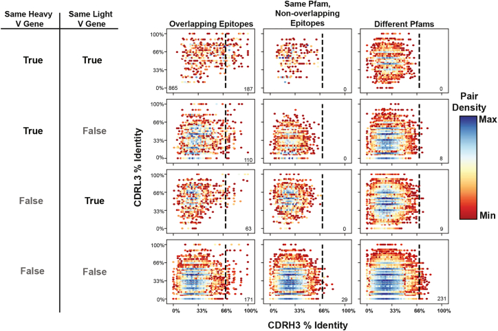 Figure 2
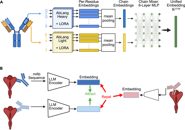 Figure 3
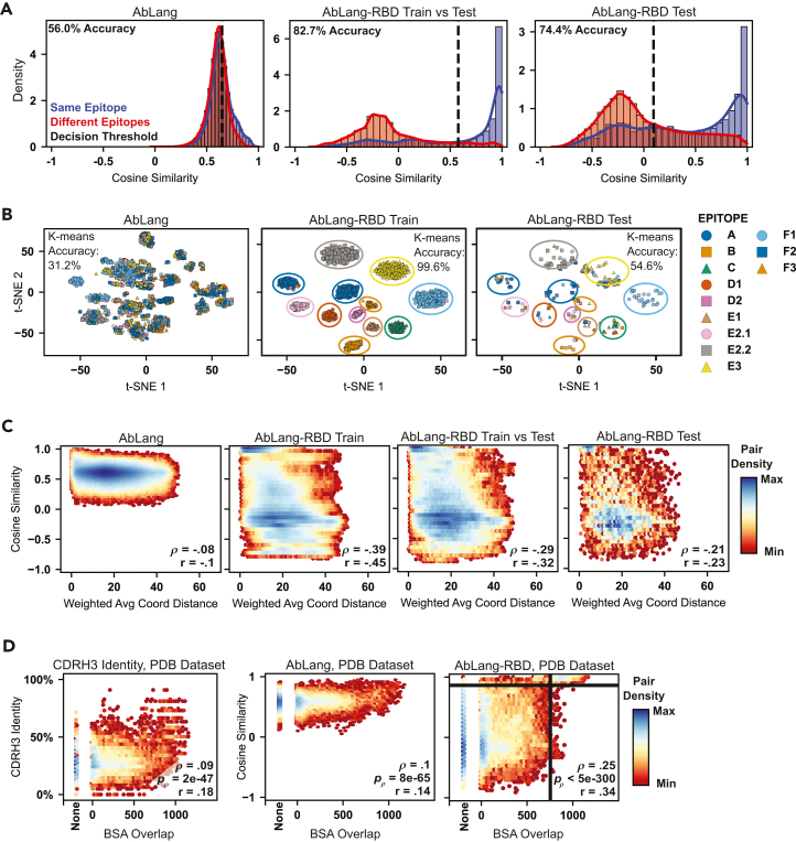 Figure 4
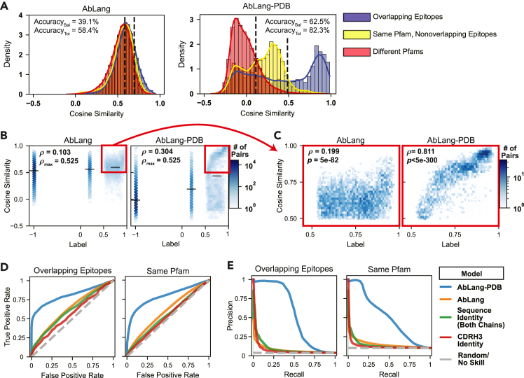 Figure 5
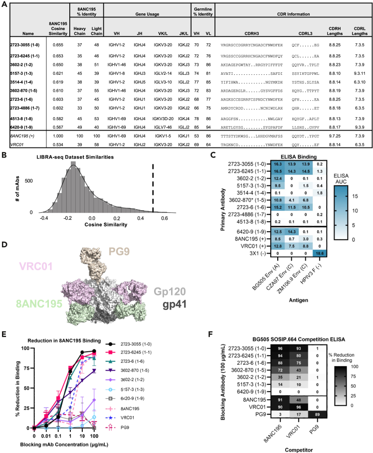 Figure 6
Peer Reviews
No public reviews on file for this paper yet. If you reviewed it on a platform where reviews are public (OpenReview, ICLR, NeurIPS, ICML), you can paste yours below so the community can read it here.
Videos
No videos yet. Explain this paper in a talk, walkthrough, or lecture? Add one.
Taxonomy
Topicsvaccines and immunoinformatics approaches · Monoclonal and Polyclonal Antibodies Research · Machine Learning in Bioinformatics
Introduction
Monoclonal antibody (mAb) therapeutics have revolutionized modern medicine since their first US Food and Drug Administration (FDA) approval in 1986, with blockbuster treatments for cancers, autoimmune diseases, and infectious diseases generating billions in annual revenue.1 Beyond therapeutics, antibodies serve as fundamental research tools and provide crucial insights into immune responses to vaccines and pathogens. Despite their clinical success, developing therapeutic antibodies remains resource intensive, with epitope characterization—identifying the specific region on an antigen where an antibody binds—posing a significant bottleneck.2 For example, in the development of broadly neutralizing antibodies (bNAbs) against HIV-1, epitope mapping is critical to ensuring efficacy across diverse viral strains.3
Epitope characterization typically proceeds through three complementary approaches: (1) structural mapping to define physical contact points between antibody and antigen, (2) functional mapping to identify binding-critical residues through mutation, and (3) competition binding experiments to group antibodies that interfere with each other’s binding. Each approach helps guide therapeutic development, whether identifying sites of vulnerability on pathogens or developing complementary antibody combinations.4^,^5^,^6
Understanding the similarities and differences (or the level of overlap) between the epitopes of different antibody candidates provides critical information that can be utilized when developing antibody therapeutics. For example, in pandemic response efforts against a newly emerging virus, the selection of two or more non-competing antibodies that synergize to form a more effective drug than either individual antibody can be critical for counteracting potential virus escape. In other cases, identifying multiple antibodies against the same functionally important epitope can provide a larger set of candidates for further evaluation, down-selection, and development.
While experimental approaches for antibody epitope characterization are undoubtedly effective, computational approaches can present an efficient and cost-effective alternative. Generally, computational approaches can interrogate the relationship between antibody sequence features and epitope similarity in order to predict the level of epitope overlap between antibody candidates. These approaches range from direct comparisons of the full amino acid sequence or just the complementarity-determining region 3 (CDR3) amino acid sequence within gene groups to comparing predicted structures or predicted antigen-binding residues.5^,^7^,^8^,^9^,^10^,^11^,^12^,^13^,^14^,^15^,^16 While the direct sequence-based methods have shown success in clustering functionally related antibodies, the antibody sequence similarity thresholds utilized by these approaches have been rigorously validated for only a few antigens and epitopes.5^,^8^,^9^,^10^,^17 The indirect approaches allow for searching a broader antibody sequence space, but levels of accuracy are low and unable to detect overlapping-epitope antibodies using distinct structural mechanisms, such as targeting the same site from different angles—an aspect that can significantly influence Fc effector functions and binding breadth.16^,^18^,^19^,^20 This limitation is particularly problematic when searching for therapeutic candidates, where expanding the candidate pool beyond highly similar structures could be necessary to overcome challenges such as low yields or suboptimal binding properties.21
To overcome the limitations of simple sequence identity thresholds, a variety of computational tools have been developed, ranging from structure-based models that predict antigen-binding residues22^,^23^,^24 to general protein language models, such as ESM-2,25 to antibody-specific language models, including Parapred,26 AntiBERTy,27 AbLang,28 AbLang2,29 IgBert,30 and BALM.31 Among these, AbLang has emerged as particularly influential, having been trained on millions of naturally occurring antibodies through masked language modeling to capture both evolutionary relationships and structural constraints within antibody sequences.28^,^32 However, like other current antibody language models, these tools face a critical limitation: their pretrained embeddings naturally cluster by sequence identity and germline gene usage,29^,^31 making them more adept at finding similar sequences than functionally similar antibodies with divergent sequences. This highlights a fundamental need for methods that can specifically learn the complex sequence patterns underlying epitope recognition beyond simple sequence similarity.
Recent advances in machine learning, particularly contrastive learning approaches, offer promising solutions to these limitations. Contrastive learning provides a framework for teaching models to recognize when two examples should be considered similar or different, even when observers see no clear patterns in their features. A useful analogy is signature recognition: while one’s signature may vary between years and with different pens, contrastive learning enables machine learning models to recognize the fundamental similarities between signatures and distinguish authentic signatures from forgeries. By applying this approach to antibody analysis, we can explicitly train models to recognize structural or functional epitope similarity even when sequence similarity is low. Using carefully curated training data from structural databases and high-throughput epitope mapping experiments, we demonstrate how this approach can enrich antibody language model embeddings with epitope-specificity information while maintaining their broad understanding of antibody sequence space.
In this work, we address three key challenges in antibody epitope prediction. First, we establish reliable sequence-based thresholds for identifying overlapping-epitope antibodies, providing a simple yet powerful tool for repertoire analysis. Second, we develop and validate a model using the well-characterized severe acute respiratory syndrome coronavirus 2 receptor-binding domain (SARS-CoV-2 RBD), where extensive epitope mapping data enable us to demonstrate how targeted training can overcome the germline bias of current language models. Finally, we present a generalized model capable of predicting epitope relationships across diverse protein families (Pfams), which we validate through the successful identification of antibodies targeting overlapping epitopes with the HIV-1 bNAb 8ANC195, a therapeutic candidate that targets a unique epitope on the HIV-1 envelope (Env) protein. These advances provide a comprehensive framework for computational epitope analysis, offering new possibilities for therapeutic antibody discovery and optimization.
Results
Sequence determinants for overlapping-epitope antibodies
To identify sequence features that reliably predict when antibodies target overlapping epitopes, we initially interrogated antibody sequence identity as a potential determinant. To that end, we focused on two key features of antibody recognition: variable (V) gene usage and CDR3 sequence similarity. We analyzed 1,909 non-redundant human antibodies from the Structural Antibody Database (SAbDab),33^,^34 generating approximately 1.8 million pairwise comparisons. These pairs were categorized based on both their V-gene sharing patterns and binding properties, specifically examining (1) pairs binding overlapping epitopes, (2) pairs binding non-overlapping epitopes within the same Pfam,35 and (3) pairs binding different Pfams (Figures 1, S1A, and S1B).Figure 1V-gene usage and CDRH3 sequence identity define reliable thresholds for predicting overlapping epitopesA comprehensive analysis of antibody sequence features predictive of epitope overlap within the Structural Antibody Database (SAbDab). Scatterplots show complementarity-determining region 3 (CDR3) sequence identity relationships between antibody pairs (n = 1,909 antibodies, ∼1.8 million pairs) categorized by epitope relationship (columns) and V-gene sharing status (rows). The columns represent overlapping epitopes (left), non-overlapping epitopes on the same protein family (middle), and different protein families (right). Rows indicate V-gene sharing patterns: both heavy and light V genes shared (top), only heavy V gene shared (second), only light V gene shared (third), or neither V gene shared (bottom). The x axis shows CDRH3 amino acid sequence identity, and the y axis shows CDRL3 amino acid sequence identity. Data density is represented by hexagonal binning with color scaling from minimum (dark red) through yellow to maximum density (dark blue). Dashed vertical lines indicate the 70% CDRH3 identity threshold. Numbers in the bottom corners indicate pair counts within the half designated by the line.
Our analysis revealed a hierarchical relationship between sequence features and epitope overlap. For antibody pairs sharing both heavy- and light-chain V genes, we identified a heavy-chain CDR3 (CDRH3) amino acid identity threshold of 70% that serves as a virtually perfect predictor—all pairs exceeding this threshold bound overlapping epitopes within the same Pfam (Figure 1, top row). This predictive power persisted when antibodies shared only one V gene, albeit with an important caveat (Figure 1, rows 2 and 3): while pairs exceeding the CDRH3 threshold and targeting the same Pfam consistently bound overlapping epitopes, 17 of 190 antibody pairs that exceeded this threshold bound antigens from entirely different Pfams. This distinction suggests that when both the heavy- and light-chain germline V genes are shared, this provides additional constraints on antigen specificity beyond epitope recognition patterns.
While these sequence-based rules provide a clear framework for predicting epitope overlap, they also have two major limitations. First, the most predictive rule applies only to the small subset of antibody pairs sharing both V genes. Second, even within this subset, the threshold fails to identify 82% of antibody pairs that do bind overlapping epitopes, resulting in a high false negative rate. These limitations suggest that while sequence identity can provide absolute confidence in some cases, more sophisticated computational approaches may be needed for broader applicability in therapeutic antibody discovery.
To address these limitations, we next explored the ability of antibody large language models to learn the rules of epitope specificity. We focused on two domains: learning discrete epitope bins within one antigen and learning continuous epitope information across diverse Pfams. These approaches, detailed in the following sections, demonstrate how modern computational methods can overcome the constraints of simple sequence-based rules.
Contrastive learning enables epitope-specific encoding of SARS-CoV-2 RBD antibodies
While sequence-based thresholds provide reliable predictions in specific cases, their limited applicability motivated us to develop more sophisticated approaches for predicting epitope relationships. We leveraged the extensive epitope mapping data available for SARS-CoV-2 RBD antibodies36^,^37^,^38 to develop and validate a contrastive learning framework that could encode epitope-specificity information directly into antibody sequence embeddings (Figures 1 and 2).Figure 2. Contrastive learning framework for encoding epitope-specificity information in antibody sequence embeddings(A) Model architecture for AbLang-RBD and AbLang-PDB and their embedding mechanism. This framework uses frozen AbLang heavy and light models with unfrozen low-rank adaptation parameters, as well as an added 6-layer multi-layer perceptron (MLP) network to create a unified 1,536D embedding for each antibody fed into the model.(B) Representation of the contrastive learning approach for the “LLM encoder” framework depicted in (A). During training, embeddings of antibodies binding overlapping epitopes (two blue antibodies) are pulled closer together in representation space, while embeddings of antibodies binding non-overlapping epitopes (pink compared to blue) are pushed apart.
Our model, AbLang-RBD, builds upon the established AbLang-Heavy and AbLang-Light chain language models through targeted fine-tuning using a supervised contrastive learning framework.28^,^39^,^40^,^41 The architecture processes paired antibody sequences through a dual-stream transformer network—with 12 separate transformer blocks per chain—followed by a six-layer multi-layer perceptron that generates unified sequence embeddings (Figure 1A). We optimized these embeddings using supervised contrastive learning as described by Khosla et al.,41 which simultaneously processes multiple positive examples within each training batch, allowing the model to learn from groups of antibodies targeting the same epitope rather than individual pairs (Figure 1B). By training on same-epitope antibodies that fall outside our previously established V-gene and CDRH3 identity thresholds, the model learns new antibody sequence patterns indicative of shared epitope binding that are missed by our outlined V-gene and CDRH3 thresholds.
We trained the model using a previously characterized set of 3,041 SARS-CoV-2 RBD antibodies binned into 12 epitopes based on deep mutational scanning (DMS) results.37^,^38 Antibodies were clustered based on heavy V-gene and CDRH3 identity, and clusters were split between training and test datasets using a CDRH3 cutoff of 65%. Despite these strict criteria, visualizing the cosine similarity distributions reveals that our contrastive learning approach effectively distinguishes epitope-bin information (Figure 3A). The pretrained AbLang model showed poor separation between same-epitope and different-epitope pairs (56.0% balanced accuracy), whereas AbLang-RBD improved this distinction, achieving 74.4% balanced accuracy on test set comparisons and 82.7% when comparing test antibodies to the training set.Figure 3. AbLang-RBD learns to predict epitope relationships from binned deep mutational scanning data(A) Distribution of cosine similarities between antibody pairs binding the same (blue) or different (red) epitopes. The pretrained model’s performance is shown on the left, while the fine-tuned model’s performance is shown for either the comparisons of train-to-test antibodies (middle) or comparisons of test versus test antibodies (right). Optimal decision thresholds (dashed lines) were determined using validation data.(B) t-SNE visualization of antibody embeddings colored by epitope class. These are split to show pretrained embeddings (left) or fine-tuned embeddings on train (middle) and test (right) antibodies.(C) Model performance assessed against continuous deep mutational scanning (DMS) data. Scatterplots show the relationship between antibody pair cosine similarities (y axis) and distance between weighted average spatial coordinates derived from DMS escape maps (x axis). Hexagonal bins are colored by pair density from minimum (dark red) to maximum (dark blue). Spearman’s (ρ) and Pearson’s (r) correlation coefficients are shown.(D) Validation using structural data from the Protein Data Bank (PDB). Scatterplots compare CDRH3 sequence identity (left), pretrained AbLang (middle), and AbLang-RBD (right) against buried surface area (BSA) overlap between antibody pairs. Spearman's (ρ) and Pearson's (r) correlation coefficients are shown; p values (p_ρ_) shown correspond to the Spearman correlation. Threshold lines are drawn at a cosine similarity of 0.85 and a BSA overlap of 750 Å^2^.
The effectiveness of our epitope-specific encoding was further demonstrated through dimensionality reduction analysis. t-Distributed stochastic neighbor embedding (t-SNE) visualization42 reveals that while the pretrained model’s embeddings show minimal epitope-based clustering (31.2% k-means accuracy),43 AbLang-RBD achieves near-perfect clustering of training data (99.6%) and substantially improved clustering of test data (54.6%) (Figure 3B). Notably, when test antibodies were misclassified, 43% of errors still placed them within the correct RBD epitope class (out of 4 generally accepted classes),44 suggesting the model captures meaningful spatial relationships between epitopes despite this information not being provided in training.
To validate that our model learned genuine epitope-specificity information rather than arbitrary clustering, we evaluated its performance against two continuous data sources. First, we examined correlation with the underlying DMS data by obtaining a “center of mass” for each antibody’s escape data on the RBD structure (Protein Data Bank, PDB: 8SGU).45 For each antibody, all residues were assigned a “mass” or “weight” at the location of their ⍺ carbon, and then a weighted average was taken over all residues (Figure S1C). AbLang-RBD embeddings exhibited greater correlation with these spatial coordinates (Spearman’s ρ = −0.39, p < 5e−300) compared to the pretrained model (ρ = −0.08, p < 5e−300) for the training set as well as for the test set antibodies (ρ = −0.21, p = 5.2e−149; Figure 3C). Second, we assessed performance on an independent set of 237 RBD-specific antibodies with structural epitope information from the PDB (Figures 3D and S1A). The correlation between embedding similarities and buried surface area (BSA) overlap improved dramatically (Spearman ρ: 0.1 to 0.25; p: 8e−65 to <5e−300). Our binary training paradigm (same versus different epitope bins) generated a bimodal distribution in the embedding space, enabling threshold-based classification: a cosine similarity > 0.85 distinguished highly overlapping epitopes (BSA > 750 Å^2^) from non-overlapping pairs with 97% accuracy and specificity (Figure 3D, right). This demonstrates that AbLang-RBD acquired a nuanced understanding of epitope relationships beyond discrete bin classifications and also suggests the predicted BSA overlap or relative overlap as an appropriate tunable parameter for our search framework.
These results demonstrate that supervised contrastive learning can effectively encode epitope-specificity information into antibody embeddings, establishing a powerful new framework for computational antibody analysis. While the model shows some limitations in distinguishing relative distances between non-overlapping epitopes, as evidenced by the presence of discrete bands in cosine similarity distributions rather than a continuous gradient (Figure 3C), its ability to accurately identify antibodies targeting shared epitopes represents a significant advance over existing sequence-based methods. This capability, validated against both DMS and structural data, provides a valuable new tool for therapeutic antibody discovery, particularly in cases where traditional sequence similarity metrics fail to identify functionally related antibodies. Most importantly, this framework establishes a foundation for developing even more sophisticated models that can capture the continuous nature of epitope relationships across diverse antigen families.
Developing a generalized model for continuous epitope overlap prediction
To extend beyond predictions for a single antigen, we developed AbLang-PDB, a model capable of predicting the degree of epitope overlap for antibodies targeting antigens from diverse Pfams represented in the PDB.46^,^47^,^48 Unlike AbLang-RBD’s discrete epitope-binning approach, AbLang-PDB employs a regression framework to predict relative degrees of epitope overlap. We maintained the same dual-stream transformer architecture (Figure 1A) but modified the training objective to a mean squared error loss function to optimize for accurate comparisons between unseen antibodies and those in our curated training set.
The training data encompassed 1,517 antibodies spanning 250 Pfams, with relationships between antibody pairs encoded on a continuous scale. Antibodies targeting different Pfams received a label of −1, while those binding non-overlapping epitopes within the same Pfam were assigned 0.2. For antibodies exhibiting epitope overlap, we assigned continuous labels from 0.5 to 1.0 based on their relative degree of structural overlap (Figures S1A and S1B). This labeling strategy was chosen based on the heuristics that antibodies binding the same antigen at non-overlapping sites often have more in common than antibodies that cannot recognize the same antigen; the wide range from 0.5 to 1 for overlapping-epitope antibody pairs was chosen to allow for more separation in the embedding space between those with highly or marginally overlapping epitopes, reflecting our own observations that sequence features converge more when the epitopes exhibit greater overlap.
Antibodies were split between training and test sets as done for AbLang-RBD. Analysis of sequence similarities between training and test sets confirms minimal overlap between them (Figure S2). The impact of this training approach is evident in the distribution of cosine similarities across different antibody pair categories (Figure 4A). While the pretrained AbLang model showed minimal separation between the three categories (39.1% balanced accuracy), AbLang-PDB achieved clear differentiation (62.5% balanced accuracy), with overlapping-epitope pairs predominantly exhibiting cosine similarities above 0.75. More importantly, the model demonstrated a strong correlation with ground-truth labels across all comparisons (Figure 4B). To contextualize this value, we calculated the maximum possible Spearman correlation (ρmax = 0.525) achievable given the large number of ties in our semi-continuous labeling scheme; our model therefore captures a substantial portion of the learnable sequence-epitope relationship.Figure 4. AbLang-PDB enables accurate prediction of epitope relationships across diverse protein familiesEvaluation of AbLang-PDB’s performance on the Structural Antibody Database (SAbDab), comparing antibodies in the training dataset to held-out antibodies.(A) Distribution of cosine similarities between antibody pairs categorized as overlapping epitopes (blue), non-overlapping epitopes on the same protein family (yellow), or different protein families (red). Both the pretrained (left) and fine-tuned (right) model results are shown, as well as optimal classification thresholds shown as dashed lines.(B) Relationship between model-predicted cosine similarities and ground-truth labels. Hexagonal bins colored by pair density (white to dark blue). Black bars indicate mean ± 95% confidence intervals. Spearman correlations (ρ) and maximum possible correlations (ρmax) are shown.(C) A zoomed-in view of high-confidence predictions for overlapping-epitope pairs (cosine similarity and label ≥ 0.5). The Spearman corelation (ρ) and corresponding p value (p) are shown.(D and E) Receiver operating characteristic curves (D) and precision-recall curves (E) for several classification methods, both for predicting overlapping epitopes (left) and for binding the same protein family (right).
The model’s predictive power is most pronounced for high-confidence predictions among overlapping-epitope pairs. When restricting the analysis to pairs where both the predicted cosine similarity and the ground-truth label are above 0.5, the correlation between the model’s prediction and the actual degree of epitope overlap increases dramatically to ρ = 0.811 (Figure 4C, p < 5e−300). This indicates that the model is highly reliable for ranking the extent of epitope overlap among the most promising candidates.
Comprehensive benchmarking revealed substantial improvements over existing methods (Figures 4D–4F; Table 1). For overlapping-epitope classification, AbLang-PDB achieved an area under the receiver operating characteristic curve (AUROC) of 0.81, an average precision of 0.54, and an F1-score of 0.56, significantly outperforming both the pretrained model (0.63, 0.077, and 0.12) and sequence-identity-based predictions (0.56, 0.08, and 0.08). Similar improvements were observed for Pfam prediction, with 1.23×, 2.69×, and 2.14× enhancements over the best alternative classifier in the respective categories (Figures 4D–4F). The performance profile seen for AbLang-PDB reflects our optimization strategy, where AbLang-PDB was specifically trained to maximize accuracy when comparing novel antibodies against reference sequences in the training set rather than for direct comparisons between entirely unseen antibodies. While this approach led to less substantial performance gains in test-versus-test evaluations (Table 1), the model is still the best in 5/6 categories among these evaluations, with significant improvements over the pretrained model in all of these categories. More importantly, this training paradigm directly supports our intended application of mining large sequence databases for antibodies with overlapping epitopes relative to known therapeutic references.Table 1. Benchmarked model performancePairs usedModelSARS-CoV-2 RBD DMS datasetSAbDabSame epitopeSame PfamOverlapping epitopesAUROCAvg PrecF1AUROCAvg PrecF1AUROCAvg PrecF1Train versus testAbLang-RBD0.840.64****0.590.510.150.190.580.16a0.14AbLang-PDB0.540.150.200.790.510.500.810.54****0.56AbLangPre0.57a0.180.21a0.590.150.210.630.080.13AbLang-Heavy0.560.160.210.620.180.230.650.100.14AbLang20.540.130.200.540.120.190.540.050.08IgBERT0.540.140.200.580.150.210.600.070.12BALM0.560.160.200.590.150.220.610.070.12AntiBERTy0.570.170.210.64a0.19a0.24a0.66a0.120.17aESM-20.560.180.200.550.130.190.600.080.10Parapred0.550.150.200.540.120.190.580.060.10SEQID0.530.140.200.530.120.180.560.070.08CDRH3ID0.560.19a0.210.540.140.180.610.090.12Test versus testAbLang-RBD0.730.390.390.530.210.190.610.250.22AbLang-PDB0.530.140.200.680.340.330.680.33****0.35AbLangPre0.57a0.160.200.630.250.200.680.210.19AbLang-Heavy0.560.150.200.640.230.26a0.680.190.23AbLang20.530.120.200.530.150.200.560.080.12IgBERT0.540.130.200.600.200.230.630.140.18BALM0.550.150.200.600.220.240.630.190.19AntiBERTy0.570.160.21a0.66a0.27a0.250.710.26a0.30aESM-20.560.170.200.560.190.200.600.170.20Parapred0.550.140.200.590.200.210.630.180.15SEQID0.550.18a0.190.580.230.200.69a0.260.13CDRH3ID0.530.130.200.540.200.200.640.230.17Performance metrics across all models for the SARS-CoV-2 RBD DMS dataset and the SAbDab dataset (subdivided into “same Pfam” and “overlapping epitopes” tasks). Models are evaluated under two conditions: “train versus test” (comparing antibodies in the train dataset to the test dataset) and “test versus test” (comparing held-out antibodies against each other). The bold formatting indicates the best performance in a benchmark. Baseline models include AbLang-Heavy (Olsen et al.28); AbLang-Pre, which is a concatenation of AbLang-Heavy and AbLang-Light embeddings (Olsen et al.28); AbLang2 (Olsen et al.29); IgBERT (Kenlay et al.30); BALM (Burbach and Briney31); AntiBERTy (Ruffolo et al.27); ESM-2 (Lin et al.25); and Parapred (Liberis et al.26). In these benchmarks, Parapred was used by generating antibody-specific embeddings in a custom method. The final hidden state of the model was extracted and averaged over all CDR residues in both chains to obtain the embedding. AUROC, area under the receiver operating curve; Avg Prec, average precision; RBD, receptor-binding domain, DMS, deep mutational scanning; SEQID, total amino acid sequence identity over the variable region; CDRH3ID, heavy chain complementarity-determining region 3 amino acid sequence identity.aThe second best performance in a benchmark.
These results demonstrate that our continuous learning approach successfully captures epitope relationships across diverse Pfams while maintaining high precision for overlapping-epitope predictions. The model’s ability to provide reliable confidence scores through cosine similarities makes it particularly valuable for therapeutic antibody discovery, where false positives can be costly.
Experimental validation of AbLang-PDB through epitope-targeted HIV-1 antibody discovery
To validate AbLang-PDB’s practical utility, we applied it to identify antibodies sharing epitope overlap with the HIV-1 bNAb 8ANC195.49^,^50^,^51 This antibody represents an ideal test case due to its unique binding site at the gp120-gp41 interface and its therapeutic potential, despite having somewhat limited neutralization breadth compared to other bNAbs.52 We analyzed a dataset53^,^54 of 7,056 class-switched antibodies from persons living with HIV-1, computing cosine similarities between each antibody and 8ANC195’s embedding (Figures 5A and 5B; methods). From this analysis, we identified 20 candidates with the highest cosine similarities (range: 0.567–0.655). After eliminating eight antibodies with high sequence identity to other selected candidates and two showing potential reactivity to respiratory syncytial virus or hepatitis C virus in their linking B cell receptor (BCR) to antigen specificity through sequencing (LIBRA-seq) profiles, we prioritized 10 antibodies for experimental characterization. Notably, the intermediate cosine similarity scores suggested partial rather than complete epitope overlap, consistent with 8ANC195’s unique epitope characteristics (Figure 5A).Figure 5. AbLang-PDB successfully identifies HIV antibodies that compete for binding with 8ANC195Experimental validation of AbLang-PDB predictions using HIV broadly neutralizing antibody 8ANC195.(A) Sequence characteristics of top candidate antibodies selected by cosine similarity to 8ANC195. The table shows model predictions, sequence identity metrics, gene usage, and CDR information for each antibody. Reference antibodies 8ANC195 and VRC01 are included for comparison.(B) Distribution of cosine similarities across the complete LIBRA-seq dataset (n = 7,056 antibodies), with dashed line indicating recommended threshold (0.5) for mining overlapping-epitope candidates.(C) ELISA binding profiles against HIV-1 envelope SOSIP.664 constructs (BG505, CZA97, ZM106.9) and HPIV3 F control protein. Binding strength is indicated by the area under the curve (white to blue). The 3X1 antibody was included as an HPIV3-specific control.(D) Structural representation of HIV-1 BG505 envelope showing competitor antibody epitopes: 8ANC195 (green, gp120-gp41 interface), VRC01 (pink, CD4-binding site), and PG9 (tan, V1-V2 region) from PDB: 5VJ6, 8VGW. The envelope surface is shown with gp120 (light gray) and gp41 (black).(E) Competition ELISA curves showing percentage of reduction in binding of biotinylated 8ANC195 (10 μg/mL) to BG505 SOSIP.664 in the presence of increasing concentrations of blocking antibodies. Filled symbols indicate mAbs displaying competition with 8ANC195.(F) Competition matrix showing percentage of reduction in binding at fixed concentrations (blocking: 100 μg/mL; detection: 10 μg/mL 8ANC195 and 1 μg/mL VRC01 and PG9). Values range from no competition (white) to complete competition (black).
We first assessed HIV-1 specificity through ELISA against three diverse HIV-1 Env SOSIP.664 constructs, BG505 (clade A), CZA97 (clade C), and ZM106.9 (clade C), using human parainfluenza virus 3 fusion (HPIV3 F) protein as a negative control (Figures 5C, S3A, and S3B53). Seven of the ten selected antibodies (70%) demonstrated HIV-1 Env specificity, with six (60%) showing cross-clade binding. Out of our unbiased selection, two had previously been characterized—2723-3055 and 3602-870—both of which had been shown to potently neutralize a broad panel of tier 2 HIV-1 viruses (12/12 and 11/14 viruses tested).53
To assess epitope overlap, we performed competition ELISAs against BG505 SOSIP.664 using a panel of well-characterized HIV-1 antibodies targeting distinct epitopes: 8ANC195 (gp120-gp41 interface),51 VRC01 (CD4 binding site),55 and PG9 (V1-V2 region)56 (Figures 5D–5F and S3C). Five antibodies (50%) showed competition with 8ANC195, defined as having >30% reduction in binding, with four of those displaying strong competition (>70% reduction). These same antibodies also competed with VRC01 at a slightly weaker level but not with PG9, consistent with the structural overlap between the 8ANC195 and VRC01 epitopes.
The success rate of our computational predictions—50% for identifying 8ANC195-competing antibodies and 70% for HIV-1 Env specificity—highlights AbLang-PDB’s potential to streamline therapeutic antibody discovery by accurately identifying functionally relevant candidates that conventional sequence similarity metrics can miss. This is particularly noteworthy given that the model identified two previously validated bNAbs without any prior knowledge of their functional properties. These results support AbLang-PDB’s utility for therapeutic antibody discovery, especially in cases where conventional sequence similarity metrics would fail to identify functionally related candidates.
Discussion
The identification of antibodies targeting overlapping epitopes remains a critical challenge in therapeutic antibody development. Current approaches typically rely on experimental screening57 or database searches for sequence-similar antibodies,7 both of which have significant limitations. Our work establishes three complementary computational strategies that address this challenge at different levels of complexity and applicability.
First, we provide rigorously validated sequence-identity-based thresholds for predicting epitope overlap. When antibody pairs share both heavy- and light-chain V genes and have CDRH3 amino acid identity exceeding 70%, they not only consistently bind overlapping epitopes but are also guaranteed to target the same Pfam. This constraint relaxes slightly when only one V gene is shared—while the 70% CDRH3 identity threshold still perfectly predicts overlapping epitopes when antibodies target the same Pfam; some pairs meeting this criterion may bind antigens from different Pfams altogether. While these findings appear straightforward, they represent the first systematic validation of such thresholds across more than 200 antigen specificities. These simple yet powerful criteria provide immunologists with reliable tools for initial repertoire analysis, particularly valuable when analyzing vaccine responses or comparing antibody lineages across individuals.
Second, through our first model, AbLang-RBD, we demonstrate how supervised contrastive learning can enhance language model embeddings with epitope-specificity information. Using the well-characterized SARS-CoV-2 RBD as a model antigen, we showed that this approach achieved 74.4% accuracy in predicting epitope relationships between previously unseen antibodies. The model’s ability to generalize beyond its training data is evidenced by its improved correlation with both DMS data and structural epitope measurements compared to sequence-based metrics or the pretrained model. This indicates that our contrastive learning framework indeed captures genuine epitope-specificity information rather than merely clustering similar sequences.
Third, we developed AbLang-PDB, which extends epitope prediction capabilities across diverse Pfams while capturing continuous relationships between epitopes. This model demonstrates substantial improvements over existing methods, achieving significant increases in AUROC, average precision, and F1-scores for both same-antigen Pfam pair predictions and overlapping-epitope prediction across a wide range of benchmark models (Figures 4D and 4E; Table 1). Notably, our contrastive learning approach increased average precision 7-fold relative to the pretrained model and 4.5-fold relative to the second-best-performing model, AntiBERTy (Figure 4E; Table 1). Beyond classification performance, AbLang-PDB accurately estimates epitope overlap through cosine similarity scores, with high-confidence predictions (cosine similarity > 0.5) for overlapping-epitope antibody pairs showing strong correlation (ρ = 0.811) with actual epitope overlap (Figure 4C).
Our model demonstrated therapeutic relevance through the identification of HIV-specific antibodies sharing epitope overlap with 8ANC195. Of our 10 computationally selected candidates, 70% showed HIV-1 specificity, 50% competed with 8ANC195 for binding, and 20% were broadly neutralizing. The 2 bNAbs had both been previously discovered, but of note, they were the only two previously discovered bNAbs present among the 7,056 mAbs in the database. Feature attribution analysis using integrated gradients of the five experimentally validated 8ANC195-competing antibodies revealed unexpected insights into AbLang-PDB’s decision-making process (Figure S4).58 The analysis showed that predictions were predominantly driven by light-chain features, particularly residues surrounding the CDRL3 loop. We then repeated this analysis over the broader set of all test antibodies (see methods for full details). The attribution pattern was consistent with 8ANC195-competing antibodies, showing that light-chain residues received significantly higher attribution scores than heavy-chain residues, with the exception that there was considerably greater variability in results between antibodies than seen for the set of 5 antibodies. These findings suggest that while heavy chains are traditionally considered the primary determinants of binding specificity, machine learning approaches may reveal previously underappreciated contributions of light-chain sequences to epitope recognition patterns.
Beyond epitope overlap prediction, our models can be leveraged for large-scale antigen-specificity screening applications as was seen by our high hit rate for HIV-1-specific antibodies. One implementation to capture a wider diversity of epitopes during antigen-specificity screening utilizes a multi-reference search approach. Here, a panel of antibodies with known antigen specificity is used as a reference, followed by classification of antigen specificity based on having cosine similarities of >0.2 compared to the majority of reference antibodies.
The selection between AbLang-RBD and AbLang-PDB depends on two critical considerations: existing knowledge of antigen specificity and extent of epitope overlap. For antibodies with confirmed SARS-CoV-2 RBD binding, AbLang-RBD is recommended. This is due to its specialized training on extensive RBD-specific data, achieving superior performance across all evaluated metrics (AUROC, average precision, and F1-scores) on SARS-CoV-2 datasets compared to AbLang-PDB. However, for antibodies lacking confirmed RBD specificity or when continuous epitope overlap quantification is prioritized, AbLang-PDB provides broader applicability with more correlated cosine similarity values to relative epitope overlap measures, whereas AbLang-RBD exhibits more discrete, bimodal similarity distributions.
However, our approaches have important limitations. The sequence-identity-based thresholds, while providing perfect precision for predicting overlapping epitopes, exhibit low recall—failing to identify 82% of antibody pairs that do share epitope overlap. AbLang-RBD demonstrates high performance on SARS-CoV-2 index strain RBD epitope prediction but faces two key constraints: it is not clear that it will generalize to RBD-specific antibodies incapable of binding the index strain and its training approach using epitope bins from DMS data has not been validated to show that this will work using epitope bins from the more commonly available antibody-antibody competition binding data. AbLang-PDB’s training paradigm is optimized for comparing novel antibodies against reference antibodies from its training set rather than directly comparing two previously unseen antibodies. Additionally, this training paradigm is likely influenced by the specific training labels chosen by hand (−1, 0.2, and 0.5–1.0), which represent heuristic choices that could potentially be optimized through systematic hyperparameter search approaches. Furthermore, while our HIV-1 validation demonstrates practical utility, this antigen is well represented in our training data, though notably, our validation epitope (8ANC195’s epitope) had minimal representation.
Despite these limitations, our work provides a comprehensive framework for computational epitope analysis that will be of significance for the field of therapeutic antibody discovery. The combination of simple sequence rules and sophisticated machine learning models offers researchers a tiered approach to identifying overlapping-epitope antibodies, from rapid initial screening to detailed prediction of epitope relationships. Looking forward, these methods can be further enhanced through integration with emerging structural prediction tools and expanded training datasets, potentially enabling even more accurate prediction of antibody-antigen interactions.
Methods
Data curation
We curated a comprehensive dataset from the SAbDab33^,^34 (February 19, 2024, cutoff date) for training and validating the AbLang-PDB model. Starting with 16,105 antibody-antigen complexes, we applied the following filtering criteria: resolution ≤ 4.5 Å, human antibodies with both chains present, and ≥5 amino acid differences between antibodies. This yielded 1,909 non-redundant complexes, of which 184 had no same-Pfam pairs and 485 had no overlapping-epitope pairs.
Antigen classification utilized pfam_scan35^,^59 software to group antigens by domain architecture using hidden Markov models. Multiple Pfam assignments were consolidated such that any shared Pfam between antigens classified their respective antibodies as targeting the “same Pfam” and thus a machine learning label of 0.2 or greater. When no overlap was present, these pairs were assigned a machine learning label of −1. For quantifying epitope overlap, we employed two complementary approaches.
Approach 1: BSA
Here, we calculated BSA per residue using the Shrake-Rupley60 algorithm within the Dictionary of Secondary Structure Predictions (DSSP 4.0)61^,^62 tool. This algorithm calculates solvent accessibility in a method equivalent to rolling a ball the size of a water molecule along a protein surface and calculating the square angstroms for each residue that are accessible to this water molecule. To translate the accessible surface area (ASA) into antibody BSA, the amount of surface area at each residue for the antigen is calculated either in complex with the antibody or after synthetically removing the antibody from the structure file, such that . Per-residue BSA overlap between two antibody-antigen complexes was calculated as
where residue_i refers to equivalent residues in each of the 2 complexes as defined by a pairwise sequence alignment of the antigen sequences. Pairwise sequence alignments of the antigen sequences were accomplished using the BLOSUM62 matrix63 and Needleman-Wunsch64 algorithm. Antibody pairs with total BSA overlap summed over all residues ≤20 Å^2^ were labeled as non-overlapping (Figure S1A, label = 0.2).
Approach 2: Distance and volume overlap
In this approach, we defined epitopes as antigen heavy atoms within 4.5 Å of antibody atoms, and these selections were accomplished using PyMOL.65 For all antibody-antigen complexes sharing at least one Pfam label, the antigens were aligned in PyMOL. Next, the volume overlap of epitope atoms was calculated using PyMOL’s overlap function, with pairs showing overlap of ≤5 Å^3^ labeled as non-overlapping (label = 0.2).
For overlapping epitopes, final labels were assigned on a continuous scale from 0.5 to 1.0 using the formula
where rBSA_OVERLAP and rATOM_OVERLAP represent overlap relative to the smaller of the two self-overlap values seen for each epitope pair. For partitioning antibodies between datasets, antibodies sharing both heavy and light V genes and CDRH3 amino acid identity >65% were assigned to the same clone group. These groups were then distributed across training (80%), validation (10%), and test (10%) sets, ensuring no clone group appeared split between multiple sets. Additionally, pairs with >92.5% sequence identity in either chain were excluded to maintain diversity.
For AbLang-RBD, we utilized published DMS data comprising 3,195 antibodies from 2 papers,37^,^38 of which only the 3,093 that demonstrated binding to the SARS-CoV-2 index strain were kept. These antibodies were clustered based on heavy-chain V-gene usage and CDRH3 amino acid identity >70%, with clusters distributed across training (80%), validation (10%), and test (10%) sets such that no antibodies in the same cluster existed in the training and test sets. A separate test set was curated from the PDB by selecting RBD-specific antibodies from the Coronavirus Antibody Database (CoV-AbDab)36 that demonstrated index strain binding and were unique from those in the DMS dataset. This left 237 antibodies and 27,345 pairs.
Model architecture
We built our approach upon the pretrained AbLang framework, which comprises separate heavy- and light-chain transformer models for antibody sequence analysis. We utilized the published AbLang model weights from Huggingface (qilowoq/AbLang_heavy and qilowoq/AbLang_light), accessed through the transformers library (AutoModel and AutoTokenizer).28^,^66 The base architecture follows RoBERTa67 with modifications for antibody sequence processing: each chain is processed through 12 transformer blocks containing 12 attention heads, with a hidden dimension of 768 and an intermediate dimension of 3,072. A learned positional embedding layer handles sequences up to a length of 160.
For sequence processing, antibody amino acid sequences were first tokenized using the transformers module. Heavy- and light-chain sequences were processed independently through their respective models to generate chain-specific embeddings. For each chain, the final hidden layer outputs (768D vectors) from all non-masked positions were mean pooled. Whereas for the pretrained AbLang model, we simply concatenated these chain embeddings, the architecture for AbLang-RBD and AbLang-PDB introduces additional processing layers to enable cross-chain information flow. Specifically, the concatenated 1,536D vector (768 dimensions per chain) is processed through a 6-layer multi-layer perceptron with rectified linear unit (ReLU) activation between layers, except for the final layer. The normalized output of this network serves as the unified antibody embedding.
To enable efficient fine-tuning while preserving pretrained weights, we employed QLORA (quantized low-rank adaptation) with rank R = 16, alpha = 32, and dropout = 0.3.39 This dual-stream architecture—with 12 transformer blocks per chain, followed by the cross-chain mixing network—allows the model to capture both chain-specific features and relationships between heavy- and light-chain sequences.
AbLang-RBD training
The AbLang-RBD model was trained using a supervised contrastive learning approach to differentiate antibody embeddings based on their epitope label. Specifically, we employed the supervised contrastive loss function, as introduced by Khosla et al.41 This loss function is designed to pull embeddings with the same label closer together in the embedding space while pushing apart those with different labels.
Training was performed with a batch size of 256 antibodies. Optimization was performed using the AdamW optimizer with a learning rate of 1e−5. During training, we froze all pretrained weights except for the QLORA adaptation parameters and the six “mixing” layers that enable crosstalk between heavy- and light-chain embeddings. The detailed mathematical formulation of the loss function is outlined below.
Contrastive loss mathematical formulation
Notation:
- (1)Batch size of antibodies: .
- (2)Embeddings: zi ∈ R^1,536^ for i = 1, …, B.
- (3)Epitope labels: yi ∈ Z for i = 1, …, B.
- (4)Temperature parameter: τ = 0.5.
- (5)Set of positive pairs for anchor antibody i: .
Step 1: Similarity matrix. Compute the similarity matrix S ∈ R^B×B^, where each element represents the scaled cosine similarity between two antibody embeddings:
Step 2: Numerical stability. For numerical stability during exponentiation, subtract the maximum value from each row of the similarity matrix:
Step 3: Denominator calculation. For each antibody i, calculate the denominator Di by summing the exponentiated similarities over all other antibodies in the batch:
Step 4: Final loss calculation. The total loss is the average of the negative log likelihood over all positive (same epitope label) pairs in the batch.
The loss contribution from a single positive pair (i,j) is .
The final loss averages these terms:
Training proceeded for 400 epochs on a single NVIDIA A6000 GPU, requiring approximately 5 h, including inter-epoch evaluations. Model selection was based on AUROC performance on the validation set (weighted by epitope class size), with the epoch 280 checkpoint achieving optimal performance.
Histogram generation and pairwise accuracy or F1 calculation
Distributions of antibody pair relationships were visualized and analyzed using histograms implemented in Python 3.8.18 with seaborn 0.13.1. All histograms were generated using probability density normalization with 30 uniform-width bins. Classification thresholds were determined differently for AbLang-RBD and AbLang-PDB versus the pretrained model. For AbLang-RBD and AbLang-PDB, thresholds were optimized to maximize balanced accuracy on the validation dataset. The pretrained model threshold in Figure 4A was similarly optimized using train versus validation parameterization, while in Figure 3A, it was optimized for maximal balanced accuracy across the complete dataset (note: this approach overestimates model performance).
For three-category classification, optimal decision boundaries were determined via grid search across 90,000 threshold combinations (300 × 300 cosine similarity values). The threshold pair yielding maximum balanced accuracy across all three categories (overlapping epitopes, non-overlapping epitopes within the same Pfam, and different Pfams) was selected. Balanced accuracy was calculated as the mean of individual category accuracies, in contrast to total accuracy, which can be biased by class imbalance.
Calculation of a representative DMS coordinate
DMS escape data were obtained for 1,375 SARS-CoV-2 RBD antibodies from the publicly available Bloom laboratory database.37^,^38^,^68^,^69 To translate high-dimensional DMS data into a single, representative 3D coordinate for each antibody’s epitope, we calculated a weighted average of the SARS-CoV-2 RBD’s atomic coordinates. The goal was to find the center of mass for each epitope, where the “mass” of each residue is determined by its importance for antibody binding.
For each antibody, we first obtained its complete DMS escape map, which details how every possible mutation to the RBD affects antibody binding. For each residue position on the RBD, we summed the escape scores of all possible mutations at that site. This sum, wi, serves as a weight representing the overall importance of residue i to the antibody’s epitope. We then used the 3D coordinates (xi) of the ⍺ carbon of each residue from the SARS-CoV-2 RBD structure (PDB: 8SGU). The final 3D epitope coordinate (x) was calculated as the weighted average of these ⍺ carbon positions:
This method provides a single 3D point for each antibody’s epitope, allowing for the calculation of simple Euclidean distances between epitopes for comparison with our model’s predictions (Figure S1C).
Regression analysis
Statistical analyses were performed using SciPy (v.1.10.1) for correlation calculations and significance testing.70 Spearman’s rank correlation (spearmanr) and Pearson correlation (pearsonr) coefficients were calculated for various pairwise comparisons. In Figure S1B, the relationship between relative BSA (rBSA) and training labels was fit using linear regression (scipy.stats.linregress), excluding pairs with labels below 0.5. For correlation analyses in Figures 4D, 5B, and 5C, Spearman correlations were calculated with associated p values; p values below the numerical precision limit of 64-bit floating point numbers are reported as p < 5e−300.
For Figure 4B, we calculated the maximum achievable Spearman correlation (ρmax) by considering the optimal ranking scenario where (1) all antibody pairs with a label of −1 rank below those with a label of 0.2, (2) all pairs with label 0.2 rank below those with labels of ≥0.5, and (3) pairs with labels between 0.5 and 1.0 are perfectly rank ordered. Mean values with 95% confidence intervals were calculated for discrete label categories (−1 and 0.2) and for the continuous range of labels ≥0.5 (plotted at x = 0.75). For Figure 4C, analysis was restricted to pairs with both predicted cosine similarities and ground-truth labels between 0.5 and 1.0 to assess performance on high-confidence predictions.
t-SNE analysis and k-means accuracy calculation
Dimensionality reduction and clustering analyses were performed using scikit-learn (v.1.3.2). For t-SNE visualization, 1,536D antibody embeddings were reduced to two dimensions using the following parameters: principal-component analysis (PCA) initialization, automatic learning rate determination, a perplexity of 30, and a maximum of 1,000 iterations with a learning rate of 1,000. For AbLang analysis, the complete dataset was visualized in aggregate. For AbLang-RBD, while dimensionality reduction was performed on the complete dataset, training and test sets were subsequently visualized separately to assess generalization performance.
Clustering analysis was performed using k-means with cosine similarity as the distance metric. The algorithm was initialized with 12 clusters using the k-means++ strategy for greedy centroid initialization and allowed to run for a maximum of 300 iterations. Clustering accuracy was assessed by assigning the most highly represented epitope class within each cluster as the cluster’s representative epitope. Antibodies within each cluster were considered accurately clustered if they matched this epitope and incorrectly clustered otherwise. This approach, while disadvantaging underrepresented epitopes due to class imbalance, provides a conservative estimate of clustering performance.
For visualization clarity, we cycled through three marker shapes (circles, squares, and triangles), as well as ten distinct colors.
AbLang-PDB training
The AbLang-PDB model was trained using the architecture described in the model architecture section, utilizing the curated structural antibody dataset. During training, we maintained the pretrained weights of the base model, modifying only the QLORA adaptation parameters and the six mixing layers responsible for cross-chain information integration. Training employed the AdamW optimizer with a learning rate of 1e−5 and a mean squared error loss function, using a batch size of 16.
To address class imbalance in the training data, we implemented a balanced sampling strategy where each epoch processed 15,270 antibody pairs, evenly distributed across three categories: overlapping epitopes, non-overlapping epitopes within the same Pfam, and pairs targeting different Pfams. While this approach ensured equal representation of each category during training, it resulted in more unique pairs from the non-overlapping-epitope classes being trained on.
Training proceeded for 500 epochs on an NVIDIA A6000 GPU, requiring approximately 36 h, including inter-epoch evaluations. Model selection was based on AUROC performance comparing training and test sets, with the epoch 240 checkpoint achieving optimal performance.
Feature attribution analysis
To interpret which sequence features AbLang-PDB uses to make predictions, we employed the integrated gradients method using the Captum library for PyTorch.58 We calculated per-residue attribution scores, which quantify how much each amino acid contributed to the final cosine similarity prediction. A pad-token embedding was used as the baseline for all calculations. Two primary analyses were conducted: (1) we calculated attributions for the five experimentally validated 8ANC195-competing antibodies against 8ANC195 itself and (2) we computed attributions for every antibody in the test set against its nearest neighbor (by cosine similarity) in the training set. For visualization, mean attribution scores were mapped onto the 8ANC195 structure (PDB: 7KDE) and plotted by international immunogenetics information system (IMGT) position for both the heavy and light chains.
Receiver operating characteristic, precision-recall, and F1-score calculation
Model performance was evaluated using multiple complementary metrics implemented through scikit-learn. For receiver operating characteristic (ROC) analysis, we calculated true positive and false positive rates across 2,001 equally spaced thresholds spanning the range of possible prediction values (cosine similarity from −1 to 1 for model predictions and from 0 to 1 for sequence identity comparisons). The area under the ROC curve was computed using scikit-learn’s trapezoidal rule implementation. For AbLang-RBD, AUROC values were calculated separately for each of the 12 epitope classes and combined using a weighted average based on class size. For AbLang-PDB, the calculation used a binary classification scheme where overlapping-epitope pairs constituted the positive class and non-overlapping pairs the negative class.
Precision-recall characteristics were assessed using scikit-learn’s precision_recall_curve and average_precision_score functions. For F1-score calculations, we utilized the previously determined optimal threshold that maximized balanced accuracy. In the case of Pfam classification, F1-scores were calculated considering all antibody pairs targeting the same Pfam as positives, regardless of their specific epitope overlap status.
LIBRA-seq dataset curation
Antibody sequence datasets for 8ANC195-like antibody mining came from previous in-house LIBRA-seq experiments.53 LIBRA-seq is a high-throughput technology that enables simultaneous identification of antigen specificity and BCR sequences at single-cell resolution. In this approach, B cells are exposed to oligonucleotide-barcoded antigens, allowing quantitative assessment of antigen binding through unique molecular identifiers during subsequent single-cell sequencing. From these experiments, 7,056 class-switched antibody sequences were compiled using peripheral blood mononuclear cells (PBMCs) from persons living with HIV-1.
The dataset comprised 21 LIBRA-seq experiments where antigen-specific B cells were isolated from PBMCs using fluorescence-activated cell sorting (FACS). While this experimental design enriched for HIV-1-specific antibodies in the dataset, the majority of sequences are not expected to be HIV-1 specific. Analysis included only functional, single-cell records from 10× Genomics VDJ sequencing where cells had undergone FACS using PE-labeled antigens, including at least one HIV Env protein and one unrelated control antigen. Each antibody was assigned a unique identifier containing a 4-digit sequencing run prefix, with most run prefixes corresponding to unique donors except for runs 2723 and 3514, which both originated from donor 45 (source of VRC01).53^,^71 Nucleotide sequences were processed through IMGT HighV-Quest to determine amino acid sequences, germline gene assignments, CDR3 sequences, and percent identity to germline.72^,^73^,^74^,^75 The resulting amino acid sequences were embedded using the AbLang-PDB model, and cosine similarities were calculated between each antibody and 8ANC195. Selection of the top 20 candidates was performed blind to all functional annotations and specificity data, including suspected antigen specificity toward positive control and negative control antigens, enabling unbiased identification of antibodies with potential epitope overlap based solely on sequence features learned by the AbLang-PDB model.
Antibody production
Antibody heavy and light chains were synthesized as cDNA by Twist Bioscience or Genscript. Variable genes were inserted into either bicistronic plasmids encoding the constant regions of the H chain and either the kappa or lambda light chain or into separate heavy- and light-chain plasmids. mAbs made in house were transiently expressed using the ExpiFectamine transfection reagent (Thermo Fisher Scientific) in Expi293F cells in FreeStyle F17 media supplemented with 0.1% poloxamer 188 and 20% 4 mM L-glutamine (Thermo Fisher Scientific). Transfected cultures were incubated shaking for 5 days at 37°C with 8% CO_2_ saturation. After 5 days, cultures were harvested and centrifuged at a minimum of 4,000 rpm for 20 min. Supernatant was then filtered with Nalgene Rapid-Flow disposable filter units with a polyethersulfone (PES) membrane (0.45 or 0.22 μm). Filtrate was run over phosphate-buffered saline (PBS) equilibrated columns containing protein A resin. Columns were then washed with PBS, and purified antibodies were eluted using 10 mL of 100 mM glycine HCL at pH 2.7 into 1 mL of 1 M Tris-HCl (pH 8). These were then buffer exchanged into PBS. The remaining mAbs were synthesized by Genscript in their 10 mL TurboCHO high-throughput antibody expression system.
Antigen production
HPIV3 prefusion stabilized F ectodomain (PDB: 6MJZ)76 was expressed in Expi293F cells through transient transfection using ExpiFectamine transfection reagents (Thermo Fisher Scientific) in Freestyle F17 expression media (Thermo Fisher Scientific) with the addition of 0.1% pluronic acid F-68 and 20% 4 mM L-glutamine.
Upon transfection, cultures were grown at 37°C and 8% CO_2_ saturation levels. Six days after transfection, cultures were centrifuged at 4,000 × g for 20 min and filtered with Nalgene Rapid-Flow disposable filter units with a PES membrane (0.45 or 0.22 μM). Protein was purified through nickel affinity chromatography using an equilibrated, 1 mL, prepacked HisTrap HP column (GE Healthcare, Chicago, IL). The column was equilibrated with 15 mL binding buffer (20 mM sodium phosphate, 0.5M NaCl, and 0.3 M imidazole [pH 7.4]). Purified protein was eluted from the column with 15 mL binding buffer supplemented with 0.5 M imidazole. Concentrated protein was buffer exchanged into PBS. The HisTrap purified protein was further purified by size exclusion on Superose 6 Increase 10/300 GL on the AKTA fast protein liquid chromatography (FPLC) system. Fractions containing pure trimeric HPIV3 were identified through SDS-PAGE and the molecular mass. Antigenicity was confirmed with binding to 3X1. Protein concentration was quantified using UV-visible spectroscopy and frozen at −80°C until use.
HIV-1 Env proteins (BG505, CZA97, and ZM106.9) were designed using the SOSIP platform to yield soluble Env proteins stabilized in the prefusion conformation. These SOSIP constructs incorporated several stabilizing mutations: an intermolecular disulfide bond between gp120 and gp41 (A501C and T605C), a trimer-stabilizing mutation (I559P), a truncated gp41 transmembrane region at position 664, and an I201C/A433C mutation to inhibit CD4-induced movement of Env. Additionally, a flexible serine-glycine linker was inserted between gp120 and gp41 (positions 507 and 512) to create single-chain constructs.77
HIV-1 Env proteins were expressed in a highly similar fashion but with the following caveats. Post-culture and centrifugation, the filtered supernatant was applied to an affinity column of agarose-bound Galanthus nivalis lectin (Vector Laboratories) at 4°C. After washing with PBS, proteins were eluted with 30 mL of 1 M methyl-α-D-mannopyranoside. The eluate was buffer exchanged three times into PBS and concentrated using either 30 or 100 kDa Amicon Ultra centrifugal filter units.
Final purification was achieved by size-exclusion chromatography using either a Superose 6 Increase 10/300 GL or Superdex 200 Increase 10/300 GL column on an AKTA FPLC system. Fractions corresponding to correctly folded trimeric Env proteins were collected and validated by SDS-PAGE for molecular weight determination and by ELISA for antigenicity using Env-specific mAbs.
Indirect ELISA
In a 96-well plate, 100 μL of antigen was coated at 2 μg/mL overnight at 4°C. The plates were then washed three times with PBS supplemented with 0.05% Tween 20 (PBS-T) and blocked using 5% bovine serum albumin in PBS. Plates were incubated for 1 h at room temperature and then washed three times using PBS-T. Primary antibodies were diluted in 1% bovine serum albumin in PBS-T starting at 10 μg/mL with a 1:5 dilution. After incubating at room temperature for 1 h and washing with PBS-T, 100 μL of goat anti-human immunoglobulin G (IgG) conjugated to peroxidase was added at a 1:10,000 dilution in 1% bovine serum albumin in PBS-T. These were incubated for 1 h at room temperature, washed three times with PBS-T, and then developed using 3,3′,5,5′ tetramethylbenzidine dihydrochloride (TMB) substrate. Plates were developed for 10 min at room temperature and were then stopped using 1 N sulfuric acid. Absorbance was then measured at 450 nm.
Competition ELISA
Wells of a 96-well plate were coated with 100 μL of 2 μg/mL purified BG505 N332T SOSIP and left at 4°C overnight. Plates were then washed three times using PBS-T, and each well was blocked using 100 μL of 5% bovine serum albumin in PBS for 1 h. After washing three times using PBS-T, primary antibodies were diluted 10-fold starting at 100 μg/mL using 1% bovine serum albumin in PBS-T, and 75 μL was added to each well. After incubating for 1 h at room temperature, without washing, 25 μL of biotinylated antibody was added to each well to final concentrations of 1 and 0.1 μg/mL. This was incubated at room temperature for 1 h and washed three times using PBS-T. Then, 100 μL of streptavidin-horseradish peroxidase (HRP) at a dilution of 1:10,000 in 1% bovine serum albumin in PBS-T was added to each well and incubated for 1 h at room temperature. These plates were then washed three times, and bound antibodies were detected using TMB substrate and sulfuric acid. Competition ELISAs were repeated at least 2 times. Data are displayed as the percentage change in binding relative to the binding of an antibody when no competitor is present.
Structural representation of HIV reference antibodies
A composite image of VRC01, 8ANC195, and PG9 binding BG505 Env was generated by first loading PDB: 5VJ6 into open-source PyMOL Schrodinger, v.2.4.0.56^,^65 The antibody-antigen complex was represented as a surface, and PG9 was colored wheat, 8ANC195 light green, gp120 light gray, and gp41 dark gray. PDB: 8VGW was then loaded into PyMOL, and the gp120 structures from one protomer were aligned to that of one gp120 protomer in 5VJ6. VRC01 was then colored pink and shown as a surface without visualization of the Env protein present in its native complex. Finally, ray tracing was performed with default parameters.
Benchmarking baseline models
AUROC, average precision, and F1-scores were calculated as described previously. For the following baseline models, the final residue-level hidden state was averaged over just the heavy chain to generate embeddings: AbLang-Heavy28 and AntiBERTy.27 For the following models, the hidden state was averaged over both the heavy and light chains: AbLang2,29 IgBERT,30 BALM,31 ESM-2,25 and Parapred.26 For Parapred, this averaging was restricted to CDR residues, with each CDR fed into the model separately, as is standard for this model.26 For ESM-2, the 650 M parameter model was used, and the heavy and light chains were fed simultaneously, separated by two classification (CLS) tokens as done in Burbach and Briney.31 For AbLang-Pre, we separately averaged the residue-level embeddings from AbLang-Heavy and AbLang-Light28 and concatenated them sequentially.
Resource availability
Lead contact
Further information and requests for resources and reagents should be directed to and will be fulfilled by the lead contact, Ivelin S. Georgiev ([email protected]).
Materials availability
Materials will be made available upon request under a completed materials transfer agreement (MTA).
Data and code availability
- •Sequences for antibodies identified and characterized in this study have been deposited to GenBank with names and corresponding accession IDs as such: 2723-3055 (GenBank: MN580560 and MN580589), 2723-6245 (GenBank: MN580555 and MN580584) 3602-2 (GenBank: PX907376 and PX907377), 5157-3 (GenBank: PX907378 and PX907379), 3514-4 (GenBank: PX907380 and PX907381), 3602-870 (GenBank: MN580654 and MN580665), 2723-6 (GenBank: PX907382 and PX907383), 2723-4886 (GenBank: MN580564 and MN580593), 4513-8 (GenBank: PX907384 and PX907385), and 6420-9 (GenBank: PX907386 and PX907387).
- •Associated code for AbLang-RBD and AbLang-PDB is available at https://github.com/IGlab-VUMC/AbLangRBD1 and https://github.com/IGlab-VUMC/AbLangPDB1. Model weights are available at https://huggingface.co/clint-holt/AbLangRBD1 and https://huggingface.co/clint-holt/AbLangPDB1%20 or at Figshare: https://doi.org/10.6084/m9.figshare.29647952.78
- •Any additional data or code reported in this paper will be shared by the lead contact upon request.
Acknowledgments
We thank Perry Wasdin for his help in curating antigen-specific training sets that helped to interrogate the efficacy of different models and loss functions and Alexandra Abu-Shmais for her insights on drawing conclusions from pairwise antibody comparisons. We additionally thank Andrea Shiakolas, Ian Setliff, Kelsey Pilewski, Rohit Venkat, and Lauren Walker for LIBRA-seq data. This research was funded, in part, by the 10.13039/100023015Advanced Research Projects Agency for Health (ARPA-H 1AY2AX000077), 10.13039/100000002NIH R01AI175245, and the 10.13039/100001229G. Harold and Leila Y. Mathers Charitable Foundation (MF-2107-01851). The funders had no role in the conceptualization or execution of any studies or drafting of the manuscript. The views and conclusions contained in this document are those of the authors and should not be interpreted as representing the official policies, either expressed or implied, of the US government.
Author contributions
Conceptualization, C.M.H. and I.S.G.; data curation, C.M.H.; formal analysis, C.M.H. and T.M.M.; software, C.M.H. and T.M.M.; methodology, C.M.H. and A.K.J.; investigation, C.M.H., I.S.G., A.K.J., P.A., T.M.M., and P.J.J.; visualization, C.M.H., I.S.G., and A.K.J.; writing – original draft, C.M.H.; writing – review & editing, C.M.H., I.S.G., A.K.J., P.A., T.M.M., and P.J.J.; funding acquisition, I.S.G.; project administration, I.S.G.; supervision, I.S.G.; validation, A.K.J. and P.A.; resources, P.J.J.
Declaration of interests
I.S.G. is listed as an inventor on patents filed describing antibodies characterized here. I.S.G. is listed as an inventor on the patent applications for the LIBRA-seq technology. I.S.G. is a co-founder of AbSeek Bio. I.S.G. has served as a consultant for Sanofi. The Georgiev laboratory at VUMC has received unrelated funding from Merck and Takeda Pharmaceuticals.
The reference list from the paper itself. Each links out to its DOI / PubMed record.
- 1Lu R.-M.Hwang Y.-C.Liu I.-J.Lee C.-C.Tsai H.-Z.Li H.-J.Wu H.-C.Development of therapeutic antibodies for the treatment of diseases J. Biomed. Sci.272020110.1186/s 12929-019-0592-z 31894001 PMC 6939334 · doi ↗ · pubmed ↗
- 2Chames P.Van Regenmortel M.Weiss E.Baty D.Therapeutic antibodies: successes, limitations and hopes for the future Br. J. Pharmacol.157200922023310.1111/j.1476-5381.2009.00190.x 19459844 PMC 2697811 · doi ↗ · pubmed ↗
- 3Mahomed S.Broadly neutralizing antibodies for HIV prevention: a comprehensive review and future perspectives Clin. Microbiol. Rev.372024 e 00152-2210.1128/cmr.00152-22PMC 1132403638687039 · doi ↗ · pubmed ↗
- 4Labrijn A.F.Janmaat M.L.Reichert J.M.Parren P.W.H.I.Bispecific antibodies: a mechanistic review of the pipeline Nat. Rev. Drug Discov.18201958560810.1038/s 41573-019-0028-131175342 · doi ↗ · pubmed ↗
- 5Saphire E.O.Schendel S.L.Fusco M.L.Gangavarapu K.Gunn B.M.Wec A.Z.Halfmann P.J.Brannan J.M.Herbert A.S.Qiu X.Systematic analysis of monoclonal antibodies against Ebola virus GP defines features that contribute to protection Cell 1742018938952.e 1310.1016/j.cell.2018.07.03330096313 PMC 6102396 · doi ↗ · pubmed ↗
- 6Zost S.J.Gilchuk P.Case J.B.Binshtein E.Chen R.E.Nkolola J.P.Schäfer A.Reidy J.X.Trivette A.Nargi R.S.Potently neutralizing and protective human antibodies against SARS-Co V-2Nature 584202044344910.1038/s 41586-020-2548-632668443 PMC 7584396 · doi ↗ · pubmed ↗
- 7Olsen T.H.Abanades B.Moal I.H.Deane C.M.KA-Search, a method for rapid and exhaustive sequence identity search of known antibodies Sci. Rep.1320231161210.1038/s 41598-023-38108-7PMC 1035415537463925 · doi ↗ · pubmed ↗
- 8Chen E.C.Gilchuk P.Zost S.J.Suryadevara N.Winkler E.S.Cabel C.R.Binshtein E.Chen R.E.Sutton R.E.Rodriguez J.Convergent antibody responses to the SARS-Co V-2 spike protein in convalescent and vaccinated individuals Cell Rep.36202110960410.1016/j.celrep.2021.109604 PMC 835265334411541 · doi ↗ · pubmed ↗