DNA methylation and lncRNA control asynchronous DNA replication at specific imprinted gene domains
Yui Imaizumi, François Charon, Caroline Surcis, Christel Picard, Pol Arnau-Romero, Jean-Christophe Andrau, Daan Noordermeer, Benoit Moindrot, Jean-Charles Cadoret, Robert Feil

TL;DR
The study shows that DNA methylation and long non-coding RNA control asynchronous DNA replication at specific imprinted gene domains in stem cells.
Contribution
The paper reveals that DNA methylation and lncRNA regulate allelic replication asynchrony at imprinted gene domains.
Findings
RT asynchrony at Dlk1-Dio3 and Snrpn domains is parent-of-origin dependent and lost during neural differentiation.
Differential DNA methylation and the lncRNA Meg3 mediate asynchronous replication at the Dlk1-Dio3 domain.
Replication timing is not linked to TAD organization in the studied imprinted domains.
Abstract
Besides genome-wide patterns of replication timing (RT), some genes display allelic replication asynchrony in stem cells, brought about by stochastic events and genetic polymorphisms. Whether epigenetic modifications control asynchronous replication remains unclear. Here, we explore domains controlled by genomic imprinting, where parental DNA methylation imprints mediate allele-specific gene expression. Our genome-wide and locus-specific assays in mono-parental and hybrid mouse ESCs reveal pronounced RT asynchrony—which is parent-of-origin dependent and lost upon neural differentiation—at the Dlk1-Dio3 and Snrpn domains, which both comprise lncRNA polycistrons. Generating a range of mutant lines, we find that asynchronous replication at Dlk1-Dio3 is mediated by differential DNA methylation, and that the lncRNA Meg3 controls early replication across parts of the domain on the maternal…
Genes, proteins, chemicals, diseases, species, mutations and cell lines named across the full text — each resolved to its canonical identifier and authoritative record.
Click any figure to enlarge with its caption.
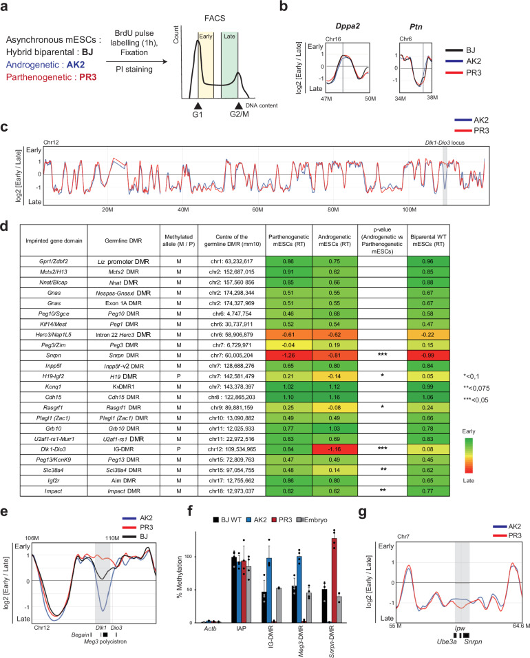 Figure 1
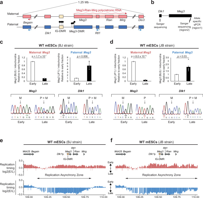 Figure 2
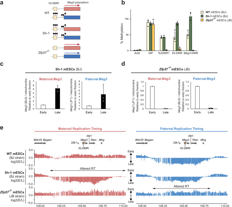 Figure 3
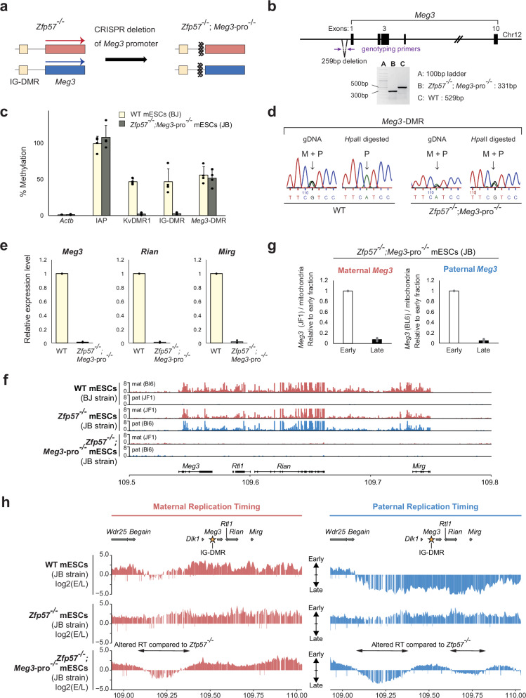 Figure 4
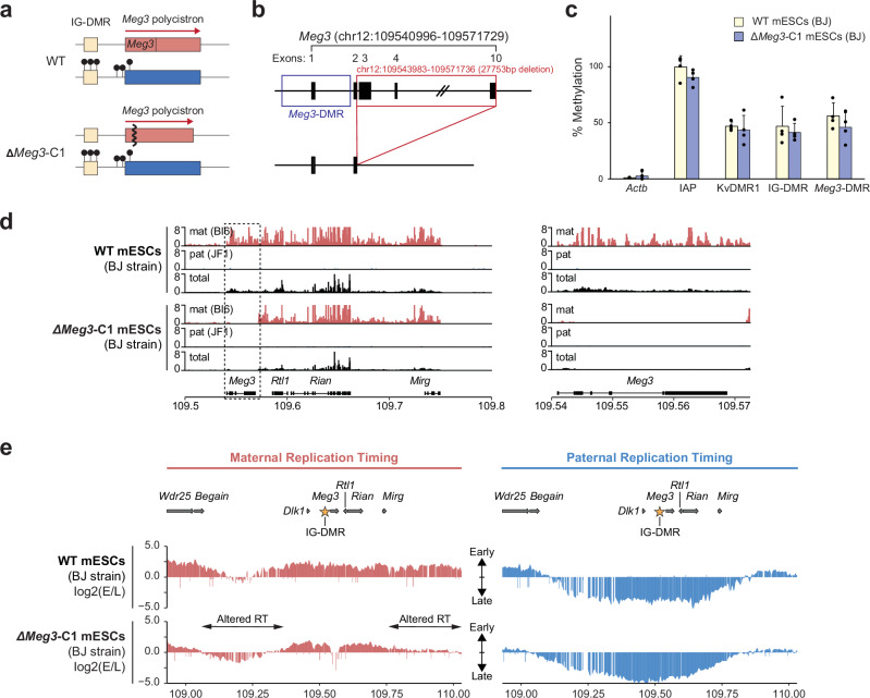 Figure 5
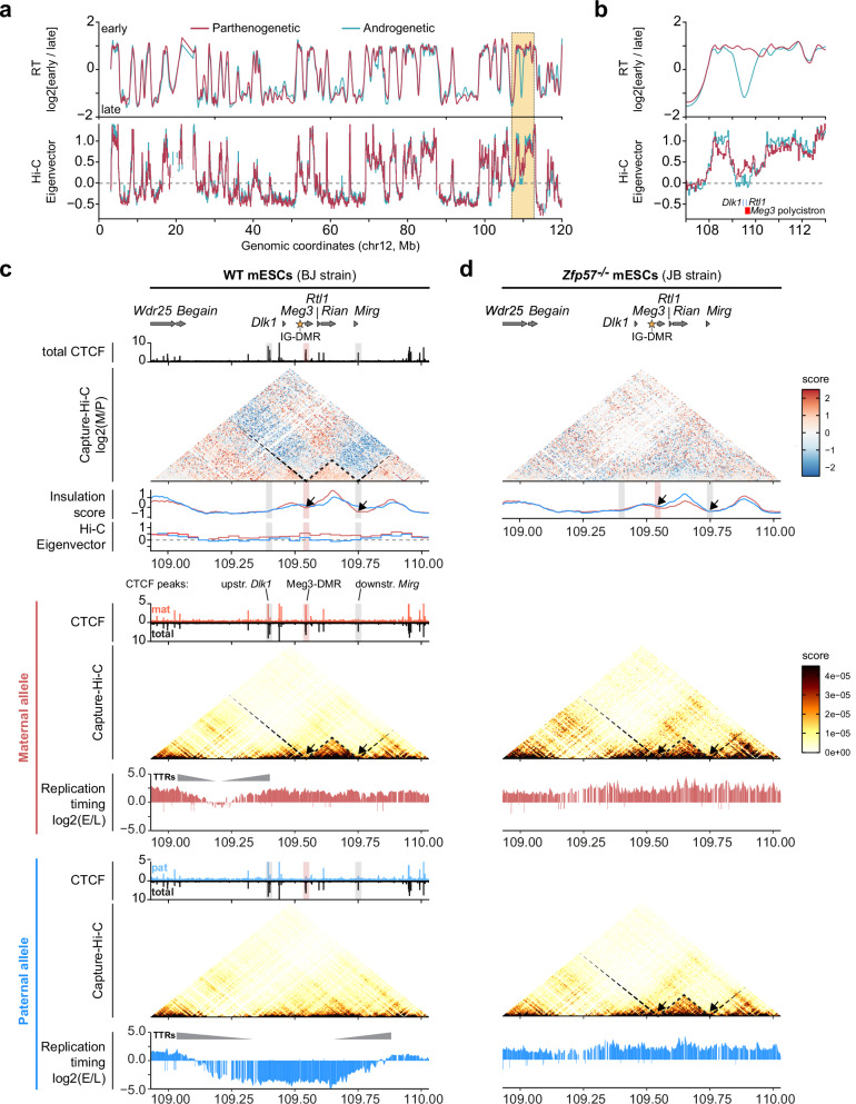 Figure 6
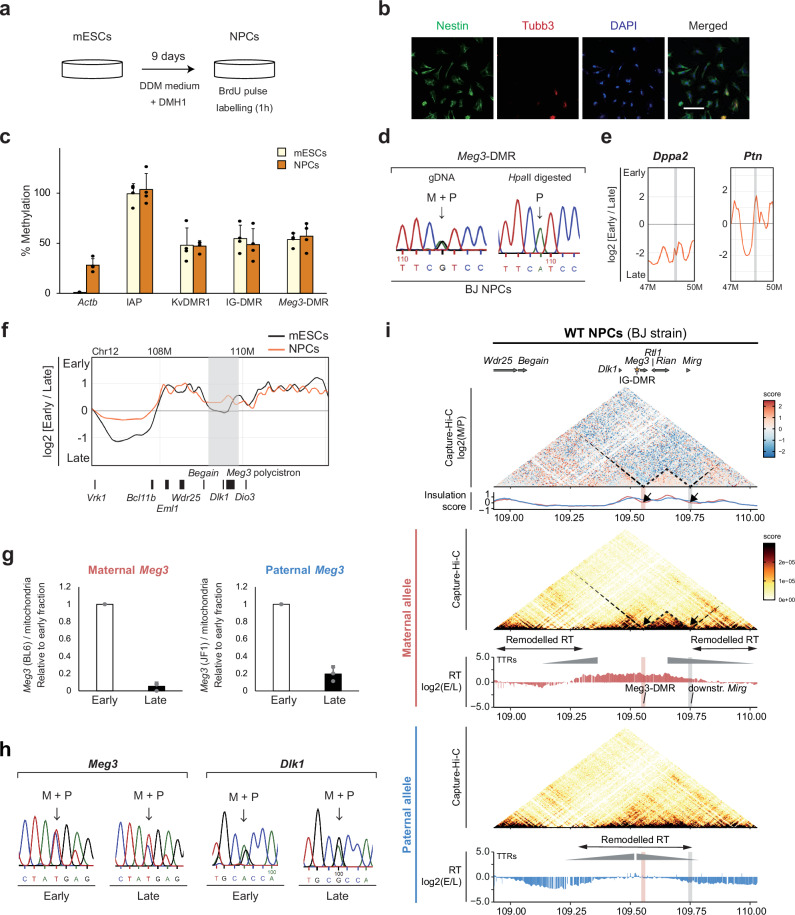 Figure 7
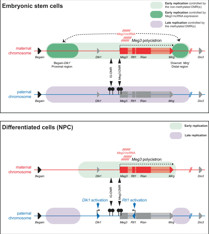 Figure 8
- —https://doi.org/10.13039/501100001665Agence Nationale de la Recherche (French National Research Agency)
- —Fondation de Recherche Médicale (FRM)-France grant reference number: EQU202103012763
Peer Reviews
No public reviews on file for this paper yet. If you reviewed it on a platform where reviews are public (OpenReview, ICLR, NeurIPS, ICML), you can paste yours below so the community can read it here.
Videos
No videos yet. Explain this paper in a talk, walkthrough, or lecture? Add one.
Taxonomy
TopicsGenetic Syndromes and Imprinting · Epigenetics and DNA Methylation · DNA Repair Mechanisms
Introduction
Eukaryotic genomes are organized in 400–800-kb regions that replicate early or late in S-phase named CTRs (Constant Timing Regions), separated by transition zones called TTRs (Transitional Timing Regions)^1,2^. Replication timing (RT) domains have been associated with gene expression states and GC content^3^, and their boundaries coincide with the boundaries of Topologically Associating Domains (TADs)^4,5^. Gene-rich and transcriptionally-active regions tend to replicate early and are within the structurally-defined A-compartment of the genome. Regions with low transcriptional activity are mostly in the B-compartment and replicate mostly late in S-phase^6–10^. Although RT is maintained along a substantial part of the genome, specific regions alter their RT during stem cell differentiation^11,12^. These RT switches correlate with changes in gene activity, sub-nuclear localization, and chromatin organisation^3,9,13–16^. Despite these emerging links, the molecular regulation of differential RT remains poorly understood.
One powerful technology for RT consists of determining the copy number of DNA along the genome in individual cells^9,10,17^. Another, more broadly used approach quantifies the incorporation of chemically-tagged nucleotides into newly-synthesized DNA, in early- versus late S-phase fractions^18^, or compares the DNA content between S-phase and G1-phase cells^19^. With such approaches, asynchronous replication has been detected at a few percent of genes in clonally-derived mouse embryonic stem cells (mESCs)^15,20^. This kind of random asynchrony, in which RT is different between alleles and the early-replicating allele is different between cells, is often associated with mono-allelic transcription states^16,20,21^. Diverse studies on haematopoietic stem cells have shown stochastic replication asynchrony at specific gene loci as well, including at immune system genes^22,23^. During X-chromosome inactivation, similarly, the inactive X chromosome becomes reorganised to replicate in late-S^24^.
Imprinted gene domains provide an attractive RT paradigm. They are controlled by parental DNA methylation imprints at differentially methylated regions (DMRs), which are up to several kilobases in size, but there is no ubiquitous canonical mechanism through which these imprints mediate in-cis the allelic expression of genes. At imprinted domains, the parental chromosomes display opposite patterns of DMR methylation, gene expression, TAD structure, and binding of the CTCF architectural protein^25^, features that can all potentially influence RT^23^.
DNA fluorescence in situ hybridisation (FISH) studies on mESCs have shown a ‘doublet plus singlet’ signal in a relatively high percentage of interphase nuclei at the imprinted Igf2-H19, Snrpn, Igf2r, and Dlk1-Dio3 domains, suggestive of asynchronous replication^26–29^. However, FISH monitors chromatid separation and does not always reflect the timing of DNA replication^30,31^. Furthermore, recent genome-wide studies on hybrid mESCs did not report asynchronous replication at imprinted domains^20^, but here the emphasis was not on imprinted genes, and maintenance of methylation imprints was not monitored. It remains therefore uncertain to what extent imprinted domains might replicate asynchronously between the parental chromosomes. To strictly prevent aberrant alterations in DNA methylation at imprinted loci, here we explored mono-parental and hybrid mESCs grown under serum-free conditions in the presence of ascorbic acid^32^. In agreement with genome-wide studies^20^, we find that the parental chromosomes replicate synchronously along most of the genome, including at most imprinted domains. Pronounced replication asynchrony was detected at specific imprinted loci; however, most strongly along the Dlk1-Dio3 domain on chromosome 12. Using CRISPR-based approaches to test candidate mechanisms, for the first time, our study reveals an essential role for differential DNA methylation and for lncRNA expression in asynchronous replication. These data evoke a model in which differential DNA methylation in conjunction with developmental factors confers replication asynchrony in pluripotent cells by overriding inherent mechanisms that dominate elsewhere in the genome.
Results
A genome-wide survey in mESCs reveals replication asynchrony at imprinted domains
To assess allele-specific RT, we compared mESCs with two maternal genomes (parthenogenetic line PR3) with mESCs with two paternal genomes (androgenetic line AK2). Biparental male mESCs that were hybrid between M. m. domesticus strain C57BL/6J and M. m. molossinus strain JF1 were studied as well. Specifically, we studied mESC line ‘BJ’, in which the maternal genome is C57BL/6J and the paternal genome JF1, and line ‘JB’, which has the reciprocal genotype.
Our assay consisted of a 1 hour exposure of asynchronous cells to BrdU, followed by cell-stage-dependent FACS, precipitation of BrdU-enriched DNA, and micro-array hybridisation^11,33^ (Fig. 1a and Supplementary Fig. 1a). An ‘early fraction’ comprised of late-G1 and the first ~40% of S-phase, was compared with a ‘late fraction’ covering the last ~40% of S-phase and early G2 (Supplementary Fig. 1b). Previously, we reported that this approach gives similar results as multi-fraction Rep-seq^34^. Based on triplicate experiments, ratios between early- and late hybridisation signals [log-2 (Early/Late)] were plotted along the genome. Control developmental genes, including Dppa2 [active and early-replicating^35^] and Ptn [repressed and late-replicating^36^], confirmed correct cell-cycle-stage fractionation (Fig. 1b and Supplementary Fig. 1c). Our assay has a resolution of ~13-kb, and yielded the expected genome-wide pattern of early and late replicating regions, as shown for chromosome 12 (Fig. 1c). The androgenetic and parthenogenetic mESCs had a comparable genome-wide RT pattern (Fig. 1c), and showed a similar high overlap (~90% identity) with the biparental mESCs (Supplementary Table 1). The genome-wide RT patterns were similar to those recently reported in the inner cell mass (ICM) of the blastocyst^10,37^, from which they derived (Supplementary Fig. 1d).Fig. 1. Genome-wide RT assays on mono-parental mESCs pinpoint asynchronous replication.a Schematic of the array-based RT analysis. Following BrdU incorporation in androgenetic (AK2), parthenogenetic (PR3), and biparental hybrid (BJ) mESCs, BrdU-precipitated DNA from early and late cell-cycle fractions is used for array hybridisation, followed by data analysis. b RT (log2 of early/late hybridization signals) at Dppa2 (early replication in mESCs) and Ptn (late replication in mESCs) in BJ, AK2, and PR3 mESCs. Vertical bars indicate positions of the indicated genes. c RT along chromosome 12 in AK2 (blue) versus PR3 (red) mESCs. The grey rectangle indicates a distal region with pronounced asynchrony. d Heatmap of RT at imprinted domains in PR3, AK2, and BJ mESCs. For the domains’ gDMRs (ICRs), M indicates methylation on the maternal allele, and P indicates methylation on the paternal allele. The colour range indicates RT—red being late and green being early—determined based on the earliest and latest values obtained from the smooth curves generated by START-R. Locus-specific values are indicated, determined using the Mean method, with a sliding window length of 60 and with an overlap size of 30, as described^34^. Significance levels (AK2 versus PR3) are indicated. e, RT profile of PR3 (red line), AK2 (blue line) and BJ (in black line) mESCs at the Dlk1-Dio3 locus (distal chr.12). The grey box indicates the region with asynchronous replication, determined by START-R software with the Mean method, with a p-value threshold = 0.05, a sliding windows length = 60 and an overlap size = 30, as described previously^34^. f Methylation levels in the Dlk1-Dio3 and Snrpn domains, determined by methylation-sensitive qPCR at the domains’ ICRs (IG-DMR at Dlk1-Dio3, Snrpn-DMR at Snrpn domain) and a secondary DMR (Meg3-DMR) in PR3, AK2, and BJ mESCs. Embryo (E13) DNA is included for comparison; negative (B-Actin promoter, no methylation) and positive control regions (IAPs, highly methylated) are included. Bars represent means ± SD from 4 independent experiments. g The Snrpn domain on central chromosome 7 shows a less pronounced asynchrony, with earlier replication in AK2 mESCs across a region comprising Snrpn. The light grey box indicates the region with putative asynchronous replication.
For each mESC line, we determined RT using START-R software^34^, and focused on the imprinted gene domains controlled by parental DNA methylation imprints (Fig. 1d). We centred our analysis on the germline DMRs [i.e., the imprinting control regions, ICRs] and close-by neighbouring sequences, using all data points from multiple sequential probes in triplicate experiments (Fig. 1d). In agreement with their gene-richness and, for most, their transcriptional activity in mESCs^38^, a majority of imprinted domains showed early RT, with no apparent asynchrony between androgenetic and parthenogenetic cells (Fig. 1d). The strongest asynchrony was measured at the Dlk1-Dio3 domain on distal chromosome 12 (Fig. 1d). This was apparent also from the RT profiles, with early replication in parthenogenetic and late replication in androgenetic cells, and ‘intermediate RT’ in biparental mESCs (Fig. 1e). This correlated with maintenance of normal DNA methylation levels at the domain’s DMRs (Fig. 1f).
Although for several other imprinted domains (Igf2-H19, Peg3/Zim, RasGrf1) putative RT differences were apparent (Fig. 1d), these loci displayed similar RT profiles in androgenetic and parthenogenetic mESCs and this was observed at the imprinted Igf2r, Plagl1 and Grb10 loci as well (Supplementary Fig. 1e).
At the imprinted Snrpn locus, a tendency towards late RT was apparent in both mono-parental lines. However, this gene replicated significantly earlier in the androgenetic than in the parthenogenetic mESCs (Fig. 1d, g). This correlated with maintenance of a normal methylation level at the domain’s gDMR (Fig. 1f), which suggested that differential methylation could potentially be involved.
A large zone of asynchronous replication comprises the Dlk1 and Meg3-Rian-Mirg imprinted genes
The asynchronous RT at the Dlk1-Dio3 domain covered approximately 800 kb (Fig. 1e). It includes the paternally-expressed Dlk1—a developmental regulator of signalling—and the maternally-expressed Meg3-Rian-Mirg polycistron that produces a multitude of ncRNAs, including the lncRNA Meg3, C/D-box snoRNAs (Rian) and miRNAs (Mirg)^39^ (Fig. 2a). The two paternally methylated DMRs showed the expected methylation levels. The intergenic IG-DMR—the gDMR/ICR of the domain^40^—and the Meg3-DMR were both highly methylated in androgenetic and barely at all in parthenogenetic cells, and had an expected intermediate level (40–60%) in the biparental mESCs (Fig. 1f, Supplementary Fig. 2c).Fig. 2. Capture Repli-seq and PCR-based exploration of allelic RT at Dlk1-Dio3 in hybrid mESCs.a Schematic presentation of the Dlk1-Dio3 domain on distal chromosome 12. Rectangles indicate genes on the maternal (red) and the paternal (blue) chromosome and their expression (arrows) in the embryo^78^. The allelic expression of the 220-kb Meg3-Rian-Mirg ncRNA polycistron is controlled by the IG-DMR (beige rectangle), which acts as an enhancer on the maternal, and is methylated (black lollipops) on the paternal chromosome. The Meg3-DMR is also methylated on the paternal chromosome. b Genomic locations chosen for allelic RT studies at Dlk1 (Sanger sequencing) and Meg3-DMR (Sanger sequence analysis, allelic qPCR). RT at Dlk1 and Meg3 analysed by PCR in BJ (c) and JB (d) mESCs. Top panels, qPCR-based analysis of the maternal and paternal Meg3 in the early and late samples. Signals were normalized against mitochondria, which replicate throughout the cell cycle^73^. For each experiment, values were further normalized to the early sample. Statistical significance between early and late samples was assessed using an unpaired two-tailed Student’s t-test. Bars represent means ± SD from 3 biological replicates for BJ and 4 replicates for JB. Bottom panels, Sanger sequencing-based assessment of Meg3 and Dlk1 in early and late fractions. Arrows indicate SNPs that distinguish the parental chromosomes. At both genes, the maternal chromosome (M) replicates earlier than the paternal (P) chromosome. Capture Repli-seq at the Dlk1-Dio3 locus across 1.1-Mb in BJ (e) and JB (f) mESCs. Maternal and paternal chromosomes are distinguished in the early and late fractions using sequencing reads that cover SNPs. Regions without reads correspond to repeat-rich sequences. Depicted is the log-2 early/late ratio across the domain (~1-kb resolution) on the maternal (in red) and the paternal (in blue) chromosome. Gene positions are aligned above. The asterisk indicates the IG-DMR position. Two-headed arrows indicate RT asynchrony between the maternal and the paternal chromosome. Genomic coordinates are for chromosome 12 (mm10).
Next, we directly compared the parental alleles within the hybrid mESCs. Initially, we performed PCR across single nucleotide polymorphisms (SNPs) within Meg3 (Fig. 2b). Both allele-specific PCR (top panels) and Sanger sequencing of PCR products (lower panels) showed early replication on the maternal and mostly late replication on the paternal chromosome, both in BJ and JB mESCs (Fig. 2c, d). Since our array-based studies suggested replication asynchrony at Dlk1 as well (Fig. 1e), we also performed amplification across an SNP at this developmental gene (Fig. 2b). In both JB and BJ cells, Sanger sequencing of PCR products showed early replication on the maternal and late replication on the paternal chromosome (Fig. 2c, d). We conclude that at Meg3 and Dlk1, the asynchrony reflects the parental origin, rather than genetic strain differences between the parental chromosomes.
To pinpoint the precise sequences that undergo asynchronous replication we developed ‘Capture Repli-seq’, a high-resolution replication-sequencing approach in which regions of interest were captured prior to DNA sequencing. SNPs between C57BL/6 J and JF1 [~4 per kb^41^] allowed us to distinguish the maternal- and paternal-chromosome signal at 1-kb resolution. Similar to the recent genome-wide Repli-Capture-Seq assay^42^, our approach relies on pools of custom oligonucleotides, but our design tiled specific regions, thereby allowing their enrichment at high resolution.
We first analysed a 0.6-Mb control region on chromosome 4, which showed a comparable RT pattern on the maternal and paternal chromosome (Supplementary Fig. 2a). At the captured Dlk1-Dio3 locus, there was pronounced asynchrony with >60 fold allelic-sequence enrichment across ~750 kb. This large, continuous region of asynchrony includes the Dlk1 gene and the entire 220-kb ncRNA polycistron (Fig. 2a,e,f). RT profiles were comparable between the alleles with the same parental origin in BJ versus JB, which confirms that the RT asynchrony is parent-of-origin dependent. However, we noted a difference between the reciprocal lines downstream of the ncRNA polycistron. On the paternal chromosome, a late-replication zone includes this distal region in JB cells (paternal chromosome: B6), but not in BJ cells (paternal chromosome: JF1) (Fig. 2e, f). This difference is likely linked to genetic differences between the JF1 and C57BL/6 J mouse strains.
To determine more precisely when replication occurs, we sorted cells into four fractions: ‘late-G1’, ‘early-S’, ‘late-S’ and ‘early-G2’ (Supplementary Fig. 2d). This showed that Dppa2 (active) and Ptn (inactive) replicated very early and in late-S, respectively (Supplementary Fig. 2e). At Dlk1-Dio3, replication of the maternal Meg3 and Dlk1 alleles was detected at late-G1 and the onset of S-phase (fraction A, Supplementary Fig. 2d); the paternal alleles showed replication in late-S predominantly (Supplementary Figs. 2f–h). These findings indicate that a ~ 750-kb domain comprising Dlk**1, Meg3-Rian-Mirg and Rtl1 replicates very early on the maternal chromosome, and mostly in late-S on the paternal chromosome.
Allelic analysis of replication timing at the Ig f2-H19, Kcnq1, and Snrpn domains
Using Capture Repli-seq, we analysed the imprinted Igf2-H19 domain—at which an early study on serum-grown mESCs had reported differential RT^27^ —and the neighbouring Kcnq1 domain as well. Capture Repli-seq on this distal chromosome-7 region showed that the parental chromosomes had a similar RT (Supplementary Fig. 2b). At the Kcnq1 domain, Kcnq1 and the flanking Cdkn1c gene showed early replication on both the parental chromosomes, while the Igf2-H19 domain showed an intermediate RT on both the parental chromosomes. This lack of asynchrony correlated with unaltered methylation levels (40-60%) at the Igf2-H19 ICR and at KvDMR1, the ICR of the Kcnq1 domain (Supplementary Fig. 2c).
Capture Repli-seq could not be applied with consistent coverage across the Snrpn domain because of its exceptionally high content in repeat elements (mm10, Chr7:59,400,000–59,950,000). Instead, we performed PCR across SNPs. This showed that at Snrpn (regions 1 and 2), the early fraction showed replication of the paternal chromosome predominantly, whereas the maternal chromosome was enriched in the late fraction. A third, further-downstream region, at Ipw, did not show allelic differences in RT (Supplementary Fig. 1f). Upon sorting into 4 fractions, similarly, the paternal Snrpn allele was detected in late-G1 and early-S mostly, whereas the maternal allele was enriched in early-G2 (Supplementary Fig. 2i, j). These data show that at Snrpn, the paternal chromosome replicates on average earlier than the maternal chromosome, similar as reported in human ESCs^43,44^.
In the hybrid mESCs, we also analysed the imprinted genes H19, Peg3, Rasgrf1, and Slc38a4, which had shown putative differences between the androgenetic and parthenogenetic mESCs (Fig. 1d, Supplementary Fig. 1e). This did not show allelic enrichments in the early and/or late fractions, and therefore no evidence for replication synchrony (Supplementary Fig. 1g).
Differential DNA methylation is essential for asynchronous replication at the Dlk1-Dio3 and Snrpn domains
The Dlk1-Dio3 locus encompasses two essential DMRs: the germline DMR called the IG-DMR^40^, and the somatically-acquired Meg3-DMR, both unmethylated on the maternal allele (Fig. 2a). On the maternal chromosome, the IG-DMR acts as an enhancer that drives the expression of the close-by Meg3-Rian-Mirg ncRNA polycistron^26,45^. To determine whether DNA methylation is important for the allelic RT across the domain, we analysed BJ-derived mESCs in which methylation was either biallelic (line Sh-1) or absent from both parental alleles (line Zfp57^-/-^) at the IG-DMR and Meg3-DMR (Fig. 3a). The Sh-1 mESCs were derived previously by directing shRNAs against enhancer RNAs expressed from the maternal IG-DMR, which induced biallelic DNA methylation at both the IG-DMR and Meg3-DMR^26^. mESCs deficient in ZFP57 (Zfp57^-/-^ cells)—a KRAB-domain zinc finger protein essential for imprint maintenance^46^—were generated recently^47^. They showed some residual IG-DMR methylation and hardly any methylation at the Meg3-DMR (Fig. 3b). Concordantly, Sh-1 mESCs did not express Meg3, Rian, and Mirg, while the Zfp57^-/-^ cells showed increased Meg3, Rian and Mirg expression (Supplementary Fig. 3a). In the Sh-1 mESCs, Dppa2 and Ptn showed the expected early and late replication, respectively (Supplementary Fig. 3c). Both parental Meg3 alleles showed late replication (Fig. 3c and Supplementary Fig. 3c), indicating an early-to-late switch on the maternal chromosome. In the Zfp57^-/-^ mESCs, conversely, Meg3 replicated early on both the parental chromosomes (Fig. 3d and Supplementary Fig. 3d), indicating a late-to-early switch on the paternal chromosome.Fig. 3. Differential DNA methylation controls asynchronous replication at *Dlk1-Dio3.*a Schematic presentation of IG-DMR and Meg3-DMR methylation (black lollipops) and Meg3-Rian-Mirg polycistron expression in WT, Sh-1 and Zfp57^-/-^ mESCs. b Methylation-sensitive qPCR at the IG-DMR, Meg3-DMR, KvDMR1 (control ICR), ActB promoter (low-methylation control), and IAP elements (high-methylation control). Bars represent means ± SD from 4 experiments. Replication timing at Meg3 on maternal and paternal chromosomes in Sh-1 (c) and Zfp57^-/-^ (d) mESCs analysed by allele-specific qPCR. Bars represent means ± SD from 3 independent experiments. e Allelic Capture Repli-seq in WT (BJ), Sh-1 and Zfp57 ^-/-^ mESCs at the Dlk1-Dio3 domain. Depicted is the log-2 early/late ratio across the domain ( ~1-kb resolution) on the maternal (in red) versus the paternal (in blue) chromosome. Gene positions are aligned above; the asterisk indicates the IG-DMR position. Two-headed arrows indicate altered RT in the Sh-1 or Zfp57^-/-^ versus BJ mESCs.
To determine where precisely RT depends on the imprinted DNA methylation, we performed Capture Repli-seq. In the Sh-1 and Zfp57 ^-/-^ mESCs, RT was unaffected at the chromosome-4 control region (Supplementary Fig. 3e). In contrast, compared to WT mESCs (Fig. 3e), at Dlk1-Dio3 the entire asynchrony zone had shifted to late replication on both parental chromosomes in the Sh-1 cells. In the Zfp57^-/-^ cells, conversely, the entire zone replicated early on both parental chromosomes (Fig. 3e). Combined, these findings indicate that allelic DNA methylation at the IG-DMR and/or the Meg3-DMR is a distinguishing determinant of asynchronous replication across the entire ~750-kb zone.
Also the ICR of the Snrpn locus (Supplementary Fig. 1f) requires the KRAB-domain zinc finger protein ZFP57^47^. In the Zfp57^-/-^ mESCs, hardly any methylation was left at this gDMR (Supplementary Fig. 3b). Allelic PCR showed loss of replication asynchrony and mostly early RT (Supplementary Fig. 3f, g). Also at Snrpn, therefore, allelic DNA methylation seems to control asynchronous replication in mESCs.
LncRNA expression controls maternal chromosome-specific early replication at the Dlk1-Dio3 domain
The RT switch in the Zfp57^-/-^ cells could be attributed to the loss of DMR-methylation itself, or to resulting changes in gene expression (Fig. 3a). To determine the relative contribution of each, we generated ZFP57-deficient cells that no longer expressed the Meg3 lncRNA polycistron. A transient CRISPR-Cas9 approach was used to delete a 198-bp region directly upstream of the Meg3 TSS, to generate Zfp57^-/-^;Meg3-pro^-/-^ mESCs (Fig. 4a,b). Because of the lack of ZFP57, the IG-DMR remained lowly methylated, but the Meg3-DMR gained partial (~50%) de novo methylation on both the non-expressed alleles (Fig. 4c, d), similarly as seen in the pre-implantation embryo on the paternal Meg3 ^48^. As expected^49^, the Zfp57^-/-^;Meg3-pro^-/-^ cells did not express Meg3 or the other ncRNAs of the polycistron, as opposed to the Zfp57^-/-^ mESCs from which they derived and that showed biallelic Meg3 polycistron expression (Fig. 4e, f). As in the Zfp57^-/-^ line (Fig. 3d), in the Zfp57^-/-^;Meg3-pro^-/-^ mESCs, the region carrying the (now inactive) Meg3 and polycistron replicated early on both the parental alleles (Fig. 4g).Fig. 4. An lncRNA polycistron contributes to asynchronous DNA replication.Generation of Zfp57^-/-^;Meg3-pro^-/-^ mESCs by CRISPR-Cas9 technology (a), with a 198-bp deletion directly upstream of the TSS (b). Agarose gel electrophoresis of PCR products shows biallelic deletion. The same result was obtained in 2 independent experiments. c Methylation-sensitive qPCR analysis in Zfp57^-/-^;Meg3-pro^-/-^ and WT mESCs at IG-DMR, Meg3-DMR, KvDMR1 (control ICR), ActB promoter (low methylation) and IAP elements (high methylation). Bars represent means ±S.D. from 4 independent experiments. d Sanger sequencing of Meg3-DMR PCR products obtained from genomic DNA (gDNA) or HpaII-digested gDNA in WT and Zfp57^-/-^; Meg3-pro^-/-^ mESCs. In the Zfp57^-/-^; Meg3-pro^-/-^ cells, the partial methylation at the Meg3-DMR (panel d) is equally present on both the parental chromosomes. e Levels of Meg3, Rian, and Mirg RNA relative to Gapdh determined by RT-qPCR in WT (BJ) and Zfp57^-/-^; Meg3-pro^-/-^ mESCs, with WT values set at 1. Bars represent means ± SD from 4 independent experiments. f RNA-seq on BJ, Zfp57^-/-^ and Zfp57 ^-/-^;Meg3-pro^-/-^ mESCs. Reads on the maternal (in red) and the paternal chromosome (in blue) are depicted along the Meg3-Rian-Mirg polycistron. Gene positions are shown below. g Replication timing at Meg3 analysed by allelic qPCR at Meg3 in Zfp57^-/-^;Meg3-pro^-/-^ mESCs. Bars represent means ± SD from 3 independent experiments. h Parental chromosome-specific RT determined by Capture Repli-seq in BJ, Zfp57^-/-^ and Zfp57^-/-^; Meg3-pro^-/-^ mESCs. Two-headed arrows indicate zones with altered RT in Zfp57^-/-^; Meg3-pro^-/-^ mESCs relative to Zfp57^-/-^ mESCs.
Using Capture Repli-seq, we assessed whether other parts of the replication zone could have become altered as a consequence of the loss of Meg3 polycistron expression. This confirmed the observed early RT over the Meg3 gene in Zfp57^-/-^ and Zfp57^-/-^;Meg3-pro^-/-^ cells (Fig. 4h). However, at a proximal region located between Dlk1 and Begain, spanning ~250 kb, the deletion of the Meg3 promoter in the Zfp57^-/-^ background caused aberrant late replication on both the parental chromosomes. RT in this region had adopted the RT pattern normally associated with the paternal chromosome. The Meg3 polycistron expression therefore appears to control the early RT on the maternal chromosome at this ‘Begain-Dlk1 region’. A less pronounced switch towards late replication was observed at an ~150-kb region comprising Mirg (‘Mirg region’) at the 3’ side of the polycistron (Fig. 4h). At the Dock7 control locus, RT was unaltered in the Zfp57^-/-^;Meg3-pro^-/-^ cells (Supplementary Fig. 4). Combined, the above data evoke a model in which the paternal DMR controls differential RT in its surroundings on both the parental chromosomes, whereas the maternal expression of the large ncRNA polycistron in turn controls in cis early replication across proximal and distal parts of the asynchronous replication zone. We conclude that the asynchronous replication zone primarily depends on the action of ZFP57 (‘maintenance of allelic DMR methylation’) around Meg3 and on the expression of the Meg3 polycistron in the ‘Begain-Dlk1 region’ (strong effect) and the ‘Mirg region’ (weaker effect).
To uncouple the role of Meg3 lncRNA from transcription over the polycistron, we generated hybrid mESCs that had a maternal 27.7-kb deletion from the 5’-end of intron-2 till directly downstream of Meg3 exon-10 (Fig. 5a,b; Supplementary Fig. 5a–c). This line, called ΔMeg3-C1, had unaltered DNA methylation levels at the IG-DMR and Meg3-DMR (Fig. 5c). RT-qPCR analysis showed that besides the expected loss of Meg3 expression, there was persistent Rian and Mirg expression (Supplementary Fig. 5d). This was confirmed by RNA-seq, which showed no reads across the deleted region, and a profile of expression elsewhere across the polycistron that was comparable to that in BJ WT mESCs (Fig. 5d). Using Capture Repli-seq, we assessed whether the differential RT across the domain had become altered as a consequence of the loss of Meg3 lncRNA (Fig. 5e). A proximal region between Dlk1 and Begain and a region distal to the polycistron showed aberrant late replication on the maternal chromosome. This finding implied that the early replication, normally observed across these regions on the maternal chromosome, requires the lncRNA Meg3 and is not a consequence of transcription across its gene body. In ΔMeg3-C1 cells, the Dock7 control locus showed unaltered early replication (Supplementary Fig. 5e).Fig. 5. Meg3 lncRNA mediates early DNA replication at proximal and distal regions.a, b Generation by CRISPR-Cas9 technology of mESCs with a 27.7-kb deletion comprising intron-2 to exon-10 of Meg3 (line ΔMeg3-C1). c Methylation-sensitive qPCR analysis in ΔMeg3-C1 and WT mESCs at the IG-DMR, Meg3-DMR, KvDMR1 (control ICR), ActB promoter (low methylation) and IAP elements (high methylation). Bars represent means ±S.D. from 4 independent experiments. d RNA-seq analysis of the WT (BJ) and Δ*Meg3-C1 mESCs, on the maternal versus the paternal chromosome. In the tracks with the total reads, levels were adjusted against genome-wide RNA expression levels. Boxes show genes; Meg3 is enlarged in the right panel. e Allelic RT determined by Capture Repli-seq. Two-headed arrows indicate zones with altered RT in ΔMeg3-*C1 relative to WT (BJ) mESCs. Gene positions are aligned above; the asterisk indicates the IG-DMR.
Functional exploration of putative replication origins within the Dlk1-Dio3 domain
Given that DMR methylation is essential for asynchrony replication at the Dlk1-Dio3 domain, we wondered whether putative replication origin zones within or close to the DMRs could be involved. To test this hypothesis, first, we refined the extent of the IG-DMR and Meg3-DMR differential methylation using published genome-wide bisulphite sequencing datasets^50^. This showed that the paternal allele-specific methylation covers ~10-kb at the IG-DMR and ~6-kb at the Meg3-DMR (Supplementary Fig. 6a). Next, we explored published data on replication origins in mESCs^51^. This pinpointed two zones enriched for putative replication origins within the DMRs: one flanking the IG-DMR on its 3’ side, and one within intron-1 of Meg3 (Supplementary Fig. 6a). To explore the role of the replication origins located 3’ of the IG-DMR, we deleted a 1.2-kb region in hybrid mESCs by CRISPR-Cas9 using two guide RNAs. One clone with biallelic deletion, called ΔOri^-/-^, was selected for replication studies (Supplementary Fig. 6b,c). In these cells, the IG-DMR and Meg3-DMR had unaltered DNA methylation, but there was reduced Meg3, Rian, and Mirg expression, which is explained by this element’s role in transcriptional regulation^52^ (Supplementary Fig. 6d, e). qPCR across SNPs showed unaltered early replication at Meg3 and Dlk1 on the maternal, and unaltered late replication on the paternal chromosome (Supplementary Fig. 6f, compare with Fig. 2c, d). To explore the role of the origins within Meg3 intron-1, we took advantage of a previously generated cell line that carries a 2.2-kb deletion in the first intron of Meg3 ^49^. These ‘Δintron-1^-/-^’ mESCs did not express the Meg3 polycistron, as previously reported^49^, and have full methylation at the Meg3-DMR (and concordant loss of Meg3, Rian and Mirg expression) and unaltered methylation at the IG-DMR (Supplementary Fig. 6d, e). Despite the Meg3-DMR methylation change and the removal of the putative replication origins, Meg3 and Dlk1 remained early-replicating on the maternal, and late replicating on the paternal chromosome (Supplementary Fig. 6g). The above data suggest that the differentially methylated regions with putative origins, one by one, are not essential for the replication asynchrony at Meg3 and Dlk1.
At several other imprinted loci (Igf2r, Grb10, Rasgrf1, Cdh15, Peg1, and Gnas) we find that the germline DMRs (Fig. 1d) comprise multiple replication origins in mESCs (data from ref. ^51^). At these loci, however, there was no apparent replication asynchrony. In general, our study does not link putative replication origins activity at imprinted DMRs to asynchronous RT.
Imprinted asynchronous replication and parental chromosome-specific TADs show distinct boundaries
To explore possible links between RT asynchrony and allele-specific chromatin structure, we performed genome-wide Hi-C on the mono-parental mESCs (80-kb bin size). The genome-wide A (active) versus B (inactive) compartments—determined using Eigenvector decomposition of the Hi-C matrix—were similar in the androgenetic and parthenogenetic mESCs, as shown for chromosome 12 (Fig. 6a). As reported by others before^53,54^, the A compartment (positive values) overlapped with early-replication domains, while negative values correlated with late-replication domains (Fig. 6a). Interestingly, the imprinted Dlk1-Dio3 domain showed intermediate values on both parental chromosomes, which were however lower in the androgenetic cells that had late replication at the domain (Fig. 6b, bins are 40-kb). At the Dlk1-Dio3 domain, asynchronous replication thus associates with minor differences in A/B compartment organization.Fig. 6. Parental chromosome-specific 3D chromatin architecture is disconnected from RT at the Dlk1-Dio3 domain in mESCs.a Eigenvector-based assessment of the A versus B compartment in androgenetic and parthenogenetic mESCs (bins are 80-kb) on chromosome 12, compared to the RT profiles determined in Fig. 1. The yellow box shows the Dlk1-Dio3 region, depicted at higher resolution (bins are 40-kb) (b). c Comparison of the 3D-chromatin organization between the parental chromosomes using allelic Capture Hi-C in hybrid mESCs (BJ). Bins are 5-kb. Stronger signal on the maternal chromosome (log2 ratio) is shown in red, a stronger signal on the paternal chromosome in blue. The dashed line outlines the maternal-specific sub-TADs described before^55,56^. Capture Hi-C data are aligned with insulation scores on the maternal (red) and the paternal chromosome (blue), with Eigenvector values as a measure of A/B compartments (inferred from Hi-C in monoparental cells; red/blue signal), with allelic (red/blue tracks) and total (black tracks) CTCF binding^56^ and with RT for both the maternal (red) and the paternal (blue) chromosomes. Grey triangles highlight potential TTRs (Timing Transition Regions). Gene positions are indicated above the panels; the asterisk indicates the IG-DMR. The boundaries of the maternal Meg3-Rian-Mirg sub-TAD are indicated with dashed lines on the heatmaps. The red bar indicates the maternal chromosome-specific Meg3-DMR CTCF binding. Grey highlights indicate CTCF peaks upstream Dlk1 and downstream of Mirg that are important for the locus’ architecture^55^. Black arrows indicate insulation score minima and the linked TAD borders. d Comparison of the 3D-chromatin organization between the maternal and the paternal chromosome in Zfp57^-/-^ mESCs. Bins are 5-kb. Capture Hi-C data are aligned with allelic insulation scores and RT for both the maternal (red) and the paternal (blue) chromosomes.
To explore whether parental-chromosome-specific replication zones could be linked to TAD architecture, we performed allele-specific Capture Hi-C, similarly as reported before^55^, supplemented with allelic ChIP-seq for the CTCF insulator protein. In BJ mESCs, in agreement with our earlier studies^55,56^, the maternal chromosome adopts a TAD organisation that is different from that on the paternal chromosome. Specifically, a maternal-specific sub-domain forms with boundaries, as defined by local minima in the insulation score^57^, that aligned with a CTCF binding site at the Meg3-DMR on one side, and on the other side with a CTCF binding site downstream of Mirg that could not be assigned to either parental chromosome due to the lack of a discriminative SNP (Fig. 6c). Comparison with the 750-kb asynchronous replication zone did not reveal apparent concurrence with the positioning of TADs. Specifically, the maternal early-replicating domain was considerably larger than the structural sub-domains detected by Capture Hi-C. As a result, the early-replicating edges of Transitional Timing Regions (TTRs) on both sides were positioned >150 kb away from the sub-TAD boundaries (Fig. 6c, arrows), exceeding the very large majority of genome-wide TRR-TAD boundary pairs in earlier research^4^. We also explored a possible link between RT domains and sites of CTCF binding. Again, there was no apparent co-localization between CTCF binding and RT domain boundaries, except for the biallelic CTCF binding site, upstream of Dlk1, which located at the extremity of the TTR on the maternal allele (Fig. 6c, grey shaded; Supplementary Fig. 7a–c).
Comparative Capture Hi-C on the Zfp57^-/-^ mESCs –-in which both the parental chromosomes showed early replication timing across the entire domain—showed that the TAD structure on the paternal chromosome had become similar to that on the maternal chromosome (Fig.6d). This finding implies that DMR methylation and lack of polycistron expression do not only contribute to differential RT, but also to allelic differences in TAD structure. Yet again, we observed no apparent link between RT domains and TAD organisation or overlap between their boundaries (Fig. 6d). Similar comparisons were performed for Igf2-H19 and Kcnq1, which showed no overt overlap between RT domains and TAD structure in the WT and Zfp57^-/-^ mESCs (Supplementary Fig 7a–f).
Imprinted replication asynchrony resolves during neural differentiation
To explore whether replication asynchrony persists during differentiation, we generated neuronal progenitor cells (NPCs) using a previously reported protocol^55^ (Fig. 7a). After 9 days of differentiation, most cells expressed Nestin, a marker of proliferative NPCs. Only a minority of cells expressed Tubulin-B3, an early marker of post-mitotic neurons (Fig. 7b). In agreement with our earlier study^55^, NPC differentiation did not alter the methylation levels at ICRs (Fig. 7c, d). Following BrdU incorporation for 1-h, ‘early’ and ‘late’ fractions were sorted as for the mESCs (Supplementary Fig. 8a). In agreement with its reported repression^58^, array hybridisation and qPCR showed that Dppa2 replicated late in the NPCs, as reported before^35^. Ptn1 becomes activated upon neural differentiation^36^, and showed early replication (Fig. 7e and Supplementary Fig. 8b). Other marker genes showed correct RT as well (Supplementary Fig. 8c). In agreement with earlier studies on NPCs^11,20^, about 37% of the genome significantly changed its RT upon neural differentiation (Supplementary Table 1), as exemplified by the RT profile of chromosome 12 (Supplementary Fig. 8d).Fig. 7. Replication asynchrony resolves during neural differentiation irrespective of TAD organisation.a Schematic of mESC differentiation into NPCs in the presence of DMH1^55^. b Immunofluorescence staining of Nestin and Tubulin-B3, with DAPI counterstaining, in the obtained day-9 NPCs. Scale bar is 50 μm. The same result was obtained in 2 independent experiments. c, Methylation qPCR analysis in mESCs and NPCs at the IG-DMR, Meg3-DMR, KvDMR1 (control ICR, Kcnq1 domain), ActB (low methylation), and IAPs (high methylation). Bars represent means ±SD from 4 independent experiments. d HpaII digestion indicates paternal methylation at the Meg3-DMR in NPCs. e Micro-array RT profiles in day-9 NPCs at the Dppa2 (late) and Ptn (early) reference loci. Vertical lines indicate the gene positions. f Micro-array RT profiles of day-9 NPCs compared to mESCs at the Dlk1-Dio3 region. Gene positions are shown underneath. Light grey indicates the region with asynchronous replication in mESCs. g RT at Meg3 analysed by allelic qPCR in early and late fractions. Bars represent means ± SD from 3 experiments. h Sanger sequencing-based allelic assessment of Meg3 (left) and Dlk1 (right) in NPC early and late fractions. Arrows indicate the SNPs used to distinguish the maternal (M) and paternal (P) alleles. i Capture Repli-seq in day-9 NPCs shows remodelled RT (indicated by two-headed arrows) compared to mESCs (Fig. 2). RT is aligned with 3D-chromatin organization maps, with the maternal-versus-paternal comparison matrix above (bins are 2.5-kb). The interrupted line outlines a maternal-specific sub-TAD, similarly as described before^55,56^. Gene positions are aligned above the panels; the asterisk indicates the IG-DMR. The red highlight indicates maternal chromosome-specific Meg3-DMR CTCF binding. The grey highlight indicates the CTCF binding site downstream of Mirg involved in the maternal chromosome-specific sub-TAD in both NPCs and mESCs^55^. Black arrows indicate insulation score minima in NPCs and the corresponding sub-TAD borders. Grey triangles highlight potential TTRs (Timing Transition Regions).
Dlk1 becomes activated on the paternal chromosomes during NPC differentiation, whereas the maternal Meg3 expression is maintained^26^. Array hybridization in the hybrid mESC-derived NPCs showed a shift towards early replication in the vicinity of Dlk1 and the Meg3 polycistron (Fig. 7f), which was confirmed by qPCR and Sanger sequencing (Fig. 7g, h). To determine the precise zone of altered RT, we performed Capture Repli-seq. The maternal chromosome remained early-replicating across the central part, from a LINE-rich segment^59^ until the Mirg gene (Fig. 7i). On the paternal chromosome, conversely, the central part of the domain notably shifted towards intermediate RT in NPCs (Fig. 7i), as compared to its late replication in mESCs (Fig. 2e, f). This late-to-intermediate switch in the central part occurred despite the maintenance of the paternal methylation imprint (Fig. 7c, d). In addition, the proximal and distal parts of the domain were no longer earlier replicating on the maternal chromosome in the NPCs. Combined, these data show extensive developmental remodelling of allelic replication timing.
The parental chromosomes broadly retained their differential TAD organisation in the NPCs, despite the marked shifts in RT (Fig. 7i, compare with Fig. 6c). Like in mESCs, we explored whether apparent co-localization could be noted between RT and TAD organization. Although the observed changes in RT upon differentiation towards NPCs could not globally be linked to TAD organization, on the paternal chromosome, a larger sub-TAD may be observed whose boundaries more closely coincide with the extent of the domain with intermediate RT. The absence of obvious valleys in the insulation score indicates a less discrete boundary organization, though. Nonetheless, this may suggest that on the paternal chromosome, upon differentiation towards NPCs and the associated activation of the Dlk1 gene, a certain coincidence between RT and TAD structure may arise.
At the Snrpn domain, the NPCs showed a shift towards earlier replication compared to the mESCs (Supplementary Fig. 8e), which could correlate with gene activation during in vitro differentiation into NPCs^60,61^. Allelic qPCR and Sanger sequencing no longer showed allelic differences in the NPCs. The parental chromosomes were equally represented in late S, during which most of the replication occurred, suggesting an attenuation of the RT asynchrony (Supplementary Fig. 8f).
Capture Repli-seq indicated that Igf2-H19 replicated early on both the parental chromosomes (Supplementary Fig. 8b, compared to’middle’ in mESCs, Supplementary Figs. 2b, 1d). The adjacent Kcnq1 domain remained early replicating on both parental chromosomes in NPCs (Supplementary Fig. 8g). This was linked to unaltered DNA methylation levels at the KvDMR1 (Fig. 7c). Capture Hi-C across this 1.2-Mb region showed a comparable Hi-C pattern as in the hybrid mESCs; yet with stronger looping on the maternal chromosome originating from the Kcnq1 gene body (Supplementary Fig. 8g, compared with Supplementary Fig. 7a–c). Although the entire region shows early replication, it is structured in several 3D-compartments and loops on both the parental chromosomes, with boundaries that do not co-localise with RT boundaries, like in mESCs (Supplementary Fig. 8g). At the control chromosome 4 region, both the parental chromosomes remained early replicating in NPCs.
We conclude that imprinted asynchronous replication, only observable at two imprinted domains in stem cells in this study, attenuates or resolves during neural differentiation, and these RT changes are not associated with concordant changes in TAD organisation.
Discussion
Our genome-wide approaches combined with high-resolution targeted assays revealed pronounced replication asynchrony between the parental chromosomes at the Dlk1-Dio3 and Snrpn imprinted domains, which both comprise a large lncRNA polycistron. For Dlk1-Dio3, we found that the RT asynchrony depended strictly on the paternal allele-specific DNA methylation at its DMRs. In addition, on the maternal chromosome, the DMR-activated Meg3 lncRNA mediates early replication in cis across proximal and distal parts of the domain (Fig. 8). Another key insight is that RT domains seem unlinked to parental-chromosome-specific TAD organization at imprinted domains, notably in mESCs, which presents an intriguing difference with genome-wide trends.Fig. 8. Model for the regulation of asynchronous replication at the Dlk1-Dio3 imprinted domain.In mESCs, asynchronous replication is observed across ~750-kb. Methylation at the domain’s DMRs confers late replication, possibly as a consequence of its resulting lack of gene expression. The unmethylated status of the DMRs, conversely, is essential for early replication. Early-replication is also conferred in the proximal and distal parts of the locus by the DMR-driven expression of the 220-kb Meg3-Rian-Mirg polycistron. This generates Meg3 lncRNA that accumulates in cis^26,55^ and is important for the early replication at these parts. The RT boundaries appeared largely independent of CTCF binding and 3D chromatin architecture. Upon differentiation into NPCs, despite the maintained DMR methylation, there is no longer late replication on the paternal chromosome in the central region encompassing Dlk1 and the Meg3-Rian-Mirg polycistron, possibly because of the developmental gene activation. On the maternal chromosome, only the central part remained early-replicated, despite maintenance of Meg3 lncRNA expression. This difference suggests that, in mESCs, in addition, pluripotency-associated factors are required for asynchronous replication at the proximal and distal regions.
Using different RT assays, our study revealed pronounced replication asynchrony at the conserved, disease-associated Dlk1-Dio3 and Snrpn domains, but not at other imprinted domains. In contrast to an earlier genome-wide study on mESCs, in which imprinted asynchronous replication was not reported^20^, we used mESCs cultured under serum-free culture conditions with ascorbic acid complementation^32^, to prevent aberrant DNA methylation at DMRs. This gave stable maintenance of methylation imprints in the various cell models and conditions used. Significantly, a recent study on human mono-parental ESCs also concluded that imprinted RT is limited to the Dlk1-Dio3 and Snrpn domains, although the molecular mechanisms involved were not explained^44^. In our study, we successfully addressed the mechanism regulating asynchronous replication. Using multiple hybrid lines, our findings demonstrate that parental methylation imprints are clearly essential at both imprinted domains. This epigenetic mark, however, is insufficient on its own, since replication asynchrony resolved upon differentiation, and was not observed along other methylation-controlled imprinted domains (Fig. 1d). At the Igf2-H19 and Kcnq1 domains, for instance, despite maintained DNA methylation, Capture Repli-seq did not provide evidence for replication asynchrony between the parental chromosomes. The Dlk1-Dio3 and Snrpn domains share that they are both Mb-sized and uniquely comprise large polycistrons that express lncRNAs and multiple snoRNAs. Several of the polycistron-expressed ncRNAs are nuclear and retained in cis at these loci**—including Meg3 lncRNA and partially processed forms of the Rian snoRNAs^26,49,62^—**and might thus be functionally involved in RT.
Based on our findings and recent studies on human ESCs^43,44^, we hypothesize that lncRNA in-cis retention controls RT, possibly through titration of nuclear factors that influence DNA replication. A similar mechanism has been suggested for human ASAR lncRNAs, which are essential for the maintenance of (synchronous) early replication on human chromosomes 6 and 15^63,64^. As a first step to test this hypothesis, we deleted part of the Meg3 promoter in ZFP57-deficient hybrid mESCs such as to completely lose the expression of the polycistron in mESCs, which also no longer had differential DNA methylation. Importantly, the altered RT in these cells pinpointed a contribution of the polycistron expression primarily in the proximal “Begain-Dlk1 region”, which could coincide with the zone of Meg3 cis-accumulation (Fig. 8)^49^. By deleting Meg3, while keeping the remainder of the polycistron (and methylation profiles) unaltered, we found that Meg3 lncRNA contributes to the early replication on the maternal chromosome at this proximal region, and to a lesser extent in a distal region close to Mirg. Although this shift from early-to-late replication along non-overlapping proximal and distal parts in the ΔMeg3-C1 cells pinpoints the involvement of the lncRNA, the precise mechanism(s) remain to be addressed in future studies.
One limitation of exploring deletions that ablate Meg3 lncRNA expression (ΔMeg3-C1 and Zfp57^-/-^;Meg3-pro^-/-^) is that these may have comprised regulatory sequences that influence RT. This could be relevant particularly for the ΔMeg3-C1 line in which we deleted a large region, from exon 2 until exon 10 of Meg3. However, since these two independent, non-overlapping deletions both led to delayed replication timing within the imprinted domain, the most parsimonious model is that the lncRNA expression contributes to the early replication on the maternal chromosome (Fig. 7). Another limitation is that the deletion lines in our study were not all of the same hybrid genotype. As highlighted by the minor differences observed between the BJ and JB mESCs (Fig. 2e,f), also this may have influenced the observed effects on replication timing.
Replication domains statistically align with TADs in mammalian cells, while early and late replication correlate with A and B compartments, respectively^4,5,10,54,65^, indicating interactions between chromatin architecture and the replication programme. This relationship is further illustrated by the enrichment for CTCF binding, a master regulator of TAD organization, at RT domain boundaries^5,66^. However, at the studied chromosomal domains with imprinted asynchronous replication, the situation seems to be different. The high-resolution Repli-seq on Dlk1-Dio3 showed that both RT and TAD structure are different between the parental chromosomes in hybrid WT mESCs, with no evident overlap between TAD boundaries and RT. In agreement with earlier allelic studies^20,43^, differentiation into NPCs resulted in the loss of asynchronous replication, whereas the allelic TAD structure was globally maintained. At the flanking Igf2-H19 and Kcnq1 imprinted domains on chromosome 7, we detected early replication across the entire captured 1.3-Mb region on both parental chromosomes, despite being structured in different TADs. We conclude that, at the domains analysed, there is no apparent link between 3D chromatin architecture and replication timing. A compelling hypothesis could be that epigenetic regulatory layers at play at imprinted domains may partially disrupt such structural links.
The question remains as to what dictates the specificity of the imprinted asynchronous replication (Fig. 8). Differential DNA methylation is clearly essential at the Dlk1-Dio3 and Snrpn domains, but replication origin zones within the DMRs of the Dlk1-Dio3 domain are not required for the replication asynchrony, at least not individually. On the maternal chromosome, we find, however, that part of the RT effect is mediated by the DMR-driven allelic expression of the lncRNA polycistron, at non-overlapping regions in cis. On the paternal chromosome, the domain’s DMRs are methylated, and the associated late replication is less well understood, but could be linked to the low levels of gene expression on this parental chromosome, at least in mESCs^36^. Irrespective of the precise downstream mechanism(s), our studies provide the first demonstration that DNA methylation functionally controls imprinted asynchronous replication. Previous studies by others reported that DNA methylation does not play a major role in replication timing in general. In mESCs triple knock-out for Dnmt1, Dnmt3a, and Dnmt3b, for instance, there were no significant changes in replication timing at the genome-wide level^67^. In human colorectal cancer cells double-KO for DNMT1 and DNMT3A—which induced strongly reduced DNA methylation levels—there was a reduced precision of RT, with more variability between cells, but only 3% of gene loci showed shifts in replication timing^68^. Further studies are required in mESCs and other model systems to better understand the role of DNA methylation in asynchronous replication and the nuclear factors involved, including ZFP57, some of which could be linked to pluripotency.
Methods
mESC derivation, maintenance, and differentiation
mESC lines hybrid between C57BL/6 J and M. m. molossinus strain JF1^69^ were derived under serum-free conditions in ESGRO 2i medium (Sigma-Aldrich, SF016-200)^26,49^, and were maintained on gelatine-coated dishes in ESGRO 1i medium (Sigma-Aldrich, SF001-500P), supplemented with 50 μg/ml L-Ascorbic acid to prevent acquisition of aberrant DNA methylation^32^. Sh-1^26^ and Δintron-1^-/-^ mESCs^49^ are of (C57BL/6 J x JF1)F1 genotype, and Zfp57^-/-^ mESCs^47^ of (JF1 x C57BL6J)F1 genotype. Mono-parental mESC lines PR3 and AK2 were derived previously^70,71^, and were cultured under serum-free conditions in ESGRO 1i medium (Sigma-Aldrich, SF001-500P) supplemented with 50 μg/ml L-ascorbic acid. mESCs were differentiated into NPCs in the presence of DMH1 on gelatine-coated dishes using a previously published protocol^55^.
CRISPR-Cas9 mediated gene targeting in mESCs
gRNAs were designed using the CRISPR-Cas9 guide RNA design checker (https://eu.idtdna.com/site/order/designtool/index/CRISPR_SEQUENCE/). They were flanked with BbsI sticky ends and cloned into pSpCas9(BB)−2A-GFP plasmid (Addgene, #48138). 2.5 μg of each plasmid with sgRNA-insertion was electroporated into 5.0×10^6^ mESCs using a Nucleofector^TM^ Transfection 2b device (LONZA) and the Alexa^TM^ Mouse ES cell nucleofector^TM^ kit (LONZA). GFP-positive cells were purified 48 h post-electroporation by flow cytometry (FACS AriaII machine, Becton Dickinson), and individual cells were seeded onto 96-well plates. After 10–12 days of culture, colonies were transferred to 6-well plates to derive clonal mESC lines. Extracted genomic DNAs were subjected to PCR-based genotyping. The gRNAs are provided in the Supplementary Table 3.
Immunofluorescence staining of cells
This was done as reported before^55^. Primary antibodies were directed against Nestin (Abcam, #ab81755, batch GR154015-3, 1:500) or Tubulin-B3 (Biolegend, #801201, batch B353040, 1:500). Secondary antibodies were goat anti-mouse Alexa fluor 488 (Thermo-Fisher, #A-11011, 1:1000) or goat anti-rabbit Alexa Fluor 594 (Thermo-Fisher, #A-11012; 1:1000). A minimum of 4 images were taken with Confocal Zeiss LSM880 FastAiryscan per experiment. Images were processed with Fiji^72^.
DNA methylation analysis
Methylation levels were analysed through digestion with the methylated-sensitive restriction endonuclease HpaII, followed by qPCR analysis at loci of interest. Briefly, 1 μg of genomic DNA was pre-digested for 3 hours with 1 μl of EcoRI (New England Biolabs, #R3101) in a 100 μl reaction volume, which was split into two Eppendorf tubes subsequently. To one tube, 1 μl of HpaII (New England Biolabs, #R0171) was added, but not to the other. Following 37 °C overnight incubation, DNAs were extracted and enzymes inactivated by incubation at 65 °C for 20 min. Samples were then subjected to qPCR; obtained values were normalized to the amplification levels of a region at Col1a2 not containing an HpaII site. The percentage of methylation for each region was calculated as [HpaII + ] / [HpaII-]. The PCR primers used are in the Supplementary Table 3.
Gene expression analysis
Total RNA samples were extracted using Trizol LS reagent (Thermo Fisher, #10296010) and Phenol-Chloroform, and were reverse-transcribed into cDNA using random hexamers (Thermo Fisher, #SO142) and Superscript II (Thermo Fisher, #18064022). In subsequent RT-qPCR analyses, measured RNA quantities were normalized to Gapdh. Primer sequences are provided in the Supplementary Data file. For RNA-sequencing, total RNAs were extracted with RNeasy-Plus Mini-Kit (Qiagen, #74136), quantified by Qubit 4 Fluorometer (Thermo Fisher), and quality-checked with the Bioanalyzer RNA 6000 Assay kit (Agilent, #5067-1513). 210 ng RNA per sample were used for DNase I digestion (Merck, #AMPD1) and ribodepletion with NEBNext® rRNA Depletion Kit v2 (Human/Mouse/Rat) (New England Biolabs, E7400S). RNA was then processed with Next® Ultra™ II RNA Library Prep Kit for Illumina® (New England Biolabs, E7770) for library preparation, without prior fragmentation. Definitive libraries were obtained after 13 PCR cycles. These were quantified by Qubit, quality-checked with BioAnalyzer High Sensitivity DNA Kit (Agilent, #5067-4626), and paired-end (100-bp) sequenced by BGI Genomics (Shenzhen, Republic of China), with an average of 33 M reads per sample.
Allele-specific analysis of RNA-seq data
Paired-end sequencing reads were trimmed with Trimmomatic and aligned to the mouse reference genome (mm10) with HISAT2. Aligned reads were sorted with samtools, and genome coverage tracks were generated with bedtools. For allele-specific analyses, trimmed reads were additionally aligned to the JF1/MsJ N-masked genome using HISAT2, and allele assignment was performed with SNPsplit. BAM files were further processed with deepTools bamCoverage to generate bigWig files, normalized as CPM in 100-bp bins.
BrdU incorporation, cell cycle-dependent FACS, sonication
Asynchronous cell cultures were incubated in medium with 50 μM bromodeoxyuridine (BrdU) for 1 hour at 37 °C, followed by fixation in ice-cold 70% ethanol. For cell cycle analysis, cells were first stained with 80 μg/mL propidium iodide (Invitrogen, P3566) at RT, in the presence of 0.4 mg/mL RNaseA (Roche, #10109169001) for 1 h to degrade RNA in the cells. Flow cytometry was then performed using an AriaII FACS machine (BD Biosciences), and early and late fractions of ~250,000 cells were sorted according to the fluorescence strength of propidium iodide. Sorted cells were recovered in a lysis buffer (50 mM Tris-HCl pH8, 10 mM EDTA, 0.5% SDS, 300 mM NaCl) as described before^33^. Genomic DNA samples were extracted through incubation with 0.2 mg/mL Proteinase-K (Thermo Fisher, #EO0492) at 65 °C for 2 h, followed by Phenol-Chloroform extraction and EtOH precipitation, and were sonicated into 100-500-bp fragments using a Bioruptor Pico machine (Diagenode), followed by denaturation at 95 °C for 5 minutes.
Genome-wide analysis of RT by microarray hybridization
Denatured DNA samples were incubated with anti-BrdU antibody (10 μg mouse anti-BrdU (BD Biosciences, #347580, batch 3016583) for 5 h in IP buffer (10 mM Tris, pH 8, 1 mM EDTA, 150 mM NaCl, 0.5% Triton X-100, 7 mM NaOH), followed by a 6 h incubation with Dynabeads Protein G (Invitrogen; #10004D). The beads were then washed with Wash Buffer (20 mM Tris, pH 8.0, 2 mM EDTA, 250 mM NaCl, 1% Triton X-100) and elution was carried out at 37 °C for 2 h in a solution containing 1% SDS and 0.5 mg/mL Proteinase-K, followed by 6-h incubation at 65 °C after bead removal. Immuno-precipitated BrdU-labelled DNA was purified using phenol–chloroform and precipitated with cold ethanol. Control qPCRs were performed using oligonucleotides specific to mitochondrial DNA^73^ as well as early and late replicating control regions. Whole-genome amplification was conducted using the SeqPlex Enhanced DNA Amplification Kit, according to the manufacturer’s protocol (Sigma-Aldrich; SEQXE). The amplified DNA was purified using a PCR Purification Kit (Macherey-Nagel; #740609.50). Importantly, qPCR was performed again, as for the first step of quality control, to ascertain that the ratio between early and late replication regions was preserved. Purified early and late nascent-DNA fractions were labelled with Cy3-ULS and Cy5-ULS, respectively, using the ULS arrayCGH Labelling Kit (Kreatech; EA-005). Equal amounts of early- and late-labelled DNA were hybridized at 65 °C onto mouse DNA micro-arrays [SurePrint G3 mouse CGH Arrays (4x180K); Agilent Technologies, G4826A] that cover the whole genome with one probe every ~13 kb. The following day, the micro-arrays were scanned with an Agilent C-scanner using a resolution of 2 μm and the autofocus option. Feature extraction was performed with the Feature Extraction 9.1 software (Agilent Technologies). Data analysis was performed using the START-R suite^34^. Differential analysis, based on two independent experiments with two technical replicates each, was conducted using the START-R Analyzer and visualized with START-R Viewer^34^.
Allele-specific RT analysis by qPCR and Sanger sequencing
After BrdU-incorporation, sonication and denaturation, DNA samples were incubated overnight at 4 °C with 25 μg/mL anti-BrdU antiserum (BD Biosciences, #555627) in IP buffer (10 mM Tris-HCl, pH 8.0, 1 mM EDTA, 150 mM NaCl, 0.5% Triton X100), adjusted to 7 μM NaOH to keep the DNA molecules single-stranded. Samples were then incubated with protein-G Dynabeads (Thermo Fisher, #10004D) for 3 h at 4 °C, and washed subsequently with IP buffer, B buffer (20 mM Tris-HCl pH 8.0, 2 mM EDTA, 250 mM NaCl, 0.5% Triton X100), and 10 mM Tris-HCl pH 8.0, sequentially. Samples were then incubated with 0.2 mg/mL Proteinase-K (Thermo Fisher, #EO0492), for 2 h at 65 °C, and for 4 h at 37 °C. DNA was purified using Phenol-Chloroform, followed by EtOH precipitation. The BrdU-incorporated DNA samples from early and late fractions were subjected to Real-time qPCR using a Roche LightCycler 480 Real-Time PCR System and a LightCycler 480 SYBR-Green detection kit (Roche); the obtained data were analysed using LightCycler software (Roche). The intensity of the target gene, normalized to the intensity of a mitochondrial gene, was calculated for each early and late fraction. The relative values of the late fraction to the early fraction were calculated for more than 3 pairs of early versus late DNA samples. Allele-specific qPCR primers were designed in a region containing two SNPs. For each allele-specific primer, the two SNPs are positioned at the 3’-end of one primer in the pair, while the other primer is a common sequence. PCR primers are provided in the Supplementary Table 3.
Capture Repli-seq
Enrichment of specific genomic regions was achieved using a custom panel of probes, designed by Agilent (SureSelect DNA Design), targeting the following genomic intervals chr12:108930000-110030000; chr7:142300000-143530000 and chr4:98670000-99260000 (mm10). Library construction was performed as described before^74^. BrdU-incorporated DNA was extracted from ~250,000 cells and sonicated into 100-500 bp fragments using a Bioruptor Pico (Diagnode). Using NEBNext Ultra II Library Prep Kit for Illumina (New England Biolabs, E7645S) and NEBNext Multiplex Oligo for Illumina (New England Biolabs, E7645S), adaptors were ligated following the manufacturer’s protocol. DNA was then Phenol-Chloroform purified, denatured (95 °C, 5 min), and subjected to BrdU immunoprecipitation as described before^33^. DNA was purified subsequently and was indexed during the amplification stage, using multiplex oligos (New England Biolabs, E7645S) following the manufacturer’s method. DNA was then purified with AMPure XP beads (Thermo Fisher, #10136224). The obtained libraries were quantified with Qubit and their quality was assessed with Bioanalyzer 2100 (Agilent Technologies). 4 to 12 Repli-seq libraries were pooled in equimolar quantities (1 µg in total) and captured simultaneously, using the Arima Capture Modules (Arima, #A311032, #A311033, #A311034) according to the SureSelect XT HS2 DNA Kits protocol provided by Agilent. Hybridization of Repli-seq libraries with the SureSelect probes was achieved in a thermal cycler with 60 cycles [1 min at 65 °C, then 3 sec at 37 °C] followed by overnight incubation at 21 °C. The hybridized pool of libraries was pulled down using Streptavidin beads, and libraries were then amplified following the manufacturer’s instructions (Agilent) with 12 PCR cycles. Finally, libraries were purified using AMpure XP beads (Beckman Coulter; 1:1 beads:sample ratio), quantified using Qubit dsDNA HS kit (Thermo-Fisher Scientific; Q33230), and their size distribution measured using the TapeStation with the D1000 kit (Agilent). The material was sequenced on an Illumina NextSeq 2000 (paired-end, 2x50 cycles) at the High-throughput sequencing facility of the I2BC (CNRS, Gif-sur-Yvette).
Capture Repli-seq data analysis
Capture Repli-seq reads were processed as described before^74^, with only minor adjustments to accommodate allele-specific analyses. First, sequencing reads were mapped using bowtie2 to a modified N-masked GRCm38/mm10 genome in which all variants for Mus musculus JF1 were replaced by the ambiguous base ‘N’ (N-masked genome prepared using SNPsplit tools; variant information obtained from the Mouse genome project). PCR duplicates were then removed using Samtools rmdup, and reads were assigned to each of the parental genotypes (Bl6 or JF1) using SNPsplit. For the rest of the analyses, we followed a published procedure^74^, except for the span of the loess smoothing parameter, which was fixed to 0.01. Plots were generated using R’s ggplot2 library.
Hi-C analysis and A/B compartment allocation in mono-parental mESCs
Hi-C experiment in mono-parental mESCs (AK2 and PR8) was performed using the Arima Hi-C+ kit (Arima Genomics), following the manufacturer’s instructions. Hi-C libraries were sequenced on the Illumina NovaSeq 6000 system (2 × 150). The reads were processed using Hi-C pro^75^. Resulting ValidPairs tables were converted into mcool files, using Hi-C pro hicpro2higlass.sh script, using --res 10000 and --norm parameters. A/B compartments were then called at 80-kb and 40-kb resolution, using the cooltools eigs-cis module and phased using GC content. Eigen-vector values were plotted using R, at an 80-kb resolution for the whole genome view, and at a 40 kb resolution for local analyses at the Dlk1-Dio3 domain.
Capture Hi-C
Experiments and data analysis were performed as reported recently^55^, using the Arima Hi-C+ kit (Arima Genomics), following the manufacturer’s instructions. Sequencing libraries were prepared using the SureSelect XT HS2 DNA System kit (Agilent), and target enrichment was achieved on pools of 4 libraries using a custom panel targeting the genomic intervals chr7:142,240,000-143,530,000; chr12:108,840,000-110,050,000; chr4:98,670,000-99,260,000 (mm10). Captured Hi-C material was sequenced on the Illumina NextSeq 2000 system (2×60 bp). The read pairs were initially processed using AGeNT Trimmer to trim the adaptors and dark bases. Trimmed read pairs were then processed using Hi-C pro^75^ to generate the Hi-C allelic matrices. 2-3 technical replicates were combined, and Hi-C matrices were displayed using R. Insulation scores were calculated using the GENOVA R package^76^ with a 20-bins window.
Analysis of ChIP-seq data
CTCF ChIP-seq raw data for BJ1 and JB1 ESCs were from Farhadova et al. 2024 (GEO record GSE207166). Reads were aligned with Bowtie 2 to a modified N-masked GRCm38/mm10 genome in which all variants for Mus m. molussinus JF1 were replaced by the ambiguous base ‘N’ [N-masked genome prepared using SNPsplit tools^77^] and variant information obtained from the Mouse genome project. PCR duplicates were removed using the Samtools markdup, and reads were assigned to their respective allele using SNPsplit. A bedgraph for uniquely mapped and paired reads was generated using bedtools for each allele. Bedgraphs were scaled to a total of 10 million uniquely mapped and paired reads on both JF1 and C57BL/6 J alleles. Bedgraph was visualized and displayed using the R ggplot2 library.
Bio-informatic tools
The following bioinformatic tools were used in this study: Trimmomatic (version 0.39), HISAT2 (version 2.2.1), bamCoverage (version 3.5.1), Bowtie2 (version 2.4.2), Samtools (version 1.6), SNPsplit (version 0.3.4), Bedtools (version 2.31.0), Awk (version 5.1.0), R (version 4.04), ggplot (version 3.4.2), preprocessCore (version 1.52.1), Agent Trimmer (version 3-2), GENOVA (version 1.0.1).
Reporting summary
Further information on research design is available in the Nature Portfolio Reporting Summary linked to this article.
Supplementary information
Supplementary Information Reporting Summary Transparent Peer Review file
Source data
Source data
The reference list from the paper itself. Each links out to its DOI / PubMed record.
- 1Rhind, N. & Gilbert, D. M. DNA Replication Timing. Cold Spring Harbor Perspectives in Biology 5, a 10131 (2013).10.1101/cshperspect.a 010132 PMC 372128423838440 · doi ↗ · pubmed ↗
- 2Oji, A., Choubani, L., Miura, H. & Hiratani, I. Structure and dynamics of nuclear A/B compartments and subcompartments. Curr. Opin. Cell Biol.90, 102406 (2024).10.1016/j.ceb.2024.10240639083950 · doi ↗ · pubmed ↗
- 3Nakatani, T. et al. Emergence of replication timing during early mammalian development. Nature 625, 401–409 (2024).10.1038/s 41586-023-06872-1PMC 1078163838123678 · doi ↗ · pubmed ↗
- 4Yu, W. J., Zhong, Q., Wen, Z., Zhang, W. H. & Huang, Y. R. Genome architecture plasticity underlies DNA replication timing dynamics in cell differentiation. Front. Genet.13, 961612 (2022).10.3389/fgene.2022.961612 PMC 947875336118849 · doi ↗ · pubmed ↗
- 5Zhao, P. A., Sasaki, T. & Gilbert, D. M. High-resolution Repli-Seq defines the temporal choreography of initiation, elongation, and termination of replication in mammalian cells. Genome Biol.21, 76 (2020).10.1186/s 13059-020-01983-8PMC 709258932209126 · doi ↗ · pubmed ↗
- 6Blumenfeld, B. et al. Chromosomal coordination and differential structure of asynchronous replicating regions. Nat. Commun.12, 1035 (2021).10.1038/s 41467-021-21348-4PMC 788478733589603 · doi ↗ · pubmed ↗
- 7Bergman, Y., Simon, I. & Cedar, H. Asynchronous Replication Timing: A Mechanism for Monoallelic Choice During Development. Front. Cell Dev. Biol.9, 737681 (2021).10.3389/fcell.2021.737681 PMC 851734034660595 · doi ↗ · pubmed ↗
- 8Bartolomei, M. S. & Ferguson-Smith, A. C. Mammalian Genomic Imprinting. Cold Spring Harbor Perspect. Biol.3, a 002592 (2011).10.1101/cshperspect.a 002592 PMC 311991121576252 · doi ↗ · pubmed ↗