EGFR inhibitors suppress house dust mite allergen Der pII induced inflammation in monocytes and macrophages
Ya-Hui Chiang, I-Lun Hsin, Ping-Ju Chen, Hui-Yi Chang, Jiunn-Liang Ko, Ko-Huang Lue, Yu-Fan Liu

TL;DR
This study shows that EGFR inhibitors can reduce inflammation caused by house dust mite allergens in immune cells, suggesting a new treatment approach for allergic asthma.
Contribution
The novel contribution is demonstrating that EGFR inhibitors suppress Der pII-induced inflammation in monocytes and macrophages.
Findings
Der pII induces IL-6, IL-8, and TNF-α in immune cells.
EGFR inhibitors reduce IL-6 and IL-8 secretion in these cells.
AZD-9291 inhibits NO production in alveolar macrophages.
Abstract
Allergic asthma, often triggered by house dust mites (HDMs), is characterized by airway inflammation, mucus hypersecretion, and airway hyperresponsiveness. Among the major HDM allergens, Der pII plays a significant role in promoting inflammation. This study investigates the role of epidermal growth factor receptor (EGFR) inhibitors in modulating Der pII-induced cytokine production and inflammation in human immune cells. Recombinant GST-Der pII protein was expressed and purified for subsequent studies. Human peripheral blood mononuclear cells (HPBMC), THP-1 monocytes, THP-1-derived macrophages, and pulmonary alveolar macrophages (NR8383) were exposed to Der pII, followed by treatment with EGFR inhibitors AZD-9291 and Tarceva. Enzyme-linked immunosorbent assay (ELISA) was used to detect the expressions of IL-6 and IL-8. Nitric oxide (NO) levels were determined using the Griess Reagent…
Genes, proteins, chemicals, diseases, species, mutations and cell lines named across the full text — each resolved to its canonical identifier and authoritative record.
Click any figure to enlarge with its caption.
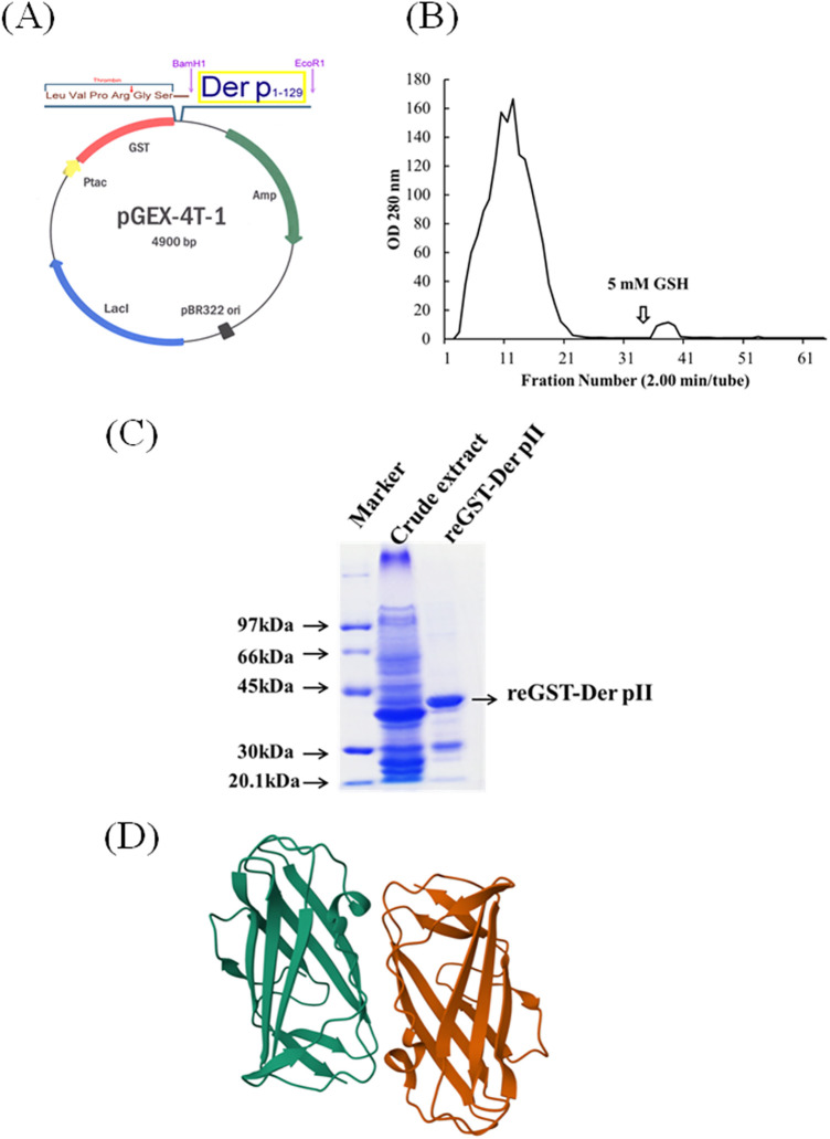 Figure 1
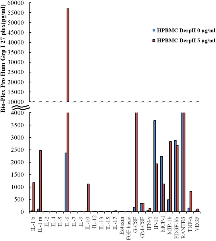 Figure 2
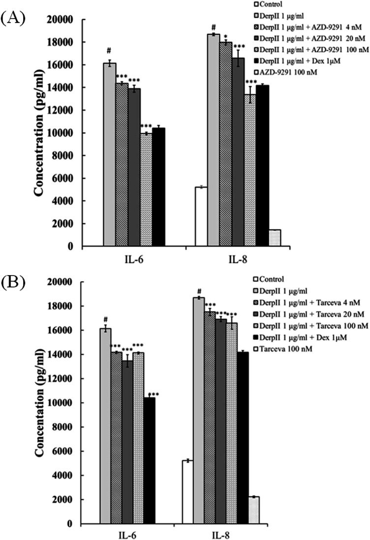 Figure 3
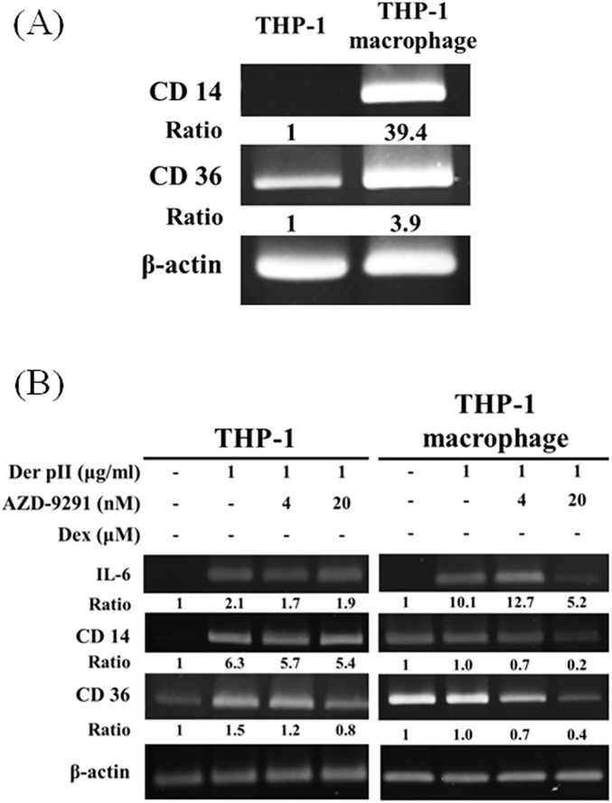 Figure 4
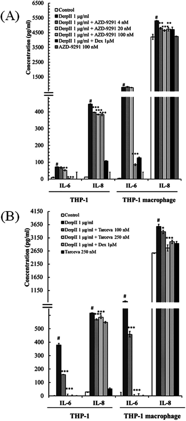 Figure 5
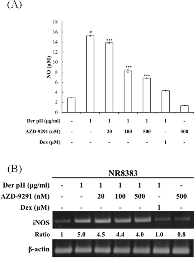 Figure 6
- —National Science and Technology Council10.13039/100020595
Peer Reviews
No public reviews on file for this paper yet. If you reviewed it on a platform where reviews are public (OpenReview, ICLR, NeurIPS, ICML), you can paste yours below so the community can read it here.
Videos
No videos yet. Explain this paper in a talk, walkthrough, or lecture? Add one.
Taxonomy
TopicsAsthma and respiratory diseases · Allergic Rhinitis and Sensitization · Psoriasis: Treatment and Pathogenesis
Introduction
1
Allergic asthma affects over 300 million people globally. Its main features include eosinophilic infiltration in the airway, excessive mucus secretion, and airway hyperresponsiveness. Literature indicates that allergic-specific T helper 2 (Th2) cells and related cytokines regulate allergic airway inflammation and stimulate mucus overproduction, further causing airway hyperresponsiveness (1, 2). Among the major causes of asthma, house dust mites are the most significant, affecting 85% of asthma patients. More than 50% of children and adolescents are sensitive to house dust mites (3, 4). Long-term exposure to mite allergens not only causes asthma but also contributes to atopic dermatitis and allergic rhinitis (5). According to the European Community Respiratory Health Survey (ECRHS), the prevalence of mite-related disease in 15 developed European countries was 21.7% (6).
According to Taiwan's Environmental Protection Administration, common allergy-causing mites thrive in household environments. These mites are tiny (∼0.2 mm), preferring temperatures between 22 and 26°C and humidity levels of 70%–80%, feeding primarily on human skin flakes. Research shows that Dermatophagoides pteronyssinus (Der p) is the most predominant dust mites in Taiwan (7). A 2006 study by Prof. Lü Kuan's team examined 462 atopic allergic children (ages 2–16) in central Taiwan. Over 80% were sensitive to various mites, especially Der m (79.5%), Der p (90.2%), and Der f (88.9%) (8). House dust mite allergens trigger antigen-presenting cells, driving Th2 activation, IgE production, and immune cell buildup in the lungs, nasal passages, and sensitive skin (9). As of now, the WHO/IUIS Allergen Nomenclature Subcommittee lists 36 allergen groups for Der p (up to Group 36), among which Groups 1, 2, and 23 are major IgE-binding proteins. Groups 1, 2, 5, 7, and 10 together account for over 80% of allergen reactivity in patient sera (10).
Monocytes and macrophages play a vital role in various inflammatory environments due to their plasticity and ability to infiltrate inflamed tissues. Bone marrow–derived blood monocytes can adhere to the vascular endothelium and migrate into tissues in response to inflammatory stimuli (11–13). Alveolar macrophages, located in the alveolar spaces of the lungs, play a key role in eliminating allergens and maintaining immune balance in the pulmonary environment (14). Chemokines such as CCL2 and MCP-1 play crucial roles in regulating the migration and infiltration of monocytes and natural killer (NK) cells (15, 16). Infected-site macrophages use membrane receptors such as CD36, CD14, and TLR4 to recognize pathogens, phagocytose them, and then eliminate internal debris (17).
Our previous research demonstrated that EGFR inhibitors alleviate Der p2-induced inflammation in human bronchial epithelial cells (18). In this study, HPBMC, THP-1 monocytes and THP-1 macrophage were used to investigate Der pII-induced inflammation. We found that EGFR inhibitors inhibit the Der pII-induced IL-6, IL-8, and NO production in HPBMC, monocytes and macrophages. This is the first study to exam the function of EGFR inhibitors on Der pII-induced inflammation in monocytes and macrophages.
Materials and methods
2
Cell culture and chemicals
2.1
THP-1 cells (ATCC, TIB-202) and NR8383 cells (ATCC, CRL-2192) were obtained from the American Type Culture Collection. THP-1 cells were cultured in RPMI 1640 Medium (GIBCO, 31800022) supplemented with 10% fetal bovine serum. NR8383 cells were cultured in Ham's F12K (GIBCO, 21127022), and supplemented with 15% fetal bovine serum. AZD-9291 MCE (HY-15772) (22289) was purchased from MedChemExpress (New Jersey). Tarceva was purchased from OSI Pharmaceuticals (Northbrook, Illinois).
Purification of GST-Der PII1-129 proteins
2.2
Escherichia coli protein expression system was used to express the recombinant GST-Der PII1-129. The detailed protocol for the purification has been described previously (18).
Enzyme-linked immunosorbent assay (ELISA)
2.3
After treatment, the medium was collected for ELISA analysis. Human IL-6 ELISA MAXTM Deluxe Set (BioLegend #430505) and Human IL-8 ELISA MAXTM Deluxe Set (BioLegend #431540) were performed to analyze the IL-6 and IL-8 expression, respectively. The expressions of IL-6 and IL-8 by ELISA were following the protocol of manufacturer's instructions.
Nitric oxide (NO) assay
2.4
Nitric oxide (NO) levels in cell culture supernatants were determined using the Griess Reagent System. Following stimulation, 50 μL of each collected supernatant was transferred to individual wells of a 96-well plate. An equal volume (50 μL) of Sulfanilamide Solution was then added to each well and incubated at room temperature for 5–10 min. Subsequently, 50 μL of NED Solution was added, followed by incubation in the dark at room temperature for another 5–10 min. If NO was present, a purple-blue color developed, and the absorbance was measured at 520 nm using an ELISA reader. All experiments were performed in triplicate, and NO concentrations were calculated by comparison with a standard curve.
Statistical analysis
2.5
One-way ANOVA and Tukey's post hoc test by Predictive Analytics SoftWare (PASW) Statistics 18 were performed to conduct the statistical comparisons between two groups. Values of p < 0.05 were considered significant. Data are presented as mean ± SD.
Results
3
Expression of GST-Der pII recombinant protein
3.1
To express the Der pII, the gene of Der pII was cloned into pGEX-4t-1 expression vector, in order to create a glutathione-S-transferase (GST) fusion protein with the N-terminus of Der pII (Figure 1A). The GST-Der pII was induced by IPTG and purified (Figures 1B,C). The protein structure of Der pII (1KTJ) from RCSB Protein Data Bank (RCSB PDB) was shown as Figure 1D.
Construction of the expression plasmid pGEX-Der pII and purified the Der pII1-129. (A) The gene mapping of pGEX-Der pII containing Der pII coding region 1–129 amino acid. (B) The fusion protein was purified by affinity chrmatograph with a Glutathione-Sepharose 4 Fast Flow column. (C) SDS-PAGE analyss of fusion protein and purified reDer pII 1–129. (D) Protein structure of Der pII (1KTJ) from RCSB Protein Data Bank (RCSB PDB).
Effect of Der pII on cytokine induction in human peripheral blood mononuclear cells
3.2
To examine the effect of Der pII on cytokine production, human peripheral blood mononuclear cells (HPBMCs) were stimulated with Der pII, and cytokine levels in the culture supernatant were assessed using the Bio-Plex Pro Human Cytokine 27-Plex Immunoassay. As shown in Figure 2, Der pII markedly induced the production of interleukin-1 beta (IL-1β), interleukin-1 receptor antagonist (IL-1ra), interleukin-6 (IL-6), interleukin-10 (IL-10), granulocyte-colony stimulating factor (G-CSF), macrophage inflammatory protein-1β (MIP-1β), and tumor necrosis factor-α (TNF-α). To explore potential interactions between EGFR and these Der pII-induced cytokines, the STRING database was utilized. Analysis revealed direct protein–protein associations between EGFR and IL-6, G-CSF, TNF-α, IL-1β, and IL-10 (Supplementary Figure S1).
Analysis the production of varies cytokines in Der pII-stimulated HPBMC cells by Bio-Plex Pro human cytokine 27-Plex immunoassay. HPBMC cells (2 × 10⁶cells/well of 24 well) were treated with GST-Der pII (5 μg/mL) for 48 h and the medium was analyzed by Bio-Plex Pro human Cytokine 27-Plex immunoassay.
EGFR inhibitors suppressed Der pII-induced IL-6 and IL-8 expression in HPBMC
3.3
To evaluate the role of EGFR in IL-6 and IL-8 induction by Der pII, AZD-9291 and Tarceva, two EGFR inhibitors, were used to inhibit the function of EGFR. As shown in Figure 3, Der pII significantly stimulated the secretion of IL-6 and IL-8. Both AZD-9291 and Tarceva significantly reduced the IL-6 and IL-8 induction by Der pII. Treatment with 100 nM AZD9291 reduced Der pII–induced IL-6 and IL-8 production by approximately 37% and 25%, respectively. 100 nM Tarceva treatment reduced Der pII–induced IL-6 and IL-8 production by approximately 13% and 11%, respectively. Dexamethasone (Dex) was used as a positive control in inhibiting IL-6 and IL-8 induction.
Analysis the production of IL-6 and IL-8 in Der pII-stimulated HPBMC. HPBMC (2 × 10⁶ cells/well of 24 well) were pre-treated with (A) AZD-9291 (B) Tarceva for 2 h followed by stimulated Der pII (1 μg/mL) for 24 h. Conditioned media were measured amounts of secreted IL-6 and IL-8 by ELISA. The data are expressed as mean ± SD. The symbols (#) indicated significant differences as compared with control, and () as compared with Der pII alone. *p < 0.05, **p < 0.01,**p < 0.001.
Effect AZD-9291 on Der pII-induced mRNA expressions in THP-1 monocytes and THP-1 macrophages
3.4
To investigate the effect of Der pII on monocyte and macrophage, THP-1, a monocyte cell line, and THP-1 derived macrophage were used to investigation. After PMA treatment, the THP-1 cells were differentiated into M0 macrophage with increasing of CD14 and CD36, the markers of M0 macrophage (Figure 4A). In THP-1 cells, Der pII increased the gene expressions of IL-6, CD14, and CD36. Der pII induced higher IL-6 expression in THP-1 macrophages compared to THP-1 monocytes (Figure 4B). AZD-9291 reduced the gene expressions of IL-6, CD14, and CD36 in THP-1 macrophages (Figure 4B).
The effects of AZD-9291 on Der pII–stimulated human macrophage. (A) Gene expressions of CD14 and CD36 in THP-1 and THP-1 macrophage. (B) THP-1 (1 × 10⁶ cells/6 cm dish) and THP-1 macrophage (1 × 10⁶ cells/3.5 cm dish) were pre-treated with AZD-9291 for 2 h followed by stimulated Der pII (1 μg/mL) for 24 h. Gene expressions of IL-6 and IL-8 were analyzed by RT-PCR.
EGFR inhibitors suppressed Der pII-induced IL-6 and IL-8 expression in THP-1 monocyte and THP-1 macrophage
3.5
To examine the involvement of EGFR in Der pII–induced IL-6 and IL-8 expression in THP-1 cells and THP-1 macrophages, EGFR function was inhibited using AZD9291 and Tarceva. In Supplementary Figure S2, we found that 100 nM AZD-9291 or 250 nM Tarceva did not alter the cell viability of THP-1 macrophage with Der pII treatment. Furthermore, AZD-9291 and Tarceva reduced Der pII-induced EGFR activation (Supplementary Figure S3). As shown in Figure 5, Der pII significantly activated the secretion of IL-6 and IL-8 in THP-1 and THP-1 macrophage. Higher expressions of IL-6 and IL-8 were induced by Der pII in THP-1 macrophages compared to THP-1 cells. AZD-9291 and Tarceva significantly reduced the IL-6 and IL-8 induction by Der pII. Treatment with 100 nM AZD9291 decreased Der pII–induced IL-6 and IL-8 production by approximately 93% and 15% in THP-1 macrophages, respectively. 250 nM Tarceva treatment decreased Der pII–induced IL-6 and IL-8 production by approximately 99% and 20%, respectively. Dexamethasone (Dex) was used as a positive control in inhibiting IL-6 and IL-8 induction.
The effects of AZD-9291 on Der pII–stimulated inflammation in human macrophage. THP-1 (1 × 10⁶ cells/6 cm dish) and THP-1 macrophage (1 × 10⁶ cell/3.5 cm dish) were pre-treated with (A) AZD-9291 (4, 20 and 100 nM) or (B) Tarceva (100 and 250 nM) for 2 h followed by stimulated Der pII (1 μg/mL) for 24 h. Conditioned media were measured amounts of secreted IL-6 and IL-8 by ELISA. The data are expressed as mean ± SD. The symbols (#) indicated significant differences as compared with control, and () as compared with Der pII alone. *p < 0.05, **p < 0.01,**p < 0.001.
AZD-9291 suppressed Der pII-induced NO production in pulmonary alveolar macrophage
3.6
To investigate the effect of AZD-9291 on Der pII-induced stimulation of pulmonary alveolar macrophages, NR8383 cells—an alveolar macrophage cell line—were used as a model to assess NO production. As shown in Figure 6A, Der pII-induced NO production was significantly inhibited by AZD-9291. However, AZD-9291 only slightly reduced the gene expression of iNOS induced by Der pII.
Effects of AZD-9291 on Der pII-induced nitrite oxide production in rat alveolar macrophages. The NR8383 cells (4 × 10⁶ cell/ 3.5 cm dish) were pre-treated with AZD-9291 (20, 100, 500 nM) or Dexamethasone (1 μΜ) for 2 h followed by stimulated Der pII (1 μg/mL) for 24 h. (A) Conditioned media was assayed level of NO by Griess reagent system. The data are expressed as mean ± SD. The symbols (#) indicated significant differences as compared with control, and () as compared with Der pII alone. *p < 0.05, **p < 0.01,**p < 0.001. (B) Gene expressions of INOS was analyzed by RT-PCR.
Discussion
4
Inflammation involves both local manifestations, such as redness and swelling, and systemic responses, including fever. Neutrophils are recruited during the early stages of inflammation, followed by monocytes and lymphocytes in the later stages. Cytokines, including IL-17, stimulate stromal cells to produce IL-6 and IL-8. IL-6 primarily mediates fever, while IL-8 promotes the recruitment and accumulation of neutrophils in the blood and inflamed tissues (19–21).
Human peripheral blood mononuclear cells (HPBMCs), comprising lymphocytes and monocytes with a round nucleus, exhibited significantly reduced IL-6 and IL-8 secretion upon Der pII stimulation when treated with AZD-9291 or Tarceva (Figure 3). This indicates that AZD-9291 may be a promising multi-target anti-inflammatory agent, potentially useful in allergic airway diseases such as asthma, even in the absence of EGFR mutations.
In the present study, Der pII induced the secretion of IL-6 and IL-8 in THP-1 macrophages. AZD-9291 and Tarceva effectively suppresses the secretion of IL-6 and IL-8 induced by Der pII (Figure 5). Interestingly, Der pII stimulation in THP-1 macrophages leads to an upregulation of CD14 and CD36, and AZD-9291 treatment significantly inhibits the expressions of these genes. CD36, a class B scavenger receptor expressed in diverse cell types such as bone marrow cells, platelets, endothelial cells, and adipocytes, plays essential roles in fatty acid transport, angiogenesis inhibition, and bacterial phagocytosis (22, 23). CD14, a glycoprotein predominantly expressed on mature cells of the monocyte lineage, has been shown to mediate LPS-induced signaling pathways in macrophage phagocytosis (24). Upon bacterial infection, LPS binds to LPS-binding proteins and then form a complex with CD14, activating the TLR4-MyD88 signaling cascade (25). This pathway triggers the translocation of NF-κB to the nucleus, leading to the transcription of pro-inflammatory genes such as TNF-α, IL-1β, IL-6, and iNOS (26).
In pulmonary alveolar macrophage models, Der pII stimulation increased NO production, a hallmark of inflammatory activation, but this was significantly reduced by AZD-9291. Interestingly, this reduction was not correlated with suppressed iNOS gene expression, suggesting that AZD9291 affects NO production at a post-transcriptional level or directly influences enzyme activity. Additionally, AZD-9291 reduced expression of macrophage activation markers CD14 and CD36 in THP-1 cells, indicating it may suppress macrophage polarization.
Conclusion
5
In this study, human peripheral blood mononuclear cells (HPBMCs), THP-1 monocytes, and THP-1–derived macrophages were used to investigate Der pII–induced inflammatory responses. We demonstrated that the EGFR inhibitors significantly suppressed Der pII–induced production of IL-6, IL-8, and nitric oxide in HPBMCs, monocytes, and macrophages. This is the first study to examine the regulatory effects of EGFR inhibitors on Der pII–induced inflammation in monocytes and macrophages.
The reference list from the paper itself. Each links out to its DOI / PubMed record.
- 1Leon B. Understanding the development of Th 2 cell-driven allergic airway disease in early life. Front Allergy. (2022) 3:1080153. 10.3389/falgy.2022.108015336704753 PMC 9872036 · doi ↗ · pubmed ↗
- 2Cho JL Ling MF Adams DC Faustino L Islam SA Afshar R Allergic asthma is distinguished by sensitivity of allergen-specific CD 4+ T cells and airway structural cells to type 2 inflammation. Sci Transl Med. (2016) 8(359):359ra 132. 10.1126/scitranslmed.aag 137027708065 PMC 5399547 · doi ↗ · pubmed ↗
- 3Zhang P Jia Y Jing Z Huang J Wu H Sun X. Efficacy and safety of house dust mite subcutaneous immunotherapy in polysensitized children with allergic asthma. Pulm Pharmacol Ther. (2023) 78:102187. 10.1016/j.pupt.2022.10218736603742 · doi ↗ · pubmed ↗
- 4Gaffin JM Phipatanakul W. The role of indoor allergens in the development of asthma. Curr Opin Allergy Clin Immunol. (2009) 9(2):128–35. 10.1097/ACI.0b 013e 32832678 b 019326507 PMC 2674017 · doi ↗ · pubmed ↗
- 5Jurkiewicz K Jutel M Smolinska S. Update on HDM allergy: principal changes over the years. Int J Mol Sci. (2025) 26(12). 10.3390/ijms 26125660 PMC 1219340440565123 · doi ↗ · pubmed ↗
- 6Calderon MA Kleine-Tebbe J Linneberg A De Blay F Hernandez Fernandez de Rojas D Virchow JC House dust Mite respiratory allergy: an overview of current therapeutic strategies. J Allergy Clin Immunol Pract. (2015) 3(6):843–55. 10.1016/j.jaip.2015.06.01926342746 · doi ↗ · pubmed ↗
- 7Wang DH Chen L Cheng L Li KN Yuan H Lu JH Fast onset of action of sublingual immunotherapy in house dust mite-induced allergic rhinitis: a multicenter, randomized, double-blind, placebo-controlled trial. Laryngoscope. (2013) 123(6):1334–40. 10.1002/lary.2393523616386 · doi ↗ · pubmed ↗
- 8Huang HW Lue KH Wong RH Sun HL Sheu JN Lu KH. Distribution of allergens in children with different atopic disorders in central Taiwan. Acta Paediatr Taiwan. (2006) 47(3):127–34.17078465 · pubmed ↗