Site-Specific Native Antibody-Conjugated Microbubbles for Molecular Ultrasound Imaging of Hepatocellular Carcinoma
James Hui, Yonghwan Shin, Janet Pham, Po-Chun Chen, Aravinth Ruppa, Veronica Hankil, Lea Guo, Hanif Saifurrahman Kalamullah, Naoki Kaneko, Jason Chiang

TL;DR
This study develops microbubbles with site-specific antibodies for precise ultrasound imaging of liver cancer, enabling noninvasive molecular profiling.
Contribution
The use of light-activated site-specific conjugation to attach antibodies to microbubbles for targeted ultrasound imaging of hepatocellular carcinoma.
Findings
LASIC chemistry enabled covalent and site-specific antibody attachment to microbubbles.
Targeted microbubbles showed significantly increased binding to GPC3-positive cells in vitro.
In vivo imaging revealed rapid and sustained tumor-specific signal enhancement with LASIC-conjugated microbubbles.
Abstract
To develop a microbubble-based contrast agent using light-activated site-specific conjugation (LASIC) for targeted molecular ultrasound imaging of glypican-3 (GPC3) positive hepatocellular carcinoma (HCC). Azide-functionalized microbubbles were conjugated with commercially available anti-GPC3 antibodies using a LASIC DBCO adapter, which enables precise site-specific labeling at the antibody’s heavy chain without compromising antigen-binding affinity. Binding specificity was confirmed through pre-blocking experiments with free anti-GPC3 antibodies in HCC cell lines. Fluorescence microscopy and quantitative image analysis were employed to evaluate cell-binding efficiency. For in vivo validation, conjugated microbubbles were intravenously administered into mice bearing orthotopic HepG2 xenografts, followed by contrast-enhanced ultrasound (CEUS) imaging at 0 seconds, 30 seconds, and 10…
Genes, proteins, chemicals, diseases, species, mutations and cell lines named across the full text — each resolved to its canonical identifier and authoritative record.
Click any figure to enlarge with its caption.
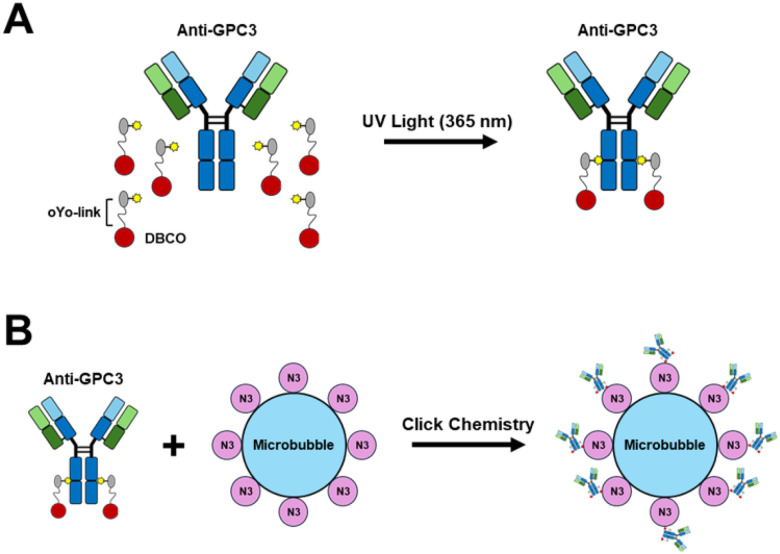 Figure 1
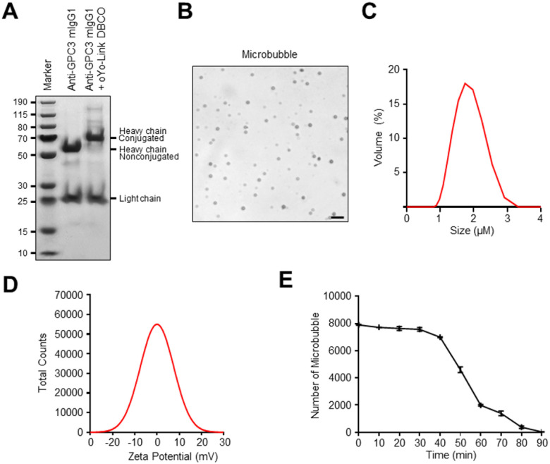 Figure 2
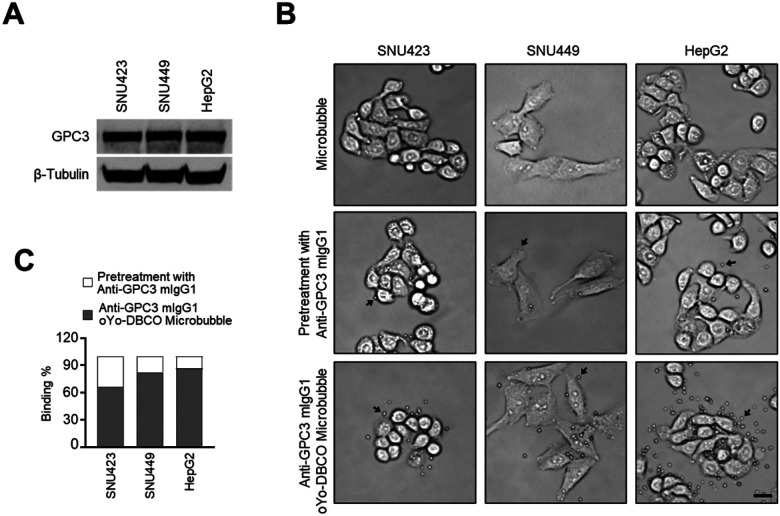 Figure 3
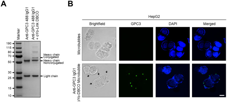 Figure 4
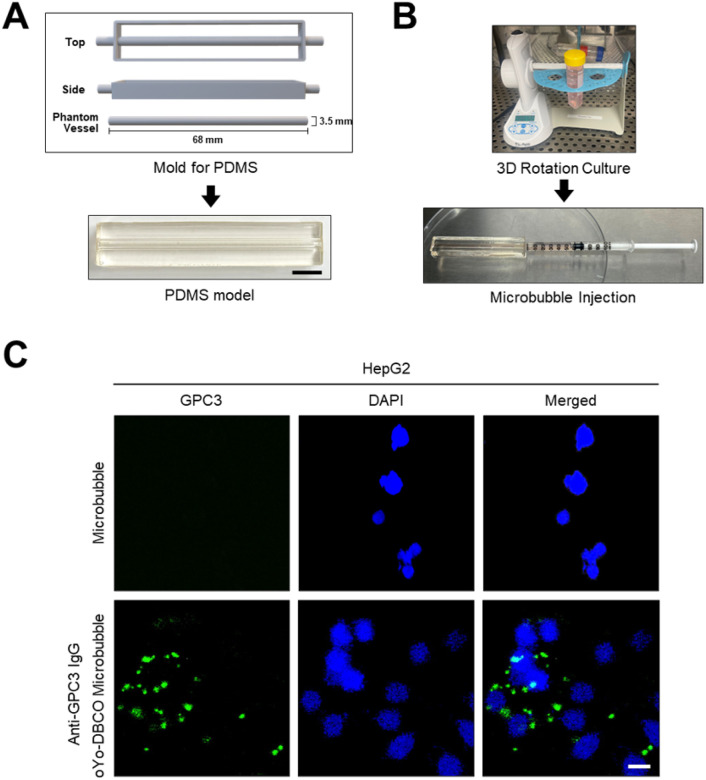 Figure 5
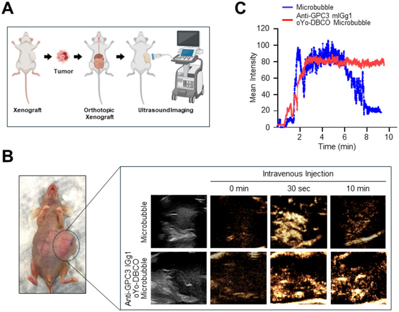 Figure 6
Peer Reviews
No public reviews on file for this paper yet. If you reviewed it on a platform where reviews are public (OpenReview, ICLR, NeurIPS, ICML), you can paste yours below so the community can read it here.
Videos
No videos yet. Explain this paper in a talk, walkthrough, or lecture? Add one.
Taxonomy
TopicsUltrasound and Hyperthermia Applications · Hepatocellular Carcinoma Treatment and Prognosis · Photoacoustic and Ultrasonic Imaging
Introduction
Hepatocellular carcinoma (HCC) is the fourth-leading cause of all cancer-related deaths worldwide, accounting for approximately 90% of primary liver cancers (1). While the standard of care diagnosis of early-stage HCC traditionally relies on contrast-enhanced CT or MRI (2), these modalities suffer from limitations including lack of real-time visualization, limited outpatient imaging availability, ionizing radiation (with CT), and motion artifact (with MRI). Contrast-enhanced ultrasound (CEUS) offers key advantages in facilitating HCC detection, including real-time evaluation, absence of ionizing radiation, and favorable safety profiles of microbubble contrast agents (3–5).
Despite its proven utility in diagnosing HCC, CEUS does not provide information about the underlying tumor molecular characteristics, which may be leveraged for prognostic purposes and determine optimal treatment strategies(6). While needle biopsies can be performed to obtain tissue in cases of suspected HCC, they are usually reserved for LR-4 or LR-M lesions, are prone to sampling heterogeneity and pose procedure-related risks(7). These limitations highlight the need for reliable ways to detect tumor markers in HCC.
Given CEUS’ favorable safety profile and ready accessibility, a compelling approach to non-invasively characterize cancer surface markers is to conjugate an antibody of interest onto contrast ultrasound microbubbles. Microbubbles that are currently being used clinically in CEUS are nontargeted, limiting them to only imaging perfusion and unable to provide molecular-level information. While there have been various precedents in developing targeted microbubbles, the techniques used for antibody attachment are often inefficient and tedious, thereby restricting their wider adoption beyond proof of principle demonstrations. For example, the most commonly used amine modification chemistry results in the antibodies that are often mis-oriented on the microbubble and indiscriminately linked via any number of lysine residues, leading to heterogeneous products with variable pharmacokinetics, toxicity and affinity for antigen(8). Another class of widely used conjugation technique - based on biotin-avidin/streptavidin binding, not only suffers from similar shortfalls since biotinylation also occurs indiscriminately on lysine groups, but what’s more is also poorly suited for clinical uses due to its immunogenicity and interference from endogenous biotins. Consequently, considerable efforts have been made to develop site-specific conjugates for a variety of antibody-based products– with the goal of achieving more precise control of the modification sites and higher production efficiency(9).
Light Activated SIte-Specific Conjugation (LASIC) is a previously developed site-specific antibodylabeling technique of native antibodies via photochemistry(10). This technique facilitates the formation of a single covalent bond between a 60mer peptide adapter containing the desired conjugate and each Fc region on IgG when exposed to gentle long wavelength UV light, leading to more homogeneous, sitespecific attachments. As this technique preserves antibody function and orientation, can be used with any off-the-shelf antibodies, and can be readily combined with the robust azide based click chemistry, it maybe well-suited for efficiently attach desired antibodies onto the fragile microbubbles. Glypican-3 (GPC3) is well recognized as a highly specific surface marker of HCC, carries prognostic values, and is the molecular target for several therapies in development (11–15). By developing targeted microbubbles against GPC3 in this study, we hope to demonstrate the efficient production of targeted, site-specifically labeled microbubbles for molecular imaging using CEUS.
Materials and Methods
Cell Culture
Human hepatocellular carcinoma (HCC) cell lines—SNU423, SNU449, and HepG2—were obtained from the American Type Culture Collection (ATCC, Manassas, VA, USA). SNU423 and SNU449 cells were cultured in either RPMI 1640 or Dulbecco’s Modified Eagle Medium (DMEM), both supplemented with 10% fetal bovine serum (FBS), 100 U/mL penicillin, and 100 μg/mL streptomycin. All cell lines were maintained at 37 °C in a humidified incubator with 95% air and 5% CO_2_.
Conjugation of the GPC3 Antibody with microbubbles
Native (Cat# BE0404, Bio X cell, Lebanon, NH, USA) and FITC-labeled Anti-GPC3 antibodies (Cat# NBP3–28387F, Novus Biologicals, Centennial, CO, USA) were site-specifically modified using oYo-link DBCO (AlphaThera, Philadelphia, PA, USA) according to the manufacturer’s instructions, specifically 20 μg of anti-GPC3 antibody was incubated with oYo-link DBCO and exposed to long wavelength (365 nm) UV light on ice for 2 hours to enable photocrosslinking. The conjugated antibody was then purified using oYo-capture magnetic beads and washed three times with PBS. Usphere^™^ labeler microbubbles containing azide-modified phospholipids (Scintica, Webster, TX, USA), which are based on a commercially available preclinical contrast agent with established in vivo dosing and imaging safety guidelines provided by the manufacturer, were agitated with the UltraMix^™^ device for 40 seconds at room temperature to generate a homogeneous suspension with a particle concentration of 1–5 × 10^10^ microbubbles/mL, according to the manufacture’s instruction. The purified oYo-link DBCO conjugated antibody was mixed with 20 μl of azide functionalized microbubbles at room temperature for at least 2 hours to allow the click chemistry covalent bond to form, resulting in stable antibody microbubble conjugates.
Dynamic Light Scattering, Zeta Potential, and Stability Measurements
Dynamic light scattering (DLS) and zeta potential measurements were performed on the Zetasizer Nano Zs analyzer (Malvern Panalytical, Malvern, UK) to characterize microbubble size distribution and surface charge. For both measurements, samples were diluted 1:10 in deionized water. Zeta potential readings were obtained using U-shaped cuvettes, while DLS measurements utilized plastic cuvettes. All analyses were performed at 25 °C and repeated in triplicate. Data were processed using Zetasizer software. Microbubble stability was evaluated by monitoring the number of intact microbubbles over time. Microbubbles were diluted 1:50,000 (v/v) in culture medium and incubated at 37 °C. At the indicated time points from 0 to 90 min, microbubble counts were quantified by light microscopy.
Western Blot Analysis
The harvested SNU423, SNU449, and HepG2 cells were washed with phosphate buffered saline (PBS) and dissolved in 20 mM Tris-HCl, pH7.5, lysis buffer. The protein samples were separated using 4–12% SDS-PAGE and transferred to a 0.2 μm nitrocellulose membrane. After blocking with 5% non-fat dry milk (Bio-Rad, Irvine, CA, USA), the membrane was incubated overnight at 4 °C with a primary antibody and washed with Tris-buffered saline with Tween 20 (TBST). The membrane was washed with TBST, incubated at room temperature with a secondary antibody for 1 h, and visualized using an ECL reagent (Thermo Fisher Scientific, San Diego, CA, USA). All experiments were performed in triplicate. Antibodies used in this study are follow: GPC3 antibody (1:5000) from Bio X Cell (Lebanon, NH, USA); β-Tubulin antibody (1:5000) from Abcam (Cambridge, MA, USA); HRP-conjugated anti-mouse antibody (1:5000) from Abcam (Cambridge, MA, USA).
Immunofluorescence
HepG2 cells were grown on cell culture slides (Thermo Fisher Scientific, San Diego, CA, USA) and treated with oYo-link DBCO-anti-GPC3 conjugated Microbubbles for 30 min. Then, these cells were stained with DAPI (Thermo Fisher Scientific, San Diego, CA, USA). The cells were washed with PBS and visualized by a Nikon Confocal Laser Scanning Confocal microscope.
PDMS Phantom Vessel Model
PDMS (Sylgard 184, 10:1 w/w base to curing agent) was cast into a rectangular acrylonitrile-butadienestyrene (ABS) mold featuring a central 3.15-mm-diameter cylindrical core, creating a phantom with vessel-like cylindrical lumen at its center. To enhance hydrophilization, the PDMS model was soaked in 10% (3-aminopropyl) trimethoxysilane (APTMS) in 100% ethanol overnight and then washed with water. The luminal surface of the model was then activated with sulfo-SANPAH followed by UV irradiation and was subsequently treated with fibronectin (40 μg/mL). HepG2 cells were cultured in DMEM supplemented with 10% FBS and 100 U/ml penicillin + 100 μg/ml streptomycin at 37 °C in a CO_2_ incubator (95% air, 5% CO_2_) then seeded into the PDMS phantom vessel model. To promote a uniform cell adhesion on the vessel luminal surface, the model was seeded in a 3D rotating instrument at 1 rotation per minute placed in the CO2 incubator. After establishing the PDMS cell culture model, oYo-link anti-GPC3 antibody-conjugated microbubbles were injected into the vessel using a 1 mL syringe, incubated to allow binding to HePG2 cells, and visualized by a Nikon Confocal Laser Scanning Confocal microscope.
HepG2 Cells-Derived Orthotopic Implantation and Ultrasound Imaging
HepG2 cells were suspended in RPMI containing 25% Matrigel (356234, Life Sciences) such that 100 μL volume contained 1×10^6^ cells to inject subcutaneously into mouse flanks. After three weeks, tumors were excised from the flanks and orthotopically implanted into the liver following a standard protocol(16). Briefly, 7-week-old nude mice (homozygous for Foxn1<nu>, male, Jackson Laboratories) were anesthetized using isoflurane, and the liver was exposed. A 70 mg tumor was attached to the left-lateral lobe of the liver using 5.0 VICRYL suture (NC2872069, Fisher Scientific, Waltham, MA, USA) and wrapped with a 1×0.5 cm piece of SURGICEL further protect the attachment and minimize bleeding. Incisions were closed using wound clips, and mice were set up for recovery with a post-surgery care protocol. After four weeks of implantation, the mice were injected into the tail vein with microbubbles or with oYo-Link DBCO-anti-GPC3 conjugated microbubbles and ultrasound imaging was performed using a linear array transducer (el 18–4). Contrast-mode imaging was conducted at a low mechanical index (MI = 0.06) to preserve microbubble integrity, with a frame rate of 23Hz. The imaging depth was set to 3.5 cm, and gain settings were kept constant across experiments. Dual-mode imaging was used to simultaneously acquire contrast-enhanced and B-mode images. The injected microbubble dose was selected based on the manufacturer’s protocol for tumor imaging in mice. All animal protocols in this study were approved by the Division of Laboratory Animal Medicine (DLAM) at the university.
Results
LASIC-based Conjugation and Characterization of GPC3 Targeted Microbubble
LASIC has previously been introduced as a versatile method to achieve site-specific antibody modification without compromising antigen binding(10). Building upon this method, we developed glypican-3 (GPC3)-targeted microbubbles using an optimized LASIC-based strategy. Specifically, long-wavelength UV light was employed to covalently attach a small peptide adapter containing a Clickchemistry compatible strained octyne moiety (oYo-link DBCO) to the Fc region of a commercially available, “off the shelf” anti-GPC3 monoclonal IgG1 antibody. This site-specific conjugation selectively modified the antibody’s heavy chains while preserving antigen recognition. Following modification, the anti-GPC3 antibodies were conjugated to commercially available azide-functionalized microbubbles via Click chemistry, resulting in stable and uniform antibody attachment to the microbubble surface (Fig. 1).
When the conjugated antibody is examined on SDS-PAGE gel, a shift in the apparent molecular weight of the heavy chain before and after photo-crosslinking with oYo-link DBCO confirmed successful site-specific labeling of antibody (Fig. 2A). Light microscopy visualized the microbubble morphology (Fig. 2B), while dynamic light scattering (DLS) analysis demonstrated consistent size distribution (Fig. 2C). Zeta potential measurements revealed a neutral surface charge, indicative of microbubble stability (Fig. 2D). Additionally, microbubble stability was assessed at 37°C in culture medium, where most microbubbles degraded within 30 minutes, on par with the in vivo stability of clinically used bubbles. (Fig. 2E), indicating reasonable stability in post-reconstitution.
Binding of GPC3 Targeted Microbubbles to HCC Cells
GPC3 is a well-established biomarker and therapeutic target in HCC (11–15, 17, 18). To validate a GPC3-targeted molecular imaging approach, we first confirmed GPC3 expression in HCC cell lines—SNU423, SNU449, and HepG2—using Western blot analysis. All three cell lines exhibited strong GPC3 expression (Fig. 3A).
Next, we evaluated the binding efficiency of microbubbles conjugated with oYo-link DBCO-anti-GPC3 antibodies. Cells were incubated under three conditions: with unconjugated microbubbles, with oYo-link DBCO-anti-GPC3-conjugated microbubbles, and with conjugated microbubbles following pretreatment with free anti-GPC3 antibodies to block surface epitopes. Bright-field microscopy revealed robust binding of the conjugated microbubbles to HCC cells, while pre-blocking with free antibodies significantly reduced binding (Fig. 3B). Quantitative analysis confirmed these observations (Fig. 3C).
To allow fluorescent assessment of microbubbles binding in models, such as the PDMS vessel model further described below, that have suboptimal bright light penetration, we used a commercial, FITC-labeled anti-GPC3 antibody to conjugate onto azide microbubbles. SDS-PAGE confirmed site-specific photo-crosslinking of oYo-link DBCO to the heavy chain of FITC-labeled anti-GPC3 IgG1 (Fig. 4A). HepG2 cells were treated with the resulting fluorescent anti-GPC3 microbubbles and analyzed via fluorescent microscopy. Strong FITC signals were seen on the cell surface in treated cells, colocalizing with microbubbles seen using bright-field microscopy, (Fig. 4B).
GPC3 Targeted Microbubble Binding in PDMS Vessel Model
To assess the targeting capability of oYo-link DBCO-anti-GPC3-conjugated microbubbles under vessellike conditions, we developed a polydimethylsiloxane (PDMS) phantom vessel model, adapted from a previously described method(19). The model featured a central cylindrical lumen, which was hydrophilized, coated with human fibronectin, and seeded with HepG2 cells. A 3D rotational culture was applied for 48 hours to promote uniform cell adhesion along the inner surface, thereby simulating a biologic vessel environment (Fig. 5A, 5B).
Following model establishment, FITC anti-GPC3-conjugated microbubbles described in the previous section were introduced via syringe injection and incubated to facilitate binding to HepG2 cells (Fig. 5B). Fluorescence imaging revealed strong FITC signals localized to the cell surfaces in samples treated with the conjugated microbubbles, confirming its ability to bind to GPC3-expressing HCC cells within a vessel-mimicking environment (Fig. 5C).
Ultrasound Imaging of Orthotopic HCC Xenograft with targeted Microbubbles
To assess the tumor-targeting ability of oYo-link DBCO-anti-GPC3 conjugated microbubbles in a physiologically relevant setting, an orthotopic HepG2 xenograft mouse model was established as previously described(16). After tumors developed in the liver, mice received intravenous injections of either unconjugated microbubbles or GPC3-targeted microbubbles, followed by CEUS imaging (Fig. 6A, 6B). CEUS revealed rapid and strong signal enhancement in tumors within 30 seconds of injecting GPC3-targeted microbubbles, persisting for up to 10 minutes (Fig. 6B). In contrast, mice injected with unconjugated microbubbles exhibited perfusion driven enhancement that diminished within 10 minutes. Quantitative analysis confirmed significantly higher and sustained signal intensity in the targeted group compared to controls (Fig. 6C). These findings demonstrate that oYo-link DBCO-anti-GPC3 conjugated microbubbles enable specific and durable imaging of GPC3-positive HCC xenografts, supporting their potential as targeted molecular ultrasound contrast agents.
Discussion
This study demonstrates the potential of LASIC—a novel site-specific conjugation technique—for advancing ultrasound imaging from purely structural assessment to molecular-level diagnostics. By targeting GPC3, a well-validated surface marker of HCC, with commercially available antibodies, LASIC-labeled microbubbles successfully detected GPC3 expression in cells and in an orthotopic HCC mouse model. This approach enables noninvasive, real-time, and widely accessible evaluation of tumor surface markers, paving the way for integrating molecular phenotyping into routine diagnostic workflows and supporting more personalized management strategies for HCC.
Although previous studies have demonstrated the feasibility of targeted microbubbles, their construction have largely relied on conventional conjugation chemistries that produce heterogeneous and often immunogenic products at low yields—making them difficult to replicate, scale, and translate clinically. These traditional methods, such as non-specific acylation of lysine residues or alkylation of cysteine thiols, have remained largely unchanged for decades and frequently result in promiscuous antibody attachment, compromising product uniformity and function(20). Recent efforts to achieve site-specific conjugation have involved complex antibody engineering—introducing ultra-reactive amines, unnatural amino acids, or peptide tags designed for bioorthogonal reactions(9, 21). While these approaches offer improved specificity, they are labor-intensive, may disrupt microbubble integrity, and require extensive customization for each target antigen(10, 22–24). The LASIC conjugation method produces homogeneous products while preserving the Fab region for antigen binding. Furthermore, by leveraging bioorthogonal click chemistry, the method used in this work maintains the structural integrity of both antibodies and microbubbles, making it ideally suited for molecular imaging applications(10, 25).
CEUS microbubbles possess favorable pharmacokinetic and safety profiles, contributing to their widespread clinical adoption in Europe and FDA approval in the United States (27, 28). The successful application of LASIC-based labeling to microbubbles presents exciting opportunities for molecular profiling across a range of solid tumors. The flexible, plug-and-play conjugation strategy described here enables the use of off-the-shelf antibodies to readily target other potential biomarkers such as HER2 in breast cancer, EGFR in lung cancer, and PSMA in prostate cancer (29, 30). Despite the advantages of ultrasound imaging—including real-time capability and safety—certain limitations remain. These include restricted soundwave penetration through bone or air-filled organs, reduced resolution in deeper tissues, and reliance on operator expertise for image acquisition. However, ongoing advancements in ultrasound technology, such as high-frequency transducers and 3D imaging, may help overcome these challenges. Finally, while LASIC-labeled microbubbles are expected to exhibit minimal immunogenicity—especially compared to biotin/avidin-based systems—further validation in animal models is necessary to confirm their safety and suitability for repeated clinical use.
This study demonstrates the potential of LASIC for efficiently labeling ultrasound microbubbles with antibodies targeting GPC3, a clinically significant surface marker in HCC. By enabling molecular-level imaging through targeted CEUS, this technique offers a powerful, noninvasive method to characterize tumor biology in real time. The anticipated improvements in specificity, stability, and imaging performance of LASIC-conjugated microbubbles may not only enhance diagnostic accuracy for HCC but also support personalized treatment planning across a range of solid tumors. Further preclinical validation and eventual clinical translation will be essential to fully realize the impact of this targeted imaging platform in oncologic radiology.
The reference list from the paper itself. Each links out to its DOI / PubMed record.
- 1Llovet JM, Kelley RK, Villanueva A, Singal AG, Pikarsky E, Roayaie S. Hepatocellular carcinoma. Nat Rev Dis Primers. 2021;7(1):6. doi: 10.1038/s 41572-020-00240-3.33479224 · doi ↗ · pubmed ↗
- 2Singal A, Volk ML, Waljee A, Salgia R, Higgins P, Rogers MA. Meta-analysis: surveillance with ultrasound for early-stage hepatocellular carcinoma in patients with cirrhosis. Aliment Pharmacol Ther. 2009;30(1):37–47. doi: 10.1111/j.1365-2036.2009.04014.x.19392863 PMC 6871653 · doi ↗ · pubmed ↗
- 3Bartolotta TV, Taibbi A, Midiri M, Lagalla R. Contrast-enhanced ultrasound of hepatocellular carcinoma: where do we stand? Ultrasonography. 2019;38(3):200–14. doi: 10.14366/usg.18060.31006227 PMC 6595127 · doi ↗ · pubmed ↗
- 4Eisenbrey JR, Gabriel H, Savsani E, Lyshchik A. Contrast-enhanced ultrasound (CEUS) in HCC diagnosis and assessment of tumor response to locoregional therapies. Abdom Radiol (NY). 2021;46(8):3579–95. doi: 10.1007/s 00261-021-03059-y.33825927 PMC 8569604 · doi ↗ · pubmed ↗
- 5Kono Y, Lyshchik A, Cosgrove D, Dietrich CF, Jang HJ, Kim TK. Contrast Enhanced Ultrasound (CEUS) Liver Imaging Reporting and Data System (LI-RADS(R)): the official version by the American College of Radiology (ACR). Ultraschall Med. 2017;38(1):85–6. doi: 10.1055/s-0042-1243369.28249328 · doi ↗ · pubmed ↗
- 6Passaro A, Al Bakir M, Hamilton EG, Diehn M, Andre F, Roy-Chowdhuri S. Cancer biomarkers: Emerging trends and clinical implications for personalized treatment. Cell. 2024;187(7):1617–35. doi: 10.1016/j.cell.2024.02.041.38552610 PMC 7616034 · doi ↗ · pubmed ↗
- 7Cannella R, Berman ZT, Tirukkovalur NV, Tohme ST, Minervini MI, Furlan A. Role of imaging-guided biopsy for hepatocellular carcinoma. Eur J Radiol. 2025;191:112271. doi: 10.1016/j.ejrad.2025.112271.40639022 · doi ↗ · pubmed ↗
- 8Wang R, Hu B, Pan Z, Mo C, Zhao X, Liu G, Antibody-Drug Conjugates (AD Cs): current and future biopharmaceuticals. J Hematol Oncol. 2025;18(1):51. Epub 20250430. doi: 10.1186/s 13045-025-01704-3.40307936 PMC 12044742 · doi ↗ · pubmed ↗