Bridging light and sound: A spironaphtopyran-rhodamine dyad with high-contrast photoswitching between fluorescence and photoacoustic signal
Nikita Kaydanov, Magdalena Olesińska-Mönch, Morgane Leite, Robert Prevedel, Claire Deo

TL;DR
This paper introduces a new photoswitchable probe that can switch between fluorescence and photoacoustic imaging, enabling dual-modality imaging with high contrast.
Contribution
The novel contribution is a spironaphtopyran-rhodamine dyad that enables reversible switching between fluorescence and photoacoustic signal with high contrast.
Findings
The spironaphtopyran photoswitches undergo reversible photoisomerization with distinct absorbing and non-absorbing states.
The FRET dyad with rhodamine shows robust and reversible switching between fluorescent and photoacoustic states in vitro.
Structure-property relationships were established, providing insights for future multimodal imaging probe development.
Abstract
Fluorescence and photoacoustic imaging are complementary modalities that provide distinct advantages for biological imaging: fluorescence microscopy offers high sensitivity and resolution, while photoacoustic imaging enables deeper penetration in complex tissue. Leveraging the strengths of both modalities through optically switchable contrast agents can offer enhanced imaging contrast and facilitate dual-modality imaging. Here, we report a photoswitchable probe capable of toggling between high fluorescence and high photoacoustic signal upon illumination, exploiting Förster Resonance Energy Transfer (FRET). We engineer novel spironaphtopyran photoswitches which undergo reversible photoisomerization between absorbing and non-absorbing states. Their photoswitching properties were systematically characterized, establishing structure-properties relationships, and providing the first…
Genes, proteins, chemicals, diseases, species, mutations and cell lines named across the full text — each resolved to its canonical identifier and authoritative record.
Click any figure to enlarge with its caption.
 Figure 1
 Figure 2
 Figure 3
 Figure 4
 Figure 5
 Figure 6
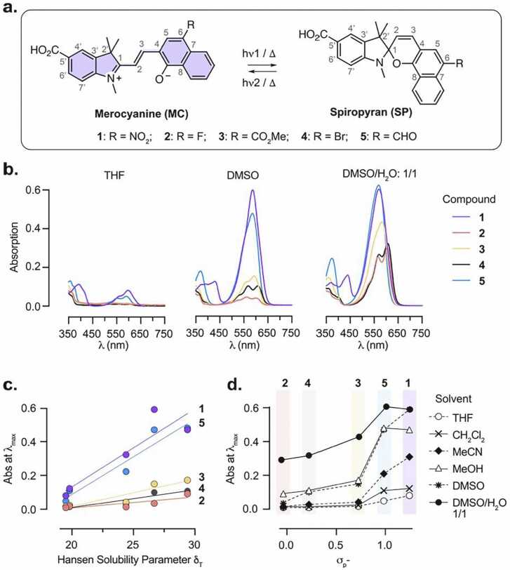 Figure 7
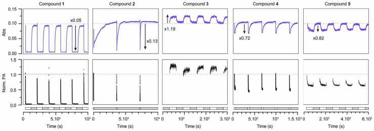 Figure 8
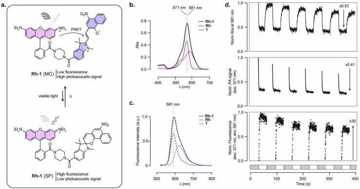 Figure 9
Peer Reviews
No public reviews on file for this paper yet. If you reviewed it on a platform where reviews are public (OpenReview, ICLR, NeurIPS, ICML), you can paste yours below so the community can read it here.
Videos
No videos yet. Explain this paper in a talk, walkthrough, or lecture? Add one.
Taxonomy
TopicsPhotochromic and Fluorescence Chemistry · Photoacoustic and Ultrasonic Imaging · Nanoplatforms for cancer theranostics
Introduction
1
Optical imaging modalities are powerful tools for the study of biological systems. Fluorescence (Fl) microscopy offers excellent sensitivity and spatial resolution but is fundamentally limited in imaging depth due to light scattering. [1] In contrast, photoacoustic (PA) imaging, which combines optical excitation with ultrasound detection, enables imaging several centimeters deep in complex biological tissues. [2], [3] Critically, the performance of these techniques depends on the availability of robust contrast agents, able to generate specific signal. [4], [5], [6] Among them, photoswitchable reporters which contrast can be modulated using light offer unique advantages. Indeed, such reporters have enabled breakthroughs in super-resolution fluorescence microscopy, [7], [8] facilitated background corrections in photoacoustic imaging, [9], [10], [11] and have been exploited in the design of photoresponsive functional tools such as biosensors. [12], [13], [14]
Extending this concept to dual-modality probes that can be optically toggled between fluorescence- and photoacoustic-dominant states can offer complementary information across scales and depths, which presents exciting opportunities for correlative imaging ranging from neuroimaging in model organisms to potential clinical applications. [15], [16], [17], [18] Achieving high contrast in both modalities, however, is challenging, as it requires precise control over how absorbed energy is distributed between radiative (fluorescence) and non-radiative (photoacoustic) decay pathways, which are inherently competitive. While several probes switch their dominant contrast modality in response to analytes, [19] reversible, light-induced modality switching remains largely unexplored.
To address this challenge, we focused on the well-established Förster Resonance Energy Transfer (FRET) mechanism. Indeed, fluorescence-quenching via FRET has been previously exploited to engineer both static and analyte-responsive probes displaying contrast in fluorescence and photoacoustic modalities. [20], [21], [22] We reasoned that coupling a photoswitchable acceptor with a fluorescent donor would enable efficient optical modulation through photoswitchable FRET (Fig. 1). In the dark state, efficient FRET between the two moieties would lead primarily to non-radiative relaxation, suppressing fluorescence, while retaining strong PA signal due to the high absorbance of both donor and acceptor. Upon illumination, photoisomerization of the acceptor to the non-absorbing isomer would abolish FRET, resulting in a large fluorescence increase and concomitant photoacoustic signal decrease, therefore efficiently modulating contrast across both modalities. To engineer such a FRET-based molecular reporter, a key requirement is an efficient photoswitchable acceptor which strongly absorbs in the dark state but becomes transparent following light activation (i.e. a “negative” photoswitch [23]). For this purpose, we focused on spiropyran derivatives, which represent a versatile class of photochromic compounds capable of reversible interconversion between a non-absorbing spiro (SP) form and a strongly absorbing merocyanine (MC) form. [24], [25], [26], [27] Importantly, depending on their structural features, derivatives of this family have been found to be stabilized as the MC isomer in the dark state [28], [29] which predispose them to negative photoswitching behavior. In addition, this class of compounds has already been successfully exploited as FRET acceptors with common fluorophores, [13], [30], [31] which makes them ideal candidates for our approach.Fig. 1. General approach for the design of dual-modality fluorescent/photoacoustic photoswitchable probes based on FRET between a rhodamine fluorophore and a spiropyran (SP)-merocyanine (MC) photoswitch. S_0_ and S1 denote the ground state and first electronic excited state of the fluorophore, respectively.Fig. 1
Here, we design and synthesize a library of spironaphthopyran derivatives and systematically characterize their photoswitching properties in absorption and photoacoustic signal using a custom-built multimodal spectroscopy platform. To the best of our knowledge, this constitutes the first photoacoustic investigation into this class of compounds. We identify a robust negative photoswitch that undergoes rapid and efficient isomerization under visible light illumination. Coupling this photoswitch with a rhodamine fluorophore yields a photoswitchable dyad that toggles reversibly between PA-dominant and FL-dominant states with fast kinetics and large dynamic range in vitro. This system provides dynamic dual-modality contrast within a single molecular probe, opening future opportunities for deep-tissue and high-resolution imaging.
Results and discussion
2
Design and synthesis
2.1
To develop a negative photoswitch suitable for FRET, we focused on the spironaphthopyran scaffold (Fig. 2a). These photochromic compounds of the spiropyran family have only been scarcely investigated, [31], [32] but can advantageously offer a red-shift in absorption in the merocyanine form due to their extended conjugation. This can provide improved spectral overlap with fluorophores in the visible range, which is an essential criterion for efficient FRET. In addition, spironaphthopyrans have been shown to be stabilized in their MC form in polar solvents, making them promising candidates for negative photoswitching. We therefore synthesized a new family of spironaphtopyrans bearing various electron-withdrawing substituents at the 6-position, which lies para to the phenolate oxygen of the MC form and is known to play a critical role in the photochromic behavior of spiropyran switches. The substituents included nitro (–NO₂), [31] halogens (–F, –Br), and carbonyl groups such as methyl ester (–CO₂Me) and aldehyde (–CHO) (Fig. 2a). To enable covalent conjugation to a fluorophore, a carboxylic acid functional group was introduced at the 5′-position of the indolenine ring. The target compounds were synthesized using established methods (Figure S1). Specifically, Duff formylation of commercially available naphthol derivatives afforded substituted naphthaldehydes, which then underwent Knoevenagel condensation with the Fischer base of indolenine S6 to provide spironaphthopyrans 1–5.Fig. 2a. Structure and photoisomerization of spironaphthopyrans 1–5 studied in this work. b. Absorption spectra of the dark state of compounds 1–5 (10 μM) in THF, DMSO and 1/1 DMSO/H_2_O mixture. c. Relationship between the Hansen Solubility Parameter δ_T_ of the solvents used and the absorbance at λ_max_ for compounds 1–5 in the dark state. d. Relationship between the Hammett constant σ_P_^-^ of the 6-position substituent and the absorbance at λ_max_ for compounds 1–5 in different solvents, in the dark state.Fig. 2
Photophysical characterization in the dark state
2.2
Spiropyran derivatives exist in equilibrium between the closed, colourless spiro (SP) and the open, highly absorbing merocyanine (MC) isomers in solution, with this equilibrium being highly dependent on the local environment. [24] To evaluate the potential of compounds 1–5 as negative photoswitches (i.e. thermally stable as the MC isomer), we measured their absorption spectra in the dark state in various solvents (Fig. 2b, Figure S2). In all cases, the compounds exhibited absorption in the visible range (λ_max_ = 585 −609 nm), corresponding to the MC isomers and consistent with their extended conjugation. As is commonly the case for photoswitchable compounds, the MC form of the switches displayed only very weak fluorescence in the visible (λ_em_ = 617 −631 nm) with Φ_FL_ ≤ 0.04 in DMSO (Figure S3). In certain solvent–compound combinations, an additional blue-shifted shoulder was observed, which can be attributed to MC isomeric forms, such as the quinoid resonance structure or other conformers. [32], [33] The relative populations of the SP and MC forms (inferred from the intensity of the MC absorption band), varied markedly with solvent polarity. In low-polarity solvents such as tetrahydrofuran (THF), the SP form was favored, whereas in highly polar solvents such as methanol and dimethyl sulfoxide (DMSO), the MC form was increasingly stabilized. This trend correlated with the Hansen solubility parameter (δ_T_) of the solvents, [34] supporting the role of solvent interactions in modulating isomer distribution (Fig. 2c). The addition of water further promoted formation of the MC form, as indicated by increased absorption. Notably, the extent of this solvent-induced shift varied among the compounds. Compounds 1 and 5 showed a substantially higher absorption in the visible across all solvents tested, which was relatively unaffected by addition of water, while compounds 2–4 exhibited a more pronounced increase in MC absorption upon polarity increase and water addition. This suggests that compounds 1 and 5 display a substantially larger proportion of the MC isomer in the dark state, which was supported by DFT calculations (see Supplementary Note), and quantified by ^1^H NMR in (CD_3_)2_SO (Figure S4). Compounds 1 and 5 exist predominantly as the MC form in the dark, with compound 1 being the more strongly shifted with 66 % of the MC isomer, while compounds 2–4 were present at > 85 % of the SP form. We note that the peaks corresponding to the MC isomer of compounds 3 and 5 are broadened, introducing uncertainty in the integration, which may arise from the presence of multiple MC conformers. [35] Overall, the SP/MC equilibrium correlated with the Hammett σ_P^-^ constants of the substituent at the 6-position, [36] providing a rational framework to tune the equilibrium position within this family of compounds (Fig. 2d). [37] Merocyanines are known to undergo rapid hydrolysis in the presence of water, which impairs their broad applicability in biological systems. [38], [39] To investigate the potential of these new compounds for future biological applications, we measured the hydrolysis rates of compounds 1–5 in water (Figure S5). Gratifyingly, these novel spironaphtopyrans showed remarkable stability in water compared to the parent nitro-spiropyran 6, with hydrolysis rate for compounds 1–5 also closely correlated with the Hammett σ_P_^-^ constants of the electron-withdrawing substituents. Overall, this suggests that these spironapthopyrans present sufficient stability in aqueous solution for biological applications.
Photoisomerization
2.3
Next, we investigated the photoisomerization behavior of compounds 1–5, focusing on the light-induced conversion of the merocyanine (MC) to spiro (SP) form under illumination with visible light. A previous study reported that compound 1 did not exhibit appreciable photoswitching, [31] which we hypothesized to be the result of rapid thermal relaxation from the SP back to the thermodynamically favored MC state. To enable precise characterization with high temporal resolution, we developed a custom-built multimodal optical platform capable of simultaneous absorption (Abs), photoacoustic (PA), and fluorescence (FL) measurements (Figure S6). Illumination was performed using the photoacoustic excitation laser tuned to the absorption maximum (λ_max_) for each compound, while absorbance and photoacoustic signal were monitored during alternating illumination/dark cycles (Fig. 3). The duration of the ON and OFF cycles was adjusted for each compound, to capture complete switching kinetics while minimizing photobleaching. All measurements were conducted in anhydrous dimethyl sulfoxide (DMSO), selected for its high polarity (favoring MC stabilization), aprotic nature (preventing protonation effects), and high boiling point (reducing evaporation-related concentration change). Compound concentrations were adjusted to maintain an optical density of 0.1 at λ_max_, ensuring comparable concentration of the MC isomer across all compounds. All five spironaphtopyrans displayed photoswitching which was fully reversible in the dark, albeit with drastically distinct behaviors (Fig. 3, Table 1). Importantly, Abs and PA signal exhibited excellent agreement in both amplitude and switching kinetics for all compounds, supporting a strictly absorption-driven switching mechanism (Fig. 3). Switching amplitude varied widely among compounds. Compounds 1 and 2 showed near-complete isomerization, with minimal residual visible absorbance at the photostationary state, while compounds 3–5 exhibited smaller switching amplitudes (18–28 %). Compounds 1, 2, 4, and 5 exhibited the expected negative switching (i.e., a decrease in Abs and PA signal upon illumination), consistent with MC-to-SP conversion. Interestingly, compound 3 exhibited an unexpected positive switching response, characterized by increased absorbance during illumination. A similar observation was made for compound 5 under prolonged light exposure, where the directionality of the switching inverted as a result of photobleaching, transitioning from turn-off to turn-on (Figure S7). This behavior evidences a more complex photoswitching process involving the formation of an additional metastable species absorbing in the visible range. In anhydrous DMSO here, formation of the protonated form of the merocyanine would be negligible. [33], [40] Therefore, we hypothesize that this third species likely arises from trans-to-cis isomerization, as cis-MC is a well-known intermediate formed during the photoisomerization process (Figure S8, see DFT and TDDFT in Supplementary Note). [41], [42], [43], [44] The occurrence of this effect only in the carbonyl-substituted compounds (3 and 5) may reflect specificity in the spectral features of the cis-MC isomer, as well as important differences in kinetics rates, where the photoconversion to the SP isomer would be unfavorable. Unfortunately, the fast relaxation kinetics prevent further experimental investigation into the nature of the species involved at the photostationnary state, and attempts to identify photobleaching products of compound 5 by HPLC-MS were unsuccessful, showing a complex mixture of numerous products.Fig. 3. Absorbance and normalized photoacoustic signal of compounds 1–5 upon cycles of illumination and dark. Excitation was performed at λ_max_ for each compound, and absorbance was measured at λ_max_+ 10 nm. Illumination and dark durations were adjusted for each compound to reach equilibrium and is given as on time/off time: compound 1: 90 s/90 s; 2: 10 s/90 min; 3: 30 s/30 s; 4: 20 s/5 min; 5: 60 s/60 s. Grey and white boxes underneath correspond to dark and illumination frames, respectively.Fig. 3. Table 1Spectroscopic properties of compounds studied in this work in anhydrous DMSO. λ_max_, λ_em_ and Φ_F_ correspond to the MC isomer.Table 1. Compoundλ_max_ (nm)λ_em_ (nm)Φ_Fl_ε_OFF_ (M^−1^.cm^−1^)aSP/MC_OFF_bAbs_ON_/Abs_OFF_PA_ON_/PA_OFF_FL_ON_/FL_OFF_τ_ON_ (s)τ_OFF_ (s)15866220.045900034/660.05 ± 0.010.05 ± 0.01n.m1.26 ± 0.14 (1.26 ± 0.09)8.21 ± 0.172560, 603625n.m.4000, 350086/140.13 ± 0.010.09 ± 0.01n.m0.31 ± 0.01 (0.34 ± 0.02)682 ± 20035936210.011500089/111.19 ± 0.011.15 ± 0.01n.m3.30 ± 0.12 (3.24 ± 0.19)2.80 ± 0.104567, 6096310.049800, 1000089/110.72 ± 0.010.63 ± 0.01n.m0.71 ± 0.01 (0.76 ± 0.01)43 ± 955856170.024700042/580.82 ± 0.010.80 ± 0.02n.m5.99 ± 0.19 (6.15 ± 0.30)5.54 ± 0.13Rh5695900.5594000------Rh-15715940.0514700023/770.53 ± 0.010.41 ± 0.0130.1 ± 1.41.19 ± 0.04 (1.14 ± 0.03)3.72 ± 0.07aExtinction coefficient of the dark state mixture,bSP/MC ratio in the dark state. Values are reported as mean ±SEM.
Photoswitching and thermal relaxation processes followed first-order kinetics. Under our experimental conditions, photoswitching was rapid (τ_ON_ < 6 s) for all compounds (Table 1, Figure S9). Thermal relaxation kinetics were widely compound-dependent: 1, 3, and 5 reverted quickly to the MC form, while halogen-substituted 2 and 4 relaxed more slowly. Interestingly, compound 2 displayed progressively faster thermal relaxation over successive switching cycles (Fig. 3). We attributed this behavior to increasing water content in the DMSO solution over time, absorbed from the surrounding water bath due to the open cuvette design. Supporting this hypothesis, measurements of the photoswitching behavior of compound 2 in DMSO/water mixtures confirmed that water significantly accelerates SP-to-MC thermal reversion, while the ON-switching process was independent of the presence of water (Figure S10). The addition of water also led to a reduced dynamic range, which could be explained by an increased stabilization of the MC isomer.
Synthesis and characterization of a photoswitchable dyad
2.4
Compound 1, exhibiting the highest proportion of the MC form in the dark state, near-quantitative photoisomerization to the SP form upon illumination, fast switching kinetics, and good photostability, appears as an ideal candidate for integration in a FRET-based multimodal photoswitchable probe. To enable energy transfer-based modulation, compound 1 was conjugated to rhodamine B, chosen as the fluorescent donor due to its high fluorescence quantum yield and excellent fluorescence spectrum overlap with the MC absorption band of 1 (Figure S11). The dyad Rh-1 was synthesized via amide coupling, with compound 1 appended to the ortho-position of rhodamine B with an amide linkage designed to abolish the open-closed equilibrium of the rhodamine, [45] which could interfere with the measurements (Figure S12, Fig. 4). As expected, the resulting probe displayed additive absorption features corresponding to both Rh and compound 1 (Fig. 4b). ^1^H NMR analysis revealed that ∼77 % of the dyad existed in the MC form in the dark state in DMSO, higher than for compound 1 alone (Figure S13). This suggests that substitution at the 5′ position influences the SP/MC equilibrium, and can further enhance MC stabilization. In the dark state, dyad Rh-1 exhibited strongly quenched fluorescence in the visible region (λ_em_ = 594 nm), with Φ_FL_ = 0.05, compared to Φ_FL_ = 0.55 for the parent fluorophore Rh (Table 1). This significant quenching indicates highly efficient FRET from Rh to the MC form of 1, with a calculated FRET efficiency > 99.9 % (see Methods). The residual weak emission observed for Rh-1 can be attributed to the small proportion of the SP isomer of Rh-1 at equilibrium in solution, and additionally to the intrinsic low fluorescence of 1 which also contributes to the overall emission spectrum (Fig. 4c). Importantly, Rh-1 exhibited highly efficient photoswitching. Upon illumination, absorption decreased by about 50 %, consistent with complete conversion to the SP form, as the residual absorption at the photostationary state can be fully attributed to the Rh moiety. As expected, the compound exhibited inverse PA and FL switching. PA signal decreased by ∼60 %, slightly larger than the absorbance modulation, which is consistent with the additional contribution of the fluorescence activation further reducing residual PA signal. Fluorescence (monitored at 591 nm which ensures negligible contribution from the fluorescence of 1 itself) increased by an impressive 30-fold upon illumination. As expected, kinetic parameters for the turn-on and turn-off processes were in the same range as parent compound 1 (Table 1, Figure S14). In contrast, rhodamine Rh alone did not show any photoswitching behavior (Figure S15), and only photobleaching was observed upon illumination, with a loss of 80 % of fluorescence over 20 ON/OFF cycles (Figure S16), confirming that the observed signal modulation in Rh-1 unambiguously stems from MC to SP conversion and concomitant photoswitchable FRET. Photobleaching was nevertheless also observed for Rh-1, and can be attributed primarily to degradation of the rhodamine fluorophore, given the fast photobleaching of parent Rh (Figure S16). Established photobleaching pathways for rhodamine derivatives include photo-oxidation in the presence of oxygen and N-dealkylations. [46], [47] While Rh-1 demonstrates robust photoswitching, further reduction of photobleaching will be necessary for long-term imaging applications. Such improvements could be achieved by incorporating more photostable fluorophore scaffolds such as next-generation rhodamines or cyanine dyes, [48], [49], [50] or through the use of antioxidants or triplet-state quenchers which would beneficially also reduce photobleaching of the merocyanine.Fig. 4a. Structure and photoisomerization of dyad Rh-1. b. Absorption spectrum of dyad Rh-1, and parent building blocks Rh and 1 (10 μM, DMSO). c. Normalized fluorescence spectrum of Rh-1 and deconvoluted contributions of the fluorescence of Rh and 1 to the full spectrum. d. Normalized absorbance, photoacoustic signal and fluorescence signal of Rh-1 upon cycles of illumination and dark. Grey and white boxes underneath correspond to dark and illumination timeframes, respectively. Illumination: 30 s, dark: 30 s.Fig. 4
Finally, we investigated the possibility to translate Rh-1 to biological conditions, by examining the effect of water on its photoswitching properties. Water addition substantially increased the photoactivation and thermal relaxation kinetics, but interestingly did not affect the switching amplitude, retaining high contrast (Figure S17). While no photoswitching of Rh-1 could be measured in fully aqueous conditions, these findings support that photoswitching still occurs efficiently but with kinetics too fast to be measured using our set-up. Further characterization of the intrinsic switching kinetics using higher temporal resolution techniques, such as transient absorption spectroscopy, would provide deeper insights. Nonetheless, the present design will require kinetic optimization for practical biological dual-modality imaging. Overall, Rh-1 demonstrates robust fluorescence/photoacoustic switching with high dynamic range and rapid kinetics, establishing it as a promising scaffold for future multimodal imaging applications.
Conclusion
3
In this work, we introduce spironaphthopyrans as a new family of photoswitches for multimodal imaging. We synthesized and characterized a small library of substituted switches and systematically investigated their photoswitching properties. We highlight key difference in SP/MC equilibrium, switching amplitude and kinetics, establishing structure-properties relationships, and identify nitro-spironaphtopyran 1 as a robust negative photoswitch. Incorporation of this compound into a rhodamine–spiropyran dyad yielded a FRET-based probe that toggles reversibly between fluorescence- and photoacoustic-dominant states, providing, to the best of our knowledge, the first demonstration of SP/MC-based probes characterized for photoacoustic imaging. This study highlights the potential of spironaphthopyrans as versatile optical switches for dynamic, dual-modality contrast, yielding a probe with rapid switching and large dynamic range. Importantly, the high contrast photoswitching in either modality alone presents interesting perspectives for this compound. [7], [51] Ultimately, this dyad offers a versatile and tunable platform that is well suited for further optimization, a prerequisite for biological applications. First, tuning the switching kinetics to slow thermal relaxation will be essential for efficient performance under aqueous conditions. Second, improved photostability will be necessary to enable prolonged imaging experiments. Finally, enhanced water solubility will facilitate probe delivery and performance in complex biological environments. These improvements may be achieved through additional structural optimization of the photoswitch moiety, including fine-tuning the electron-withdrawing strength of substituents, as well as by modifying the fluorophore FRET donor to improve photostability and introduce hydrophilic functionalities. In parallel, alternative strategies such as nanoparticle embedding [52], [53] or integration with self-labeling protein tags [54] also offer promising routes to adapt this scaffold for advanced multimodal fluorescence and photoacoustic imaging applications.
CRediT authorship contribution statement
Claire Deo: Writing – original draft, Supervision, Resources, Project administration, Methodology, Funding acquisition, Conceptualization. Robert Prevedel: Writing – review & editing, Supervision, Funding acquisition, Conceptualization. Nikita Kaydanov: Writing – original draft, Software, Methodology, Investigation. Morgane Leite: Methodology, Investigation. Magdalena Olesińska-Mönch: Writing – review & editing, Supervision, Methodology, Investigation, Conceptualization.
Declaration of Competing Interest
The authors declare that they have no known competing financial interests or personal relationships that could have appeared to influence the work reported in this paper
The reference list from the paper itself. Each links out to its DOI / PubMed record.
- 1Lichtman J.W.Conchello J.A.Fluorescence microscopy Nat. Methods 21220059109191629947610.1038/nmeth 817 · doi ↗ · pubmed ↗
- 2Ntziachristos V.Going deeper than microscopy: the optical imaging frontier in biology Nat. Methods 7820106036142067608110.1038/nmeth.1483 · doi ↗ · pubmed ↗
- 3Wang L.V.Yao J.A practical guide to photoacoustic tomography in the life sciences Nat. Methods 13820166276382746772610.1038/nmeth.3925 PMC 4980387 · doi ↗ · pubmed ↗
- 4Li C.Liu C.Fan Y.Ma X.Zhan Y.Lu X.Sun Y.Recent development of near-infrared photoacoustic probes based on small-molecule organic dye RSC Chem. Biol.2320217437583445880910.1039/d 0cb 00225 a PMC 8341990 · doi ↗ · pubmed ↗
- 5Yoon S.Cheon S.Y.Park S.Lee D.Lee Y.Han S.Kim M.Koo H.Recent advances in optical imaging through deep tissue: imaging probes and techniques Biomater. Res 2612022573627320510.1186/s 40824-022-00303-4PMC 9587606 · doi ↗ · pubmed ↗
- 6Zhou Y.Zhang W.Wang X.Li P.Tang B.Recent progress in small-molecule fluorescence and photoacoustic dual-modal probes for the in-vivo detection of bioactive molecules Chem. Asian J.17102022 e 20220015510.1002/asia.20220015535344260 · doi ↗ · pubmed ↗
- 7Olesinska-Monch M.Deo C.Small-molecule photoswitches for fluorescence bioimaging: engineering and applications Chem. Commun. (Camb.)59620236606693662278810.1039/d 2cc 05870 g · doi ↗ · pubmed ↗
- 8Jradi F.M.Lavis L.D.Chemistry of photosensitive fluorophores for single-molecule localization microscopy ACS Chem. Biol.1462019107710903099798710.1021/acschembio.9b 00197 · doi ↗ · pubmed ↗