Atypical Lung Metastases From Pancreatic Cancer
Ryoju Sato, Yasushi Fukuda, Tadashi Ishida

TL;DR
A rare case of pancreatic cancer with unusual lung metastases was diagnosed in a 77-year-old man using CT and biopsy.
Contribution
This case highlights an atypical presentation of pancreatic cancer lung metastases with a non-specific CT appearance.
Findings
Pancreatic cancer was incidentally detected during a CT scan for lung symptoms.
Lung metastases showed ground-glass opacities and consolidations on CT, which is uncommon.
Biopsy confirmed the diagnosis of pancreatic cancer with lung metastases.
Abstract
Pulmonary metastases from pancreatic cancer rarely show an alveolar pattern on computed tomography (CT). A 77‐year‐old man presented with a productive cough; CT showed ground‐glass opacities and consolidations, and a tumour in the pancreas was incidentally detected. Biopsy led to a diagnosis of pancreatic cancer with lung metastases. A 77‐year‐old man presented with a productive cough. CT showed ground‐glass opacities and consolidations. A tumour in the pancreas was incidentally detected on CT, and biopsy led to a diagnosis of pancreatic cancer with lung metastases.
Genes, proteins, chemicals, diseases, species, mutations and cell lines named across the full text — each resolved to its canonical identifier and authoritative record.
Click any figure to enlarge with its caption.
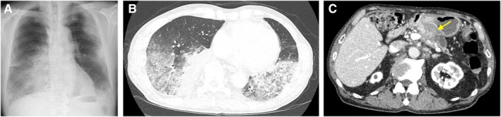 Figure 1
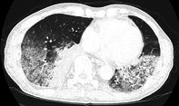 Figure 2
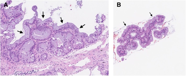 Figure 3
Peer Reviews
No public reviews on file for this paper yet. If you reviewed it on a platform where reviews are public (OpenReview, ICLR, NeurIPS, ICML), you can paste yours below so the community can read it here.
Videos
No videos yet. Explain this paper in a talk, walkthrough, or lecture? Add one.
Taxonomy
TopicsMedical Imaging and Pathology Studies · Pancreatic and Hepatic Oncology Research · Metastasis and carcinoma case studies
A 77‐year‐old man presented with a wet cough that had persisted for 3 weeks. Chest radiographs and chest computed tomography (CT) showed ground‐glass opacities and consolidations in both lung fields (Figure 1 A,B). Bronchoalveolar lavage showed an elevated lymphocyte percentage of 41% in the cell fraction, with negative cytology. Initially, interstitial lung disease was considered, but a pancreatic mass was incidentally detected on CT (Figure 1 C). A cryobiopsy of the lung and an endoscopic ultrasound‐guided fine needle aspiration (EUS‐FNA) of the pancreatic mass were performed. The diagnosis was lung metastases from pancreatic intraductal papillary mucinous carcinoma (Figure 2). Chemotherapy was performed, but the patient died 3 months after diagnosis.
Pulmonary metastases from pancreatic cancer are typically hematogenous, appearing as multiple, rounded, solid nodules on CT. However, 15%–22% of pulmonary metastases from pancreatic cancer showed an alveolar pattern on CT, and ground‐glass nodules and consolidations were each observed in 4% of cases [1, 2]. The air‐space radiological pattern corresponds on histology to lepidic tumour growth along alveolar walls [1]. In this case, the diagnosis was difficult due to ground‐glass opacities and consolidations mimicking interstitial lung disease. However, lung metastases from pancreatic cancer can appear as various types of CT findings.
Author Contributions
Ryoju Sato served as the attending physician for this patient and wrote this manuscript. Yasushi Fukuda and Tadashi Ishida supervised the patient's care and revised this manuscript.
Consent
The authors declare that written, informed consent was obtained for the publication of this manuscript and accompanying images and attest that the form used to obtain consent from the patient complies with the Journal requirements as outlined in the author guidelines.
Conflicts of Interest
The authors declare no conflicts of interest.
The reference list from the paper itself. Each links out to its DOI / PubMed record.
- 1M. Aissaoui , A. Lupo , and R. Coriat , “CT Features of Lung Metastases From Pancreatic Adenocarcinoma: Correlation With Histopathologic Findings,” Diagnostic and Interventional Imaging 102, no. 6 (2021): 371–377.33358342 10.1016/j.diii.2020.11.015 · doi ↗ · pubmed ↗
- 2M. Gaeta , S. Volta , and E. Scribano , “Air‐Space Pattern in Lung Metastasis From Adenocarcinoma of the GI Tract,” Journal of Computer Assisted Tomography 20, no. 2 (1996): 300–304.8606242 10.1097/00004728-199603000-00025 · doi ↗ · pubmed ↗