Endoscopic ultrasound-guided recanalization of pancreaticojejunostomy anastomotic stricture using a forward-viewing echoendoscope
Shota Iwata, Takuji Iwashita, Takuya Koizumi, Yosuke Ohashi, Akinori Maruta, Shinya Uemura, Masahito Shimizu

Abstract
Genes, proteins, chemicals, diseases, species, mutations and cell lines named across the full text — each resolved to its canonical identifier and authoritative record.
Click any figure to enlarge with its caption.
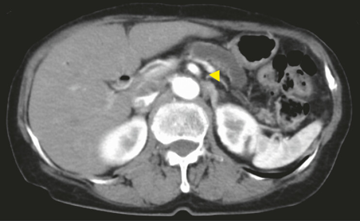 Fig. 1
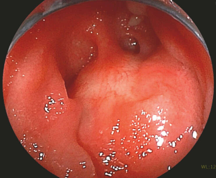 Fig. 2
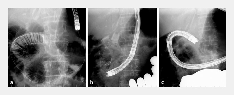 Fig. 3
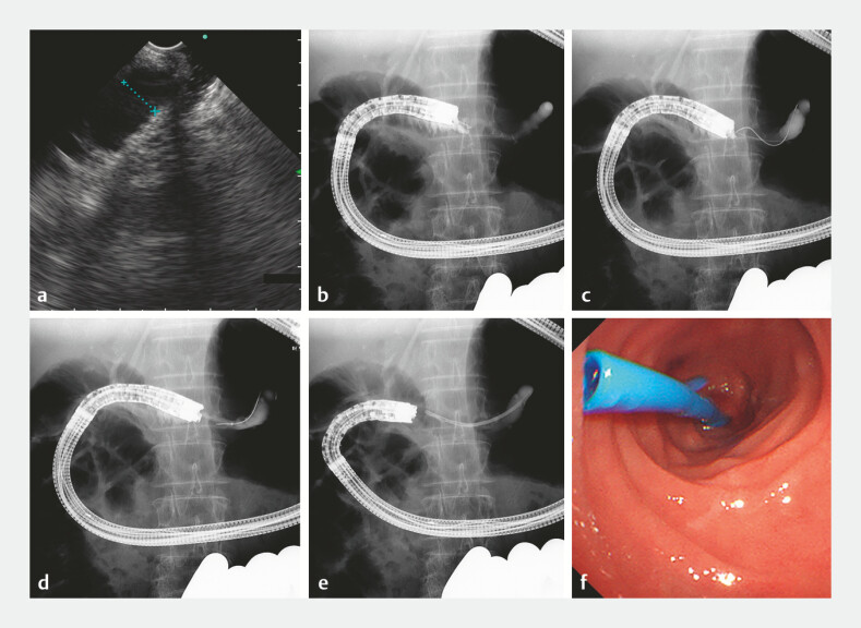 Fig. 4
Peer Reviews
No public reviews on file for this paper yet. If you reviewed it on a platform where reviews are public (OpenReview, ICLR, NeurIPS, ICML), you can paste yours below so the community can read it here.
Videos
No videos yet. Explain this paper in a talk, walkthrough, or lecture? Add one.
Taxonomy
TopicsGallbladder and Bile Duct Disorders · Pancreatic and Hepatic Oncology Research · Esophageal and GI Pathology
Endoscopic ultrasound-guided transmural pancreatic duct drainage (EUS-PDD) has become an important alternative when transpapillary or transanastomotic pancreatic duct drainage is technically challenging 1 2 . Recently, forward-viewing echoendoscope (FV-EUS)-guided drainage through the anastomotic site has been reported as a novel EUS-guided drainage technique; however, this approach has rarely been evaluated.
A 69-year-old woman presented with epigastric discomfort 4 years after undergoing subtotal stomach-preserving pancreaticoduodenectomy for a pancreatic head neuroendocrine tumor (NET G1). Computed tomography demonstrated dilation of the main pancreatic duct (15 mm) indicating a pancreaticojejunostomy anastomotic stricture (PJAS; Fig. 1 ). Double-balloon endoscopy-assisted endoscopic retrograde cholangiopancreatography was attempted; however, the opening of the pancreatic duct could not be identified although the scope reached the anastomosis ( Fig. 2 ). Therefore, EUS-guided recanalization using FV-EUS (TGF-UC260J, Olympus, Tokyo, Japan) was planned. With the aid of a guidewire placed near the anastomosis ( Fig. 3 a ) and external manual compression, FV-EUS was successfully advanced to the anastomotic site ( Fig. 3 b, c ). The dilated main pancreatic duct (9 mm) was punctured at the anastomotic site using a 19-gauge needle (Sonotip, Medi-Globe, Achenmühle, Germany), followed by contrast injection to confirm proper puncture ( Fig. 4 a, b ). A 0.025-inch guidewire was advanced into the pancreatic duct ( Fig. 4 c ). The tract was dilated using a 4-mm REN balloon (KANEKA MEDIX, Osaka, Japan; Fig. 4 d ), and finally, a 7-Fr, 7-cm plastic stent was deployed ( Fig. 4 e, f , Video 1 ). The patient’s symptoms improved after the procedure, without adverse events.
Computed tomography shows the dilation of the main pancreatic duct (yellow arrowhead).
The opening of the pancreatic duct could not be identified at the pancreaticojejunostomy site.
a A guidewire was left in place at the anastomosis, and the balloon endoscope was withdrawn. b External manual abdominal compression enabled the advancement of the echoendoscope along the guidewire. c The echoendoscope was successfully inserted at the anastomotic site.
a EUS showed the dilated pancreatic duct through the anastomosis. b The pancreatic duct was punctured through the anastomosis. c A 0.025-inch guidewire was inserted into the pancreatic duct. d The tract was dilated using a 4-mm balloon. e A 7-Fr, 7-cm plastic stent was deployed across the anastomotic stricture. f An endoscopic view of the deployed stent. EUS, endoscopic ultrasound
EUS-guided recanalization of the pancreaticojejunostomy anastomotic stricture using a forward-viewing echoendoscope, including follow-up imaging. EUS, endoscopic ultrasound.Video 1
This case indicated that EUS-guided recanalization of the PJAS using FV-EUS was feasible and safe. Compared with conventional transmural EUS-PDD, EUS-guided recanalization may offer improved safety because the access route is through the PJA rather than through the abdominal cavity and pancreatic parenchyma, if FV-EUS can reach the anastomotic site 3 4 5 . Further studies are required to clarify the safety, feasibility, and long-term outcomes of this technique.
Endoscopy_UCTN_Code_TTT_1AS_2AI
The reference list from the paper itself. Each links out to its DOI / PubMed record.
- 1Nakai Y Kogure H Isayama H Endoscopic ultrasound guided pancreatic duct drainage Saudi J Gastroenterol 20192521021710.4103/sjg.SJG_474_1830632484 PMC 6714474 · doi ↗ · pubmed ↗
- 2Teh JL Teoh AYB Techniques and Outcomes of Endoscopic Ultrasound Guided—Pancreatic Duct Drainage (EUS-PDD)J Clin Med 202312162610.3390/jcm 1204162636836161 PMC 9961828 · doi ↗ · pubmed ↗
- 3Hodo Y Shirota Y Suda T Successful EUS-guided retrograde pancreatic duct stent placement for refractory pancreaticojejunostomy stricture after pancreaticoduodenectomy with a forward-viewing echoendoscope Video GIE 2018319619810.1016/j.vgie.2018.03.00930128386 PMC 6098672 · doi ↗ · pubmed ↗
- 4Sadek A Hara K Okuno N Safety and efficacy of trans-afferent loop endoscopic ultrasound-guided pancreaticojejunostomy for post pancreaticoduodenectomy anastomotic stricture using the forward-viewing echoendoscope: a retrospective study from Japan Clin Endosc 20255831131910.5946/ce.2024.08939188116 PMC 11983134 · doi ↗ · pubmed ↗
- 5Imoto A Ogura T Higuchi K Endoscopic ultrasound-guided pancreatic duct drainage: Techniques and literature review of transmural stenting Clin Endosc 20205352553410.5946/CE.2020.17332967409 PMC 7548157 · doi ↗ · pubmed ↗