Electrohydraulic lithotripsy-assisted endoscopic retrograde appendicitis therapy for a giant appendiceal fecalith
Zhi-Yuan Zou, Qin-Qin Yi, Lu Bai, Yan-Hui Tian, Li-Sheng Wang, De-Feng Li, Sheng-Gang Zhan

Abstract
Genes, proteins, chemicals, diseases, species, mutations and cell lines named across the full text — each resolved to its canonical identifier and authoritative record.
Click any figure to enlarge with its caption.
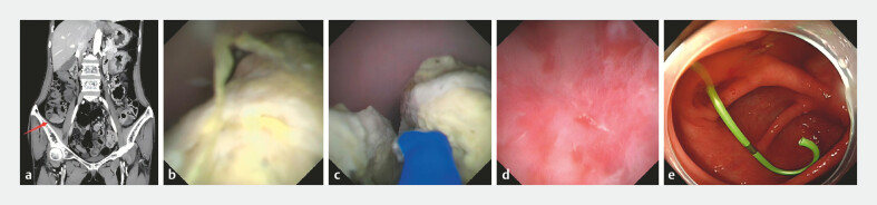 Fig. 1
- —Science and Technology Innovation Committee of Shenzhen
- —Science and Technology Innovation Committee of Shenzhen
Peer Reviews
No public reviews on file for this paper yet. If you reviewed it on a platform where reviews are public (OpenReview, ICLR, NeurIPS, ICML), you can paste yours below so the community can read it here.
Videos
No videos yet. Explain this paper in a talk, walkthrough, or lecture? Add one.
Taxonomy
TopicsAppendicitis Diagnosis and Management · Intestinal and Peritoneal Adhesions · Gallbladder and Bile Duct Disorders
A 57-year-old woman complained of recurrent pain and tenderness in the right lower quadrant for 3 months. An abdominal computed tomographic scan revealed appendicitis and a giant appendiceal fecalith (2.5 × 3 cm; Fig. 1 a ). Therefore, the patient was admitted to remove the fecaliths by endoscopic retrograde appendicitis therapy (ERAT). The EyeMax subscope (Micro-Tech, Nanjing, China) was intubated into the appendiceal cavity, and found a giant appendicolith measuring approximately 3cm obstructing the appendiceal lumen ( Fig. 1 b ). Unfortunately, it was unsuccessful to remove the appendicolith using a basket. Subsequently, it was decided to perform electrohydraulic lithotripsy (EHL) to break the appendicolith and success the removal of them ( Fig. 1 c, d and Video 1 ). After this, a 5 Fr-5cm Single Pigtail Stent was successfully implanted ( Fig. 1 e ). Excitingly, the patient was recovered quickly from abdominal pain and was discharged 2 days later.
a An abdominal CT scan revealed appendicitis and a giant appendiceal fecalith. b A giant appendicolith was found by the EyeMax subscope in the appendiceal lumen. c Electrohydraulic lithotripsy (EHL) was performed to break the appendicolith. d The appendicolith was successfully removed. e A 5 Fr-5cm Single Pigtail Stent was successfully implanted. CT, computed tomography.
The procedure of electrohydraulic lithotripsy-assisted endoscopic retrograde appendicitis therapy for a giant appendiceal fecalith.Video 1
ERAT presents a pioneering strategy in the management of appendicoliths, as a groundbreaking minimally invasive procedure specifically conceived to alleviate appendiceal obstruction 1 2 . Appendicoliths are usually dimensionally less than 1 cm, with those larger than 2 cm designated as giant appendicoliths 3 . However, it is difficult to remove the giant appendicoliths by traditional strategies, such as basket, balloon, etc. In this case, electrohydraulic lithotripsy-assisted ERAT successfully removed a giant appendiceal fecalith, which maybe a promising approach in the administration of giant appendicoliths in the future.
Endoscopy_UCTN_Code_CCL_1AD_2AJ Endoscopy_UCTN_Code_TTT_1AQ_2AJ
The reference list from the paper itself. Each links out to its DOI / PubMed record.
- 1Lin D Su Y Guo Z Direct vision endoscopic retrograde appendicitis therapy in the treatment of appendicitis with appendicolith in young women Endoscopy 202456 E 386E 38738714296 10.1055/a-2304-8243 PMC 11076136 · doi ↗ · pubmed ↗
- 2Guo M Yang W Guo YA novel loop-head guidewire-assisted endoscopic retrograde appendicitis therapy for fecalith obstruction Endoscopy 202557 E 44E 4510.1055/a-2512-375439837547 PMC 11750421 · doi ↗ · pubmed ↗
- 3Ishiyama M Yanase F Taketa T Significance of size and location of appendicoliths as exacerbating factor of acute appendicitis Emerg Radiol 20132012513010.1007/s 10140-012-1093-523179506 · doi ↗ · pubmed ↗