Full aspiration technique using a 7-Fr double-pigtail stent for endoscopic ultrasound-guided pancreatic fluid drainage of a pancreatic pseudocyst
Shunsuke Omoto, Mamoru Takenaka, Akihiro Yoshida, Kae Fukunishi, Hidekazu Tanaka, Yoriaki Komeda, Masatoshi Kudo

Abstract
Genes, proteins, chemicals, diseases, species, mutations and cell lines named across the full text — each resolved to its canonical identifier and authoritative record.
Click any figure to enlarge with its caption.
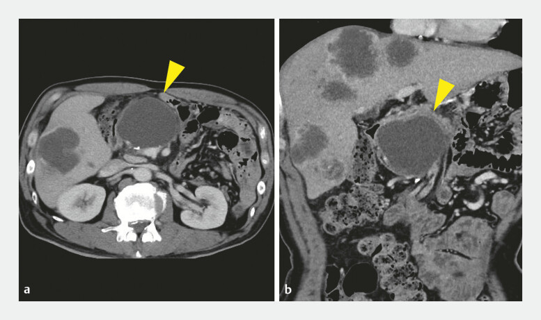 Fig. 1
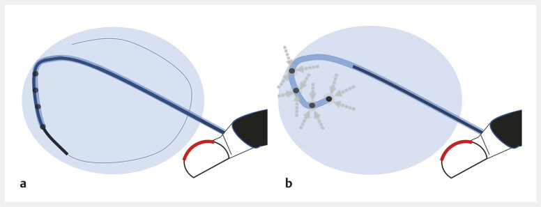 Fig. 2
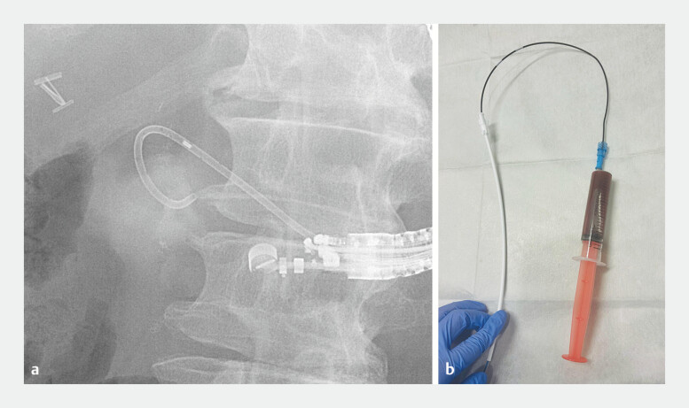 Fig. 3
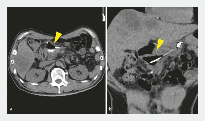 Fig. 4
Peer Reviews
No public reviews on file for this paper yet. If you reviewed it on a platform where reviews are public (OpenReview, ICLR, NeurIPS, ICML), you can paste yours below so the community can read it here.
Videos
No videos yet. Explain this paper in a talk, walkthrough, or lecture? Add one.
Taxonomy
TopicsPancreatitis Pathology and Treatment · Gallbladder and Bile Duct Disorders · Pancreatic and Hepatic Oncology Research
Endoscopic ultrasound–guided pancreatic fluid drainage (EUS-PFD) is an established treatment for pancreatic pseudocysts 1 . To prevent pseudocyst infection caused by stent occlusion, multiple plastic stents are often placed 2 . However, when fistula dilation is difficult, placing two stents is technically challenging, and a single 7-Fr stent has been associated with an increased risk of stent occlusion 3 . Large pseudocysts have also been associated with stent migration, especially when fluid drainage is insufficient 4 5 . Achieving reliable drainage with fewer plastic stents remains challenging. We developed a “full aspiration technique” to place a single 7-Fr double-pigtail stent after aspirating all cystic fluid before stent release ( Video 1 ).
EUS-guided drainage of a postoperative pancreatic pseudocyst using the “full aspiration technique,” involving complete cystic fluid aspiration before stent deployment to prevent occlusion and migration. EUS, endoscopic ultrasound.Video 1
A 54-year-old man, who had undergone distal pancreatectomy for pancreatic cancer, presented with epigastric pain. Laboratory tests showed elevated white blood cell count and C-reactive protein. MDCT revealed a 66-mm cystic lesion in the pancreatic head, diagnosed as a pancreatic pseudocyst ( Fig. 1 ).
a and b Pre-procedural CT showing a 66-mm postoperative pancreatic pseudocyst (yellow arrowhead) in the pancreatic head. CT, computed tomography.
EUS-PFD was performed. After puncture with a 19G EZ Shot 3 needle (Olympus, Tokyo, Japan), bloody fluid was aspirated. As the cyst partially shrank and shifted, tract dilation became difficult. A drill dilator (Tornus ES, Olympus, Tokyo, Japan) was used, but tract dilation remained challenging. The cavity was re-expanded by injecting 20 mL of saline through a catheter and then dilated with a 4-mm REN balloon (Kaneka Medix, Osaka, Japan). A 7-Fr/7-cm double-pigtail stent (Through & Pass, Gadelius Medical, Tokyo, Japan) was deployed halfway, the guidewire was removed, and 85 mL of fluid was aspirated ( Fig. 2 and Fig. 3 ). The stent was then released into the stomach. The procedure was completed without adverse events, and post-procedural computed tomography confirmed complete resolution ( Fig. 4 ). This novel technique allows complete aspiration of the cystic fluid before stent release, reducing the risk of stent occlusion and migration by shrinking the cyst.
a Placement of a double-pigtail catheter into the cyst cavity. b After the partial withdrawal of the inner sheath, the guidewire is removed, and the cystic fluid is fully aspirated through the inner guiding catheter lumen (full aspiration technique).
A fluoroscopic image a showing the partial deployment of a 7-Fr double-pigtail stent, and a procedural field image b demonstrating the aspiration of 85 mL of cystic fluid through the inner guiding catheter lumen.
a and b Post-procedural CT demonstrating the complete resolution of the pancreatic pseudocyst (yellow arrowhead). CT, computed tomography.
Endoscopy_UCTN_Code_TTT_1AS_2AJ
The reference list from the paper itself. Each links out to its DOI / PubMed record.
- 1Varadarajulu S Bang JY Phadnis MA Endoscopic transmural drainage of pancreatic pseudocysts Clin Gastroenterol Hepatol 20119748753
- 2Bang JY Navaneethan U Hasan MK Multiple plastic stents versus metal stents for pancreatic pseudocyst drainage Gastrointest Endosc 201888730738
- 3Pausawasdi N Kongkam P Khemnark S Long-term outcomes of endoscopic drainage for pancreatic pseudocysts using single plastic stents Endosc Ultrasound 2021104248
- 4Alzeerelhouseini HIA Madi M Azzam A Endoscopic drainage of giant pancreatic pseudocysts Cureus 202113 e 1407733868735 10.1155/2021/6610610 PMC 8035029 · doi ↗ · pubmed ↗
- 5Arvanitakis M Dumonceau JM Albert J Endoscopic management of pancreatic fluid collections: ESGE evidence-based guidelines Endoscopy 20185084385610.1055/a-0588-536529631305 · doi ↗ · pubmed ↗