Clinical, laboratory, and radiological features of community-acquired pneumonia due to Chlamydia psittaci and Legionella pneumophila confirmed using next-generation sequencing
Ran Cheng, Zhonghua Deng, Fei Lin, Biying Zhang, Jingjin Liang, Ming Lu

TL;DR
This study compares pneumonia caused by Chlamydia psittaci and Legionella pneumophila using next-generation sequencing to identify differences in symptoms and outcomes.
Contribution
The study uses next-generation sequencing to confirm and compare clinical features of two atypical pneumonia pathogens.
Findings
LPP patients showed more severe clinical and laboratory features compared to CPP patients.
Both CPP and LPP responded well to azithromycin-based therapy when diagnosed with NGS.
LPP was associated with higher rates of organ dysfunction and intensive care admission.
Abstract
Chlamydia psittaci and Legionella pneumophila are common atypical pathogens that cause severe community-acquired pneumonia (CAP). This study aimed to compare the clinical features and outcomes of Chlamydia psittaci pneumonia (CPP) and Legionella pneumophila pneumonia (LPP) identified using next-generation sequencing (NGS) for accurate identification. This retrospective study included 68 patients with CPP and 42 patients with LPP. All cases were confirmed by metagenomic or targeted next-generation sequencing (mNGS/tNGS) of bronchoalveolar lavage fluid, serum, or sputum samples. Patients with LPP had a higher prevalence of diabetes and were predominantly male. Poultry contact was common in CPP (64.7% vs. 14.3%), whereas recent travel was associated with LPP (47.6% vs. 2.9%). LPP presented with increased extrapulmonary symptoms. Inflammatory marker levels were higher in LPP, including…
Genes, proteins, chemicals, diseases, species, mutations and cell lines named across the full text — each resolved to its canonical identifier and authoritative record.
Click any figure to enlarge with its caption.
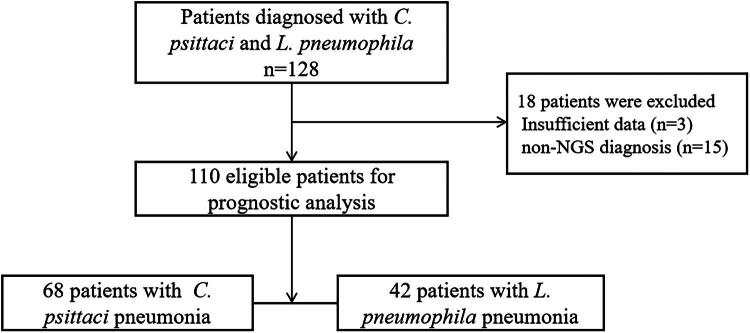 Figure 1
| Variable | Total | CPP | LPP | |
|---|---|---|---|---|
| Age, median (IQR), years | 59.27 ± 14.1 | 59.19 ± 13.6 | 59.4 ± 15.04 | 0.940 |
| Age ≥ 65 years | 44 (40.0) | 29 (42.6) | 15 (35.7) | 0.471 |
| Male, | 62 (56.4) | 33 (48.5) | 29 (69.0) | 0.035* |
| Smoking, | 34 (31.1) | 17 (25.0) | 17 (40.5) | 0.088 |
| Drinking, | 36 (32.7) | 19 (29.2) | 17 (40.5) | 0.173 |
| Travel history, | 22 (20.0) | 2 (2.9) | 20 (47.6) | 0.000* |
| History of poultry exposure, | 50 (45.5) | 44 (64.7) | 6 (14.3) | 0.000* |
| Hypertension | 52 (47.3) | 28 (41.7) | 24 (57.1) | 0.103 |
| Diabetes | 21 (19.1) | 9 (13.2) | 12 (28.6) | 0.047* |
| Heart disease | 11 (10.0) | 5 (7.4) | 6 (14.3) | 0.239 |
| Immunosuppressive disease | 9 (8.2) | 5 (7.4) | 4 (9.5) | 0.729 |
| Chronic lung disease | 5 (4.5) | 2 (2.9) | 3 (7.1) | 0.368 |
| Chronic kidney diseases | 6 (5.5) | 2 (2.9) | 4 (9.5) | 0.200 |
| Cerebrovascular diseases | 13 (11.8) | 10 (14.7) | 3 (7.1) | 0.363 |
| Malignancy | 7 (6.4) | 5 (7.4) | 2 (4.8) | 0.706 |
|
| ||||
| PaO2 | 68.17 ± 11.57 | 71.82 ± 12.41 | 62.92 ± 7.77 | 0.000* |
| Temperature, °C | 39.09 ± 3.30 | 38.95 ± 4.15 | 39.32 ± 0.79 | 0.570 |
| Fever | 110 (100) | 68 (100) | 42 (100) | 1.00 |
| Cough | 61 (55.5) | 38 (55.9) | 23 (54.8) | 0.909 |
| Sputum | 32 (29.1) | 15 (22.1) | 17 (40.5) | 0.039* |
| Dyspnea | 16 (14.5) | 11 (16.2) | 5 (11.9) | 0.557 |
| Headache | 45 (40.9) | 29 (42.6) | 16 (38.1) | 0.637 |
| Dizziness | 36 (32.7) | 24 (35.3) | 12 (28.6) | 0.465 |
| Delirium | 17 (15.5) | 9 (13.2) | 8 (19.0) | 0.413 |
| Vomit/nausea | 24 (21.8) | 17 (25.0) | 7 (16.7) | 0.304 |
| Diarrhea/abdominal pain | 13 (11.8) | 4 (5.9) | 9 (21.4) | 0.030* |
| Urinary system symptoms | 11 (10.0) | 2 (2.9) | 9 (21.4) | 0.003* |
| Laboratory findings | Total | CPP | LPP | |
|---|---|---|---|---|
|
| ||||
| White blood cell count, ×109/L | 8.09 (5.09) | 6.92 (3.85) | 10.72 (4.88) | 0.000* |
| Neutrophil count, ×109/L | 6.87 (4.88) | 5.5 (3.27) | 9.45 (4.13) | 0.000* |
| Lymphocyte count, ×109/L | 0.74 (0.56) | 0.77 (0.59) | 0.74 (0.50) | 0.627 |
| Hemoglobin, g/L | 130.5 (22.0) | 128 (18.75) | 134 (28.5) | 0.236 |
| Platelet count, ×109/L | 184.5 (78.25) | 183.5 (85) | 187 (73.75) | 0.973 |
|
| ||||
| Aspartate aminotransferase, U/L | 40.1 (47.58) | 46.5 (56.88) | 31.15 (36.55) | 0.294 |
| Alanine aminotransferase, U/L | 50 (58.5) | 53 (58.02) | 42.15 (66.55) | 0.481 |
| Lactate dehydrogenase, U/L | 305 (173) | 309 (199.5) | 299.5 (136.5) | 0.926 |
| Total bilirubin, μmol/L | 13.75 (8.85) | 12.8 (7.15) | 14.8 (11.68) | 0.169 |
| Albumin, g/L | 36.7 (8.3) | 36.6 (7.10) | 37.35 (9.55) | 0.791 |
| Serum sodium, mmol/L | 134.5 (6.73) | 134.5 (6.15) | 134.65 (8.78) | 0.866 |
| Creatinine, μmol/L | 82.5 (41.25) | 73 (34.75) | 94 (53) | 0.005* |
| Creatine kinase, U/L | 125 (460.25) | 116 (306.25) | 130 (530.25) | 0.710 |
|
| ||||
| C-reactive protein, mg/L | 186.48 ± 73.07 | 167.73 ± 68.14 | 217.73 ± 71.05 | 0.001* |
|
| ||||
| Procalcitonin, μg/L | 0.50 (1.71) | 0.37 (1.00) | 0.73 (2.99) | 0.003* |
|
| ||||
| | 0.55 (0.79) | 0.48 (0.71) | 0.63 (0.81) | 0.265 |
|
| ||||
| Lesions in isolated left lung, | 33 (30.0) | 24 (35.3) | 9 (21.4) | 0.123 |
| Lesions in isolated right lung, | 45 (40.9) | 27 (39.7) | 18 (42.9) | 0.744 |
| Lesions in bilateral lung, | 32 (29.1) | 17 (25) | 15 (35.7) | 0.229 |
|
| ||||
| Ground-glass opacity, | 60 (54.5) | 32 (47.1) | 28 (66.7) | 0.045* |
| Consolidation, | 94 (85.5) | 57 (83.8) | 37 (88.1) | 0.537 |
| Pleural effusion, | 31 (28.2) | 18 (26.5) | 13 (31.0) | 0.612 |
| Variable | Total | CPP | LPP | |
|---|---|---|---|---|
| Peripheral blood | 10 (9.1) | 6 (8.8) | 4 (9.5) | 0.938 |
| Sputum | 34 (30.9) | 22 (32.3) | 12 (28.6) | 0.671 |
| Bronchoalveolar lavage fluid | 68 (61.8) | 42 (61.8) | 26 (61.9) | 0.981 |
| No respiratory support | 23 (20.9) | 22 (32.4) | 1 (2.4) | <0.001* |
| Conventional oxygen therapy | 65 (59.1) | 34 (50.0) | 31 (73.8) | 0.003* |
| High-flow nasal cannula | 6 (5.5) | 1 (1.5) | 5 (11.9) | 0.041* |
| Non-invasive mechanical ventilation | 8 (7.3) | 6 (8.8) | 2 (4.8) | 0.252 |
| Invasive mechanical ventilation | 8 (7.3) | 5 (7.4) | 3 (7.1) | 0.721 |
| Glucocorticoids | 10 (9.1) | 7 (10.3) | 3 (7.1) | 0.741 |
| Continuous renal replacement therapy | 8 (7.3) | 2 (2.9) | 6 (14.3) | 0.041* |
| Quinolones (moxifloxacin, levofloxacin, and nemonoxacin) | 29 (26.4) | 20 (29.4) | 9 (21.4) | 0.356 |
| Tetracyclines (doxycycline, minocycline, and tigecycline) | 1 (0.9) | 1 (1.5) | 0 | 1.000 |
| Azithromycin | 3 (2.7) | 3 (4.4) | 0 | 0.285 |
| Quinolones + tetracyclines | 2 (2.9) | 1 (1.5) | 1 (2.4) | 1.000 |
| Azithromycin + tetracyclines | 2 (2.9) | 1 (1.5) | 1 (2.4) | 1.000 |
| Azithromycin + quinolones | 1 (0.9) | 0 | 1 (2.4) | 0.382 |
| β-lactam monotherapy | 41 (37.3) | 26 (38.2) | 15 (35.7) | 0.790 |
| β-lactam + quinolones | 16 (14.5) | 8 (11.8) | 8 (19.0) | 0.293 |
| β-lactam + azithromycin | 8 (7.3) | 5 (7.4) | 3 (7.1) | 1.000 |
| β-lactam + tetracyclines | 7 (6.4) | 3 (4.4) | 4 (9.5) | 0.424 |
| Quinolones (moxifloxacin, levofloxacin, and nemonoxacin) | 16 (14.5) | 3 (4.4) | 13 (31.0) | 0.000* |
| Tetracyclines (doxycycline, minocycline, and tigecycline) | 31 (28.2) | 29 (42.6) | 2 (4.8) | 0.000* |
| Azithromycin | 35 (31.8) | 20 (29.4) | 15 (35.7) | 0.491 |
| Quinolones + tetracyclines | 6 (5.5) | 6 (8.8) | 0 | 0.081 |
| Azithromycin + tetracyclines | 10 (9.1) | 10 (14.7) | 0 | 0.005* |
| Quinolones + azithromycin | 12 (10.9) | 0 | 12 (28.6) | 0.000* |
| Intensive care unit admission | 26 (23.6) | 10 (14.7) | 16 (38.1) | 0.010* |
| Hospital death | 2 (1.8) | 2 (2.9) | 0 | 0.521 |
| Time from illness onset to hospital admission | 6 (3) | 6 (3) | 5 (3.25) | 0.212 |
| Hospital stay | 7 (6) | 6.5 (6) | 7 (9) | 0.157 |
Peer Reviews
No public reviews on file for this paper yet. If you reviewed it on a platform where reviews are public (OpenReview, ICLR, NeurIPS, ICML), you can paste yours below so the community can read it here.
Videos
No videos yet. Explain this paper in a talk, walkthrough, or lecture? Add one.
Taxonomy
TopicsLegionella and Acanthamoeba research · Pneumonia and Respiratory Infections · Oral microbiology and periodontitis research
Introduction
Community-acquired pneumonia (CAP) is a major global health burden, with lower respiratory tract CAP causing an estimated 2.5 million deaths annually [1]. Among the diverse etiological agents of CAP, atypical causal pathogens have garnered increasing attention due to their diagnostic challenges and potential to cause severe disease [2]. The most common atypical pneumonia pathogens are Chlamydia pneumoniae, Mycoplasma pneumoniae, Legionella, Chlamydia psittaci (psittacosis), and Coxiella burnetii (Q fever) [3]. Legionella pneumophila, a Gram-negative facultative intracellular bacterium ubiquitously found in natural and artificial aquatic environments worldwide, is the causative agent of Legionnaires’ disease (LD) [4–6]. Chlamydia psittaci is an obligate intracellular bacterium that causes psittacosis in humans, following transmission from avian species, which serve as its primary reservoir [7]. Both pathogen cause diseases characterized by prominent extrapulmonary manifestations and a highly variable, nonspecific clinical symptom spectrum, including high fever, muscle and joint pain, nausea, and vomiting, as well as severe symptoms, including respiratory distress, respiratory failure, and multi-organ failure, which can lead to death [8,9]. Recent epidemiological studies have highlighted the considerable impact of Legionella pneumophila and Chlamydia psittaci in the context of CAP. A multicenter prospective study conducted across 17 hospitals in China revealed that L. pneumophila accounted for 11.3% of severe CAP cases, while C. psittaci was responsible for 6.8% of cases [10]. These pathogens are particularly concerning as they can cause severe atypical pneumonia that is often unresponsive to standard β-lactam therapy, leading to delayed treatment and increased risk of complications.
Given that C. psittaci and L. pneumophila detection are not included in routine microbiological testing, CPP and LPP are often underreported, misdiagnosed, or inadequately identified. This diagnostic uncertainty frequently results in empirical broad-spectrum antibiotic therapy being administered, with studies indicating that 69.8–80.9% of patients receive two or more antibiotics simultaneously before definitive diagnosis, contributing to antimicrobial resistance and unnecessary drug exposure [11]. Systematic head-to-head comparisons based on definitive pathogen identification remain scarce.
Next-generation sequencing (NGS) enables unbiased, sequence-based identification directly from clinical specimens, achieving definitive diagnosis independent of culture or antibiotic exposure [12]. Our study leveraged this technology to conduct the largest comparative analysis of CPP versus LPP to date. Our objective was to compare epidemiological patterns, clinical characteristics, laboratory results, radiologic imaging, and, more importantly, treatments and prognosis between CPP and LPP cases identified using NGS.
Methods
Study design
2.1.
We conducted a retrospective, single-center observational study between January 1, 2021, and September 30, 2025, at Peking University Third Hospital, which included patients with CAP caused by C. psittaci and L. pneumophila. Clinical data, including present and previous medical history, ongoing medication, radiology findings, and laboratory findings, were obtained from electronic medical records. Respiratory support categories were defined by their level of invasiveness: 1) No support: No oxygen therapy or conventional low-flow nasal cannula ≤2 L/min; 2. Conventional oxygen therapy: Face mask or nasal prongs delivering >2 L/min; 3. High-flow nasal cannula (HFNC): Heated humidified system delivering ≥ 30 L/min flow with FiO_2_ titration; 4. Non-invasive ventilation (NIV): Bi-level positive airway pressure (BiPAP) or continuous positive airway pressure (CPAP) via face/nasal mask; 5. Invasive mechanical ventilation (IMV): Endotracheal intubation or tracheostomy with positive pressure ventilation. All patients were classified based on the highest level of support received within 72 h of admission. The patient inclusion criteria were as follows: age ≥18 years; diagnosis of CAP according to current guidelines [13]; and positive metagenomic next-generation sequencing (mNGS) or targeted next-generation sequencing (tNGS) results from bronchoalveolar lavage fluid (BALF), serum, or sputum samples. The exclusion criteria were as follows: lack of NGS-based diagnostic evidence and missing medical records.
Ethical considerations
2.2.
The study protocol conformed to the ethical guidelines of the Declaration of Helsinki and was approved by the Institutional Review Board of Peking University Third Hospital, Beijing, China (approval no. 20250641). Written informed consent was waived due to the anonymized retrospective nature of the analysis. Researchers were blinded to the data they were analyzing.
Statistical analysis
2.3.
Continuous variables were expressed as the mean ± standard deviation and compared using Student’s t test when normally distributed, whereas non-normally distributed data were expressed as the median (interquartile range) and tested using the Mann–Whitney U test. Categorical variables were presented as frequencies and percentages and compared using the chi-square or Fisher’s exact test. Statistical analyses were performed using SPSS software (version 22.0; IBM Corp., Armonk, NY, USA). A p value < 0.05 was considered statistically significant.
NGS diagnostics protocol
2.4.
The specimens were sent to the Department of Clinical Laboratory of Peking University Third Hospital for pathogenic mNGS (PACEseq, Hugobiotech, Beijing, China) and tNGS (DRseq, Hugobiotech, Beijing, China) detection.
For the detected bacteria (Mycobacterium excluded), fungi (Cryptococcus excluded), and parasites, the positive criteria for the mNGS results were as follows: (1) genome coverage of the unique reads mapped to this microorganism ranked top10 of the same kind of microbes and the microorganism was not detected in the NTC; or (2) RPM-r (RPM_sample_/RPM_NTC)_ was > 10 (RPM_NTC_ ≠ 0).
For the tNGS results, microbial identification was performed based on three key metrics: RPM, RPM ratio (RPM-r), and detected primer ratio (number of detected primer pairs/total designed primer pairs). A positive result was confirmed when the following criteria were met: RPM of targeted pathogens in test samples exceeded 100, RPM-r > 5 (RPM_NTC_ ≠ 0); and detected primer ratio > 0.1.
Handling of co-detections and contaminants
2.5.
Patients with NGS exhibiting co-detection of other bacterial or fungal pathogens at admission were sequentially evaluated by two clinicians. Cases were excluded if co-detections were deemed clinically significant. Contaminants and low-level colonizers identified at low read depths (<5 reads) were disregarded as per our institutional NGS interpretation protocol.
Results
Comparison of demographic and clinical characteristics
3.1.
In this study, 110 patients were included in the final analysis (Figure 1): 68 with CPP and 42 with LPP, all confirmed by mNGS or tNGS. The demographic, clinical, and radiological characteristics of the patients are summarized in Table 1. Among these patients, one with CPP was pregnant. Two patients in the CPP group died from the infection, whereas the remaining patients recovered and were discharged after appropriate treatment.
Flowchart of the study.
No statistically significant differences were observed between the two groups in age or comorbidities, including hypertension, heart disease, immunosuppressive conditions, chronic lung disease, chronic kidney disease, malignancy, and cerebrovascular disease. However, patients with LPP exhibited a significantly higher prevalence of diabetes mellitus compared with those with CPP (28.6% vs. 13.2%, p = 0.047). The LPP group also had a higher proportion of male patients (29/42, 69.0%) than the CPP group (33/68, 48.5%). A marked contrast was observed in exposure history: direct contact with domestic poultry was substantially more common among patients with CPP, whereas recent travel history was far more frequent in patients with LPP (Table 1).
Fever was present in all the enrolled patients, as well as cough, headache, and dizziness (Table 1). Patients with LPP showed a markedly higher frequency of extrapulmonary symptoms, particularly gastrointestinal and urinary tract manifestations. No significant differences were observed between the two groups in the presence of fever, cough, dyspnea, headache, dizziness, delirium, or nausea/vomiting.
Laboratory and imaging findings at admission
3.2.
Laboratory findings obtained on admission are summarized in Table 2. Both groups exhibited elevated levels of aspartate aminotransferase (AST), alanine aminotransferase (ALT), lactate dehydrogenase (LDH), d-dimer, and creatine kinase (CK), as well as lymphopenia, hypoalbuminemia, and hyponatremia. However, LPP cases showed more pronounced laboratory derangements, including significantly greater leukocyte and neutrophil counts, and higher serum creatinine (Scr), C-reactive protein (CRP), and procalcitonin (PCT) concentrations (all p < 0.05 vs. CPP).
High-resolution thoracic CT showed consolidation as the dominant pattern in both groups, followed by ground-glass opacities (GGO) and pleural effusions (Table 2). Apart from the higher frequency of GGO in the LPP group (66.7% vs. 47.1%, p = 0.037), no significant between-group differences were observed in anatomical distribution or in the prevalence of consolidation and pleural effusion (all p > 0.05).
Pathogen detection and diagnostic results
3.3.
Among patients with CPP or LPP, the majority of positive NGS results were derived from BALF samples (61.8% and 61.9%, respectively). When NGS was used as the reference, conventional tests for L. pneumophila showed low concordance: only 13 of 42 NGS-positive cases were urinary Legionella antigen-positive, 6 of 42 were IgM-seropositive, and routine cultures yielded no isolates.
Treatment and clinical outcomes
3.4.
All patients received empirical antibiotic therapy prior to etiological confirmation. All patients who were initially treated with a regimen containing β-lactams had these agents discontinued upon confirmation of LPP and CPP by NGS.
Among 42 patients with LPP, 15 received empirical β-lactam monotherapy. Twenty-three exhibited inadequate responses to the initially administered empirical antibiotics, prompting regimen adjustment to fluoroquinolones, azithromycin, or combination therapy following pathogen identification. Overall, 40 patients (95.2%) exhibited improved infection and were discharged successfully. The remaining two patients (4.8%) who showed clinical improvement were transferred to specialized nursing facilities for further management.
Prior to etiological confirmation, the initial empirical antibiotic regimens varied among the 68 patients with confirmed CPP. In the CPP cohort (n = 68), 20 received empirical fluoroquinolones, 26 β-lactam monotherapy, and 8 β-lactam-fluoroquinolone combined therapy, none of which constituted first-line therapy for CPP. Fifty-one showed suboptimal responses to initial therapy and were switched to azithromycin, tetracyclines, or combination regimens after CPP was confirmed. Ultimately, 66 of 68 patients (97.1%) achieved clinical recovery and were discharged. Unfortunately, two patients (2.9%) died from disease progression despite receiving targeted antimicrobial therapy.
Patients with LPP exhibited significantly more severe impairment in gas exchange at presentation than those with CPP, as evidenced by a significantly lower partial pressure of arterial oxygen (Table 1). This greater degree of hypoxemia directly translated into higher dependence on respiratory support in the LPP cohort (Table 3). The proportion of patients who did not require respiratory support was substantially lower in the LPP group than in the CPP group. Consequently, conventional oxygen therapy and high-flow nasal cannula were used significantly more in patients with LPP. In contrast, the need for advanced ventilatory support did not differ significantly between the two groups. The rates of non-invasive mechanical ventilation (NIV) and invasive mechanical ventilation (IMV) were comparable between the two groups. The rate of intensive care unit (ICU) admission was significantly higher among patients with LPP, reflecting their initial disease severity. However, despite the differences in initial severity and ICU admission, the overall length of hospital stays did not differ significantly between the two patient populations. Patients with CPP required continuous renal replacement therapy significantly less often than those with LPP (2.9% vs. 14.3%, p = 0.041). However, the frequency of use of glucocorticoids was similar between the groups (p > 0.05).
Discussion
Our comparative study delineated significant distinctions and similarities in the demographic, clinical, laboratory, and radiological profiles of CAP caused by C. psittaci and L. pneumophila. Although both pathogens can cause severe disease, our findings consistently indicated that L. pneumophila infection presents with greater overall illness severity at admission, as reflected by more pronounced laboratory inflammatory marker levels, a higher incidence of extrapulmonary manifestations, and increased requirements for supplemental oxygen and ICU care.
Epidemiological and host-derived biomarkers of severe disease
4.1.
A higher number of males in the LPP group (69%) has been reported previously and may reflect occupational or behavioral exposure patterns (e.g. hotel stays or spa use) rather than a genuine biological predisposition [14]. The 2.2-fold higher rate of diabetes in LPP aligns with the findings of population-based studies that have identified diabetes as an independent risk factor for LD, probably through impaired macrophage bactericidal activity and heightened susceptibility to hyperinflammatory cascades [15]. Conversely, the strong poultry-exposure association in CPP (64.7%) reflects the zoonotic reservoir of C. psittaci and reinforces the dictum that a history of exposure to poultry is a strong indicator of CPP [16]. The low prevalence of recent travel in CPP (2.9%) contradicts travel-based acquisition of this disease and may guide early empirical treatment choices prior to microbiological confirmation.
Clinical presentation and organ involvement
4.2.
Clinical presentation of CPP and LPP offers key differentiating characteristics. Although both pathogens typically present as atypical CAP, LPP is distinguished by a significantly higher incidence of gastrointestinal complaints and urinary symptoms, which serve as readily discernible diagnostic cues for clinicians. Although gastrointestinal symptoms are a recognized feature of LD, the prominence of urinary symptoms in our LPP cohort is intriguing and may reflect the systemic effects of the infection or associated complications, warranting further investigation [17]. The laboratory findings further highlighted the heightened inflammatory state in LPP. The significantly higher levels of leukocytosis, neutrophilia, CRP, PCT, and organ injury markers (e.g., CK and Scr) observed in LPP suggest a more vigorous systemic inflammatory response and greater multi-organ involvement than in CPP. This pronounced physiological derangement directly explains the greater need for basic and advanced oxygen support (conventional oxygen and high-flow nasal cannula support) and the higher rate of ICU admission in the LPP group [18]. Importantly, the radiological pattern was largely nondiscriminatory, apart from the higher rate of GGO in LPP, underscoring the necessity of laboratory confirmation rather than reliance on imaging alone.
Diagnostic performance of conventional tests
4.3.
Among the NGS-confirmed LPP cases, only 31% were urinary antigen-positive, and no isolates were recovered by culture, re-emphasizing the low sensitivity of conventional tests after prior antibiotic exposure or when the infecting strain is a non-serogroup 1 type.
Novel insights from an NGS-confirmed cohort and treatment implications
4.4.
This direct comparison of NGS-confirmed LPP and CPP within a single-center cohort provides real-world evidence of disease severity and management using rapid molecular diagnostics. Guided by mNGS or tNGS results, we performed targeted escalation or de-escalation of antimicrobial therapy, and both cohorts achieved high recovery rates after pathogen-directed adjustments. These findings support the early integration of mNGS or tNGS into the diagnostic workup of severe CAP, particularly when empirical treatment fails. In this observational setting, clinical outcomes for patients with LPP treated with azithromycin monotherapy were similar to those receiving Fluoroquinolones, consistent with the findings of prior reports [19]. Similarly, for CPP, the outcomes of treatment with Azithromycin alone appeared comparable to those of Tetracycline-based regimens, though this requires validation in larger studies.
Respiratory support and resource utilization
4.5.
Despite the clear differences in initial disease severity and ICU utilization, the comparable length of hospital stays between the two groups was clear. Additionally, the similar rates of advanced ventilatory support (NIV and IMV) suggest that although LPP causes moderate hypoxemia more frequently, its propensity to progress to the most severe stages of respiratory failure does not significantly differ from that of CPP. In the present cohort, no deaths attributable to LPP occurred, yielding an in-hospital case-fatality rate of 0%, which is substantially lower than the 10–20% rate frequently reported in existing literature [18]. We attribute this favorable outcome to the routine early deployment of NGS, which permitted pathogen identification within 24–48 h of admission and ensured that antimicrobial therapy remained consistently aligned with the Legionella-directed guidelines. These data highlight the pivotal role of rapid molecular diagnostics in reducing LPP-related deaths.
Limitations
Our study had certain limitations. First, its single-center retrospective design may have introduced selection bias and limited the generalizability of the findings. Second, although substantial for these relatively rare pathogens, the sample size may have been insufficient to detect subtle differences in clinical outcomes. Third, due to sample size constraints and risk of model overfitting, multivariable adjustment for potential confounders (age, sex, diabetes, and chronic lung disease) was not performed. Therefore, the observed associations should be interpreted as unadjusted comparisons that may reflect confounding rather than causal relationships. Finally, although highly sensitive, the reliance on NGS as the primary diagnostic tool did not allow for antimicrobial susceptibility testing.
Conclusion
In conclusion, this detailed comparative analysis demonstrated that LPP typically presents with greater initial severity than CPP, as evidenced by more marked inflammatory responses and higher oxygen requirements. However, key epidemiological factors and specific clinical features can aid in differentiating these infections. The critical role of rapid molecular diagnostics, such as mNGS or tNGS, is highlighted by the frequent need to adjust therapy following pathogen identification. Azithromycin demonstrated excellent efficacy as a targeted therapy for both LPP and CPP. This finding substantiates the current guideline recommendation of combining a β-lactam with a macrolide for the empirical treatment of hospitalized patients with CAP^13^. This regimen provides critical coverage against these elusive atypical pathogens, thereby improving the outcomes of empirical therapy.
The reference list from the paper itself. Each links out to its DOI / PubMed record.
- 1Age-sex differences in the global burden of lower respiratory infections and risk factors, 1990-2019: results from the Global Burden of Disease Study 2019. Lancet Infect Dis. 2022;22(11):1626–1647.35964613 10.1016/S 1473-3099(22)00510-2PMC 9605880 · doi ↗ · pubmed ↗
- 2Cillóniz C, Ewig S, Polverino E, et al. Microbial aetiology of community-acquired pneumonia and its relation to severity. Thorax. 2011;66(4):340–346. doi: 10.1136/thx.2010.143982.21257985 · doi ↗ · pubmed ↗
- 3Cunha BA. The atypical pneumonias: clinical diagnosis and importance. Clin Microbiol Infect. 2006;12 Suppl 3(Suppl 3):12–24. doi: 10.1111/j.1469-0691.2006.01393.x.16669925 PMC 7128183 · doi ↗ · pubmed ↗
- 4Fraser DW, Tsai TR, Orenstein W, et al. Legionnaires’ disease: description of an epidemic of pneumonia. N Engl J Med. 1977;297(22):1189–1197. doi: 10.1056/NEJM 197712012972201.335244 · doi ↗ · pubmed ↗
- 5Lévesque S, Lalancette C, Bernard K, et al. Molecular typing of Legionella pneumophila Isolates in the Province of Quebec from 2005 to 2015. P Lo S One. 2016;11(10):e 0163818. doi: 10.1371/journal.pone.0163818.27706210 PMC 5051737 · doi ↗ · pubmed ↗
- 6Gonçalves IG, Simões LC, Simões M. Legionella pneumophila. Trends Microbiol. 2021;29(9):860–861. doi: 10.1016/j.tim.2021.04.005.33994277 · doi ↗ · pubmed ↗
- 7Hogerwerf L, DE Gier B, Baan B, et al. Chlamydia psittaci (psittacosis) as a cause of community-acquired pneumonia: a systematic review and meta-analysis. Epidemiol Infect. 2017;145(15):3096–3105. doi: 10.1017/S 0950268817002060.28946931 PMC 9148753 · doi ↗ · pubmed ↗
- 8Cunha BA, Burillo A, Bouza E. Legionnaires’ disease. Lancet. 2016;387(10016):376–385. doi: 10.1016/S 0140-6736(15)60078-2.26231463 · doi ↗ · pubmed ↗