Assessing the robustness of an artificial intelligence segmentation model for quantitative cardiovascular magnetic resonance imaging across cardiac phenotypes
Hadil Saad, Clemens Ammann, Thomas Hadler, Yashraj Bhoyroo, Philine Reisdorf, Jana Veit, Teodora Chitiboi, Jens Wetzl, Christian Geppert, Jeanette Schulz-Menger

TL;DR
This study evaluates an AI model for cardiovascular MRI segmentation, showing it performs reliably across various heart conditions and imaging parameters.
Contribution
The novel contribution is the development and validation of Nick, an AI model for robust cardiovascular MRI segmentation in clinical settings.
Findings
Nick showed high agreement with manual segmentations for LV and RV volumes and LVM with R² values ≥0.93.
T1 and T2 mapping values demonstrated excellent agreement with manual references (R² ≥ 0.92) and minimal biases.
Despite some underestimations in volumes, clinically acceptable biases were observed, and less than two slices per case required correction on average.
Abstract
To introduce an artificial intelligence-based cardiovascular magnetic resonance segmentation algorithm (Nick) for automated quantification of function and parametric mapping across cardiac phenotypes reflecting clinical routine. Nick was compared to manual gold standard (GS) segmentations in 359 multi-centre cases at 1.5T and 3T, consisting of 104 healthy individuals and 255 diseased patients with various cardiac phenotypes. Left and right ventricular (LV, RV) volumes and LV mass (LVM) were derived from short-axis segmentations. For parametric mapping, the LV myocardium was segmented to quantify T1 and T2 relaxation times. Statistical analysis comprised mean differences, correlation coefficients (R²), Bland-Altman analysis, tolerance range assessments, and paired boxplots. The number of slices and contours requiring manual correction was estimated based on slice-level differences.…
Genes, proteins, chemicals, diseases, species, mutations and cell lines named across the full text — each resolved to its canonical identifier and authoritative record.
Click any figure to enlarge with its caption.
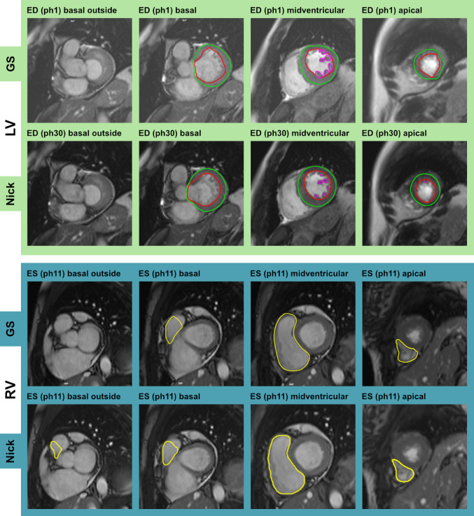 Figure 1
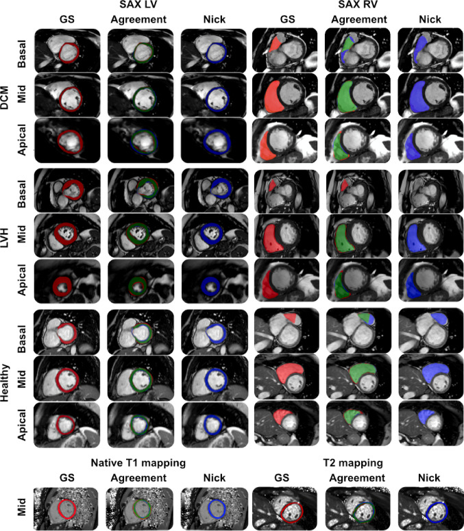 Figure 2
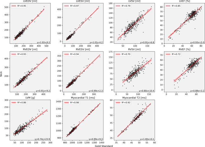 Figure 3
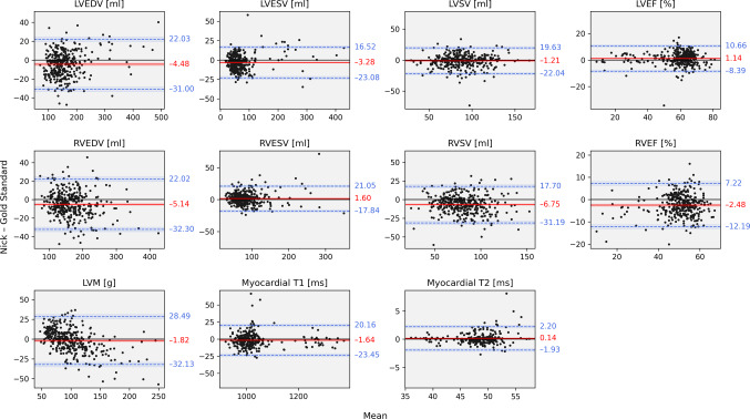 Figure 4
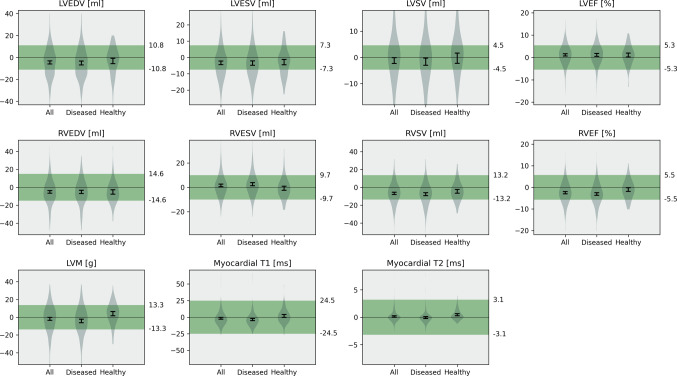 Figure 5
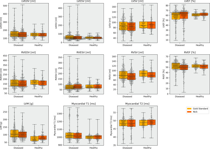 Figure 6
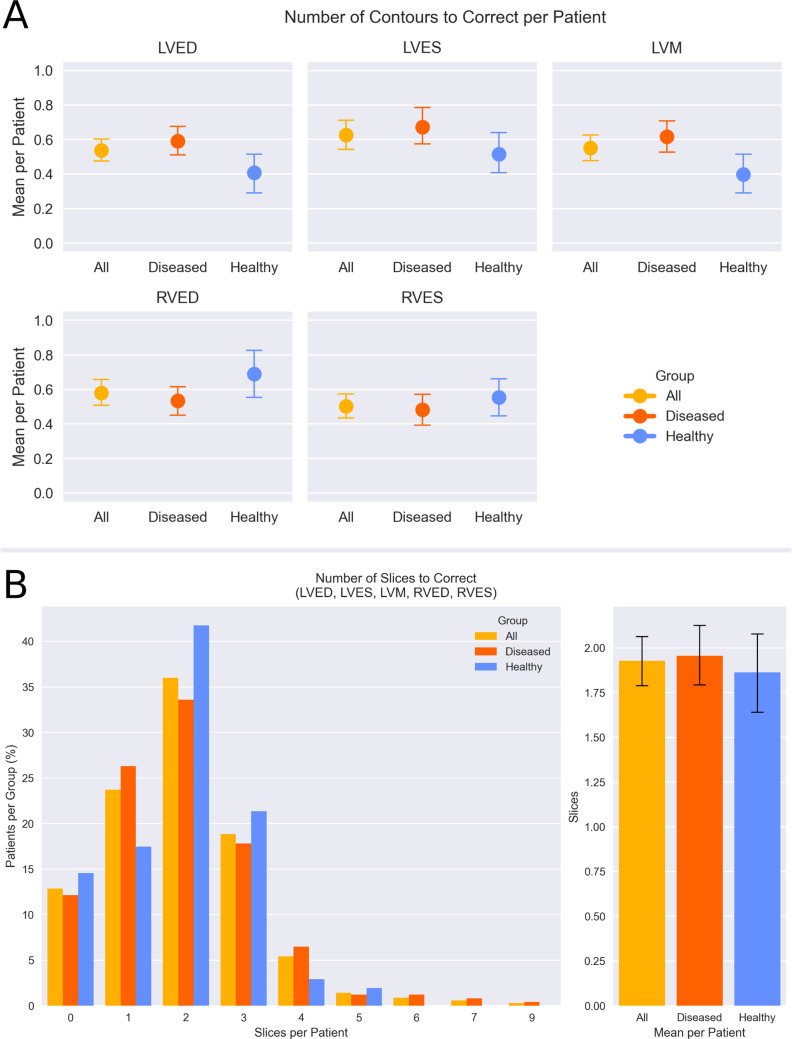 Figure 7
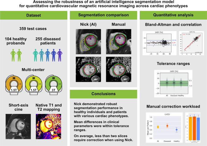 Figure 8
Peer Reviews
No public reviews on file for this paper yet. If you reviewed it on a platform where reviews are public (OpenReview, ICLR, NeurIPS, ICML), you can paste yours below so the community can read it here.
Videos
No videos yet. Explain this paper in a talk, walkthrough, or lecture? Add one.
Taxonomy
TopicsCardiac Imaging and Diagnostics · Cardiovascular Function and Risk Factors · Advanced MRI Techniques and Applications