The feasibility and accuracy of real-time intra-operative confocal tissue diagnosis in brain and spine cancer surgery
William S. Bolton, Oluwaseyi Adebola, Piravin K. Ramakrishnan, Dharsshini Reveendran, Vassili Crispi, Richard Digby, Rohitashwa Sinha, Arundhati Chakrabarty, Ryan K. Mathew

TL;DR
This study evaluates a fast confocal microscope for real-time brain and spine tumor diagnosis during surgery, showing promising results for quick and accurate intraoperative decisions.
Contribution
The study introduces and validates the use of an ultra-fast confocal scanner for real-time intraoperative tissue diagnosis in brain and spine cancer surgery.
Findings
94% of 47 tissue specimens produced interpretable images within 60 seconds using the Histolog® system.
Histolog® matched gold standard diagnoses in 68% of cases, comparable to existing intraoperative methods.
The system enabled real-time tumor cell identification in all specimens.
Abstract
Real-time intra-operative brain tumour tissue analysis can reduce turnaround times and enable repeated sampling, enhancing diagnostic accuracy and guiding resection. We investigated the use of an ultra-fast confocal microscopy scanner (Histolog®, SamanTree Medical SA) for real-time brain tumour tissue diagnosis. This study aims to demonstrate Histolog®’s diagnostic accuracy in a cohort of brain or spinal cord tumour patients. A single-group, observational study included adult patients undergoing tissue biopsies or tumour debulking surgery. Multiple, freshly excised tissue samples were stained and imaged within 60 seconds using Histolog® alongside standard diagnostic methods. A Consultant Neuropathologist performed a blinded concordance analysis. Of 47 specimens, 94% produced interpretable images during surgery. Histolog® images enabled real-time tumour cell identification in all…
Genes, proteins, chemicals, diseases, species, mutations and cell lines named across the full text — each resolved to its canonical identifier and authoritative record.
Click any figure to enlarge with its caption.
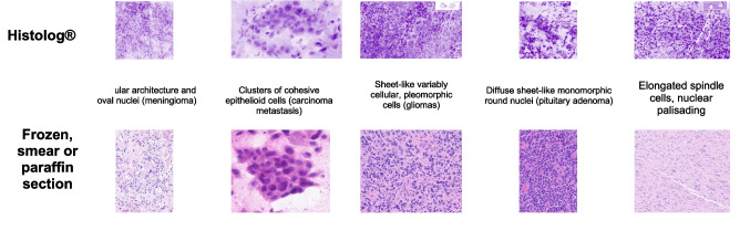 Figure 1
 Figure 2
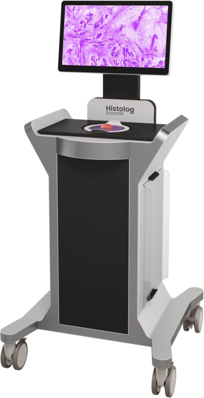 Figure 3
Peer Reviews
No public reviews on file for this paper yet. If you reviewed it on a platform where reviews are public (OpenReview, ICLR, NeurIPS, ICML), you can paste yours below so the community can read it here.
Videos
No videos yet. Explain this paper in a talk, walkthrough, or lecture? Add one.
Taxonomy
TopicsGlioma Diagnosis and Treatment · Brain Tumor Detection and Classification · AI in cancer detection