Diagnosing growth in low-grade gliomas with and without artificial intelligence-measured longitudinal volume measurements: A retrospective observational study
Hassan M Fathallah-Shaykh, Houman Sotoudeh, Markus Bredel, Alex Whitley, Jinsuh Kim, Fanny E Morón, Fabio Raman, Nidhal Bouaynaya, Hayat Rahal

TL;DR
This study shows AI-assisted volume analysis can detect brain tumor growth earlier than traditional methods, with physician oversight improving accuracy.
Contribution
The study introduces AI-assisted volumetric analysis for early detection of low-grade glioma growth, demonstrating its clinical potential with physician review.
Findings
AI segmentation with human review detected tumor growth 21 months earlier in progressing cases compared to visual inspection.
In stable cases, AI identified growth 23 months earlier than the last MRI scan.
AI without human review had a 25% false positive and 8.33% false negative rate.
Abstract
Low-grade or grade 2 diffuse gliomas (LGG) infiltrate the brains leading to significant neurological morbidity. This retrospective observational study evaluates the ability of AI-assisted volumetric analysis to correctly detect tumor growth in longitudinal studies of LGG as compared to the standard clinical method. A total of 56 gliomas and 7 stable FLAIR lesions were included; gliomas were classified as clinical progression (n = 34), or clinically stable (n = 22). All gliomas were from radiation-naïve patients; only 2 patients had completed treatment with temozolomide. The dates of tumor growth were gathered from clinical notes. Longitudinal tumor volumes were calculated by the MRIMath FLAIR AI. Golden truths were obtained by physician reviews using the MRIMath Smart contouring system. Growth by significant shifts in tumor volumes was detected by using the statistical method of online…
Genes, proteins, chemicals, diseases, species, mutations and cell lines named across the full text — each resolved to its canonical identifier and authoritative record.
Click any figure to enlarge with its caption.
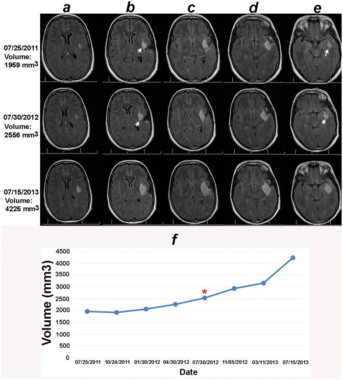 Figure 1
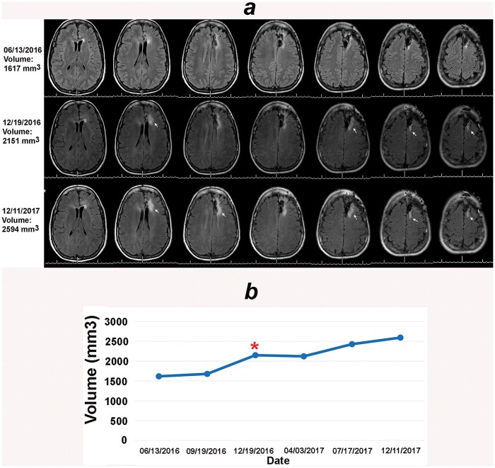 Figure 2
Peer Reviews
No public reviews on file for this paper yet. If you reviewed it on a platform where reviews are public (OpenReview, ICLR, NeurIPS, ICML), you can paste yours below so the community can read it here.
Videos
No videos yet. Explain this paper in a talk, walkthrough, or lecture? Add one.
Taxonomy
TopicsGlioma Diagnosis and Treatment · Advanced MRI Techniques and Applications · Cerebrospinal fluid and hydrocephalus