The protocol for mesoscopic wide-field optical imaging in mice: from zero to hero
Evgenia N Kislukhina, Natalia V Lizunova, Alexander M Surin, Zanda V Bakaeva

TL;DR
This paper provides detailed protocols for mesoscopic wide-field optical brain imaging in mice, enabling visualization of brain activity and metabolism in awake animals.
Contribution
The paper introduces a novel protocol with a wide cranial window, skull thinning, and UV-curable coating for long-term transparent imaging in awake mice.
Findings
The cranial window remains transparent for at least three months, allowing long-term imaging.
The protocol enables simultaneous measurement of hemodynamics and intracellular parameters like FAD and Ca2+.
A lightweight headplate ensures stable fixation without alignment during data analysis.
Abstract
This article provides protocols that enable researchers to master mesoscopic wide-field optical brain imaging from scratch. The protocols describe surgery for wide-field cranial window creation in mice, as well as the imaging process and setup. The protocols for components of the imaging system selection and assembly, creation of a headplate for fixation, and training mice are also provided. The final section briefly outlines methods for data processing. The described procedure can be used to visualize the dorsal cortex using wide-field optical imaging and laser-speckle contrast imaging methods. The distinguishing features of our protocol include: a wide cranial window (up to 60% of the entire cortex), skull thinning (without craniotomy), a UV-curable transparent coating (gel polish), and the ability to perform measurements in awake, behaving mice. During the surgery, a…
Genes, proteins, chemicals, diseases, species, mutations and cell lines named across the full text — each resolved to its canonical identifier and authoritative record.
Click any figure to enlarge with its caption.
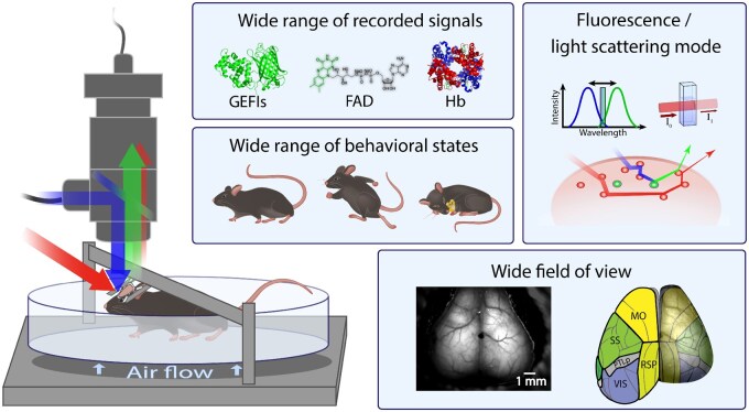 Figure 1
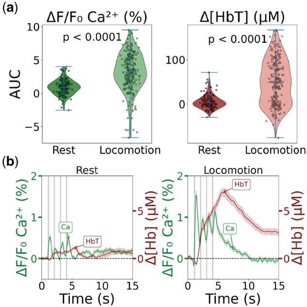 Figure 2
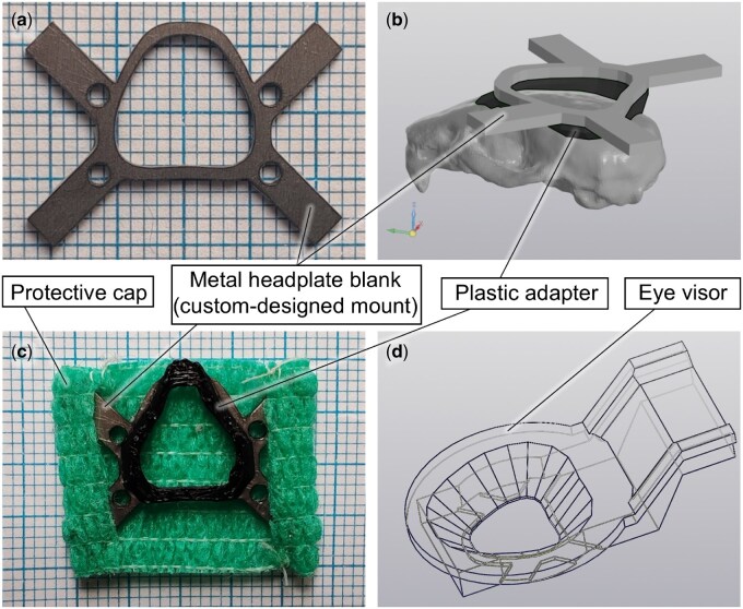 Figure 3
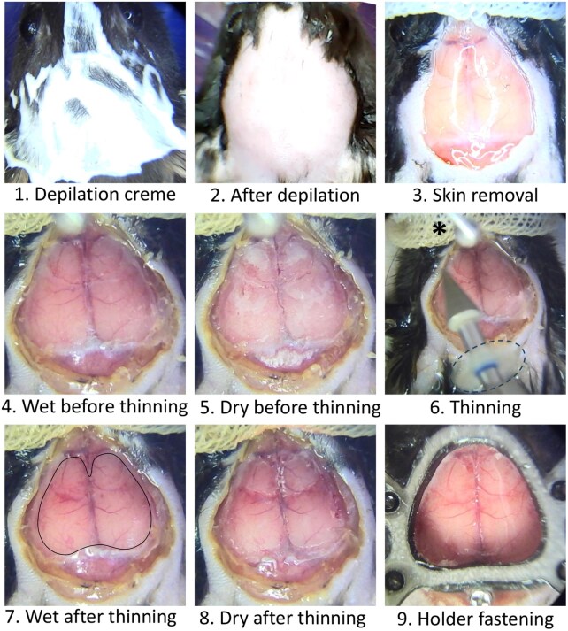 Figure 4
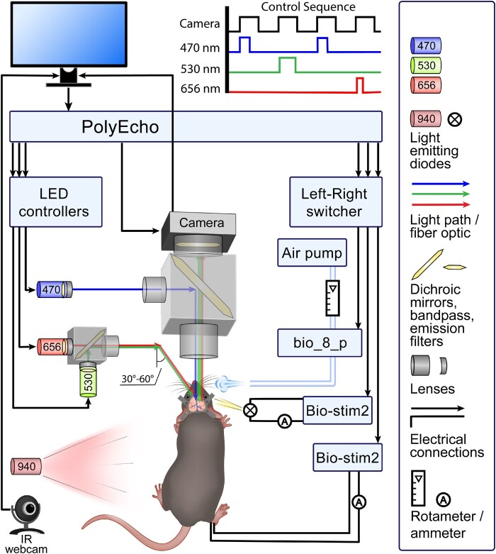 Figure 5
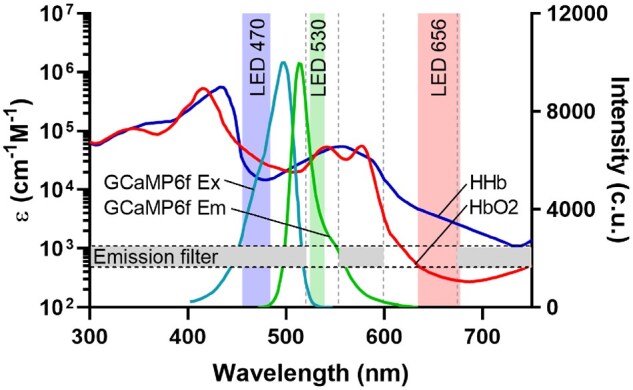 Figure 6
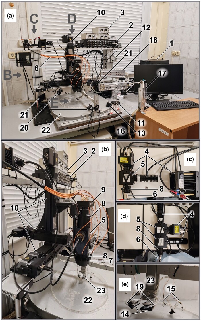 Figure 7
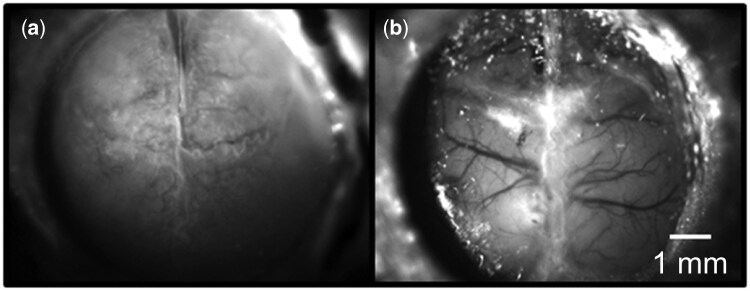 Figure 8
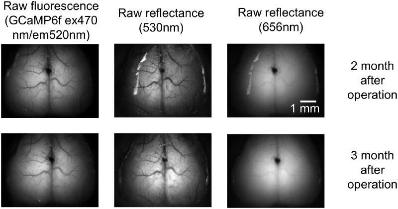 Figure 9
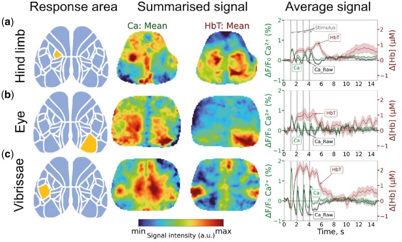 Figure 10
Peer Reviews
No public reviews on file for this paper yet. If you reviewed it on a platform where reviews are public (OpenReview, ICLR, NeurIPS, ICML), you can paste yours below so the community can read it here.
Videos
No videos yet. Explain this paper in a talk, walkthrough, or lecture? Add one.
Taxonomy
TopicsOptical Imaging and Spectroscopy Techniques · Photoacoustic and Ultrasonic Imaging · Advanced Fluorescence Microscopy Techniques