A novel projection data domain material decomposition method for dual-energy CT and its impact on the accuracy of attenuation values
Viktor Haase, Frédéric Noo, Karl Stierstorfer, Andreas Maier, Michael McNitt-Gray

TL;DR
This paper introduces a new method for improving the accuracy of CT scans by decomposing materials in the projection data domain, reducing artifacts and errors in attenuation values.
Contribution
A novel projection data domain material decomposition method with object-specific scatter correction is proposed for dual-energy CT.
Findings
The method significantly improves attenuation value accuracy, especially at low energies (<70 keV).
It reduces beam hardening artifacts and provides more uniform quantitative error across non-water inserts.
Object-specific scatter correction prevents major artifacts and performs well in abdominal phantom imaging.
Abstract
Objective. Despite major advances in dual-energy computed tomography (CT), obtaining accurate attenuation values for quantitative applications remains a technical challenge. To address this topic, we introduce a novel projection data domain material decomposition method that is an extension of an approach we recently proposed for beam hardening correction in single energy CT. Approach. The proposed method employs object-specific scatter correction and an analytical energy response model. We compare its performance to image-based material decomposition on accuracy of attenuation values using the American College of Radiology (ACR) CT accreditation phantom, scanned with consecutive low and high energy axial scans in centered and off-centered positions. Accuracy is assessed across the five inserts, and the images are analyzed for beam hardening artifacts and noise. Additionally, we assess…
Genes, proteins, chemicals, diseases, species, mutations and cell lines named across the full text — each resolved to its canonical identifier and authoritative record.
Click any figure to enlarge with its caption.
 Figure 1
 Figure 2
 Figure 3
 Figure 4
 Figure 5
 Figure 6
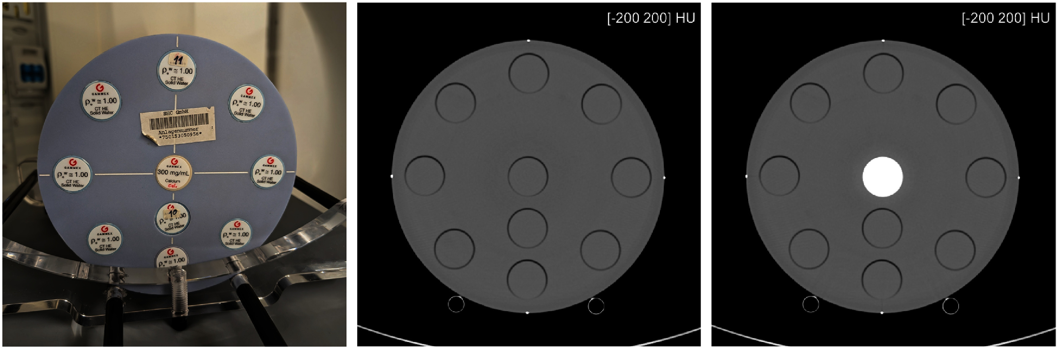 Figure 7
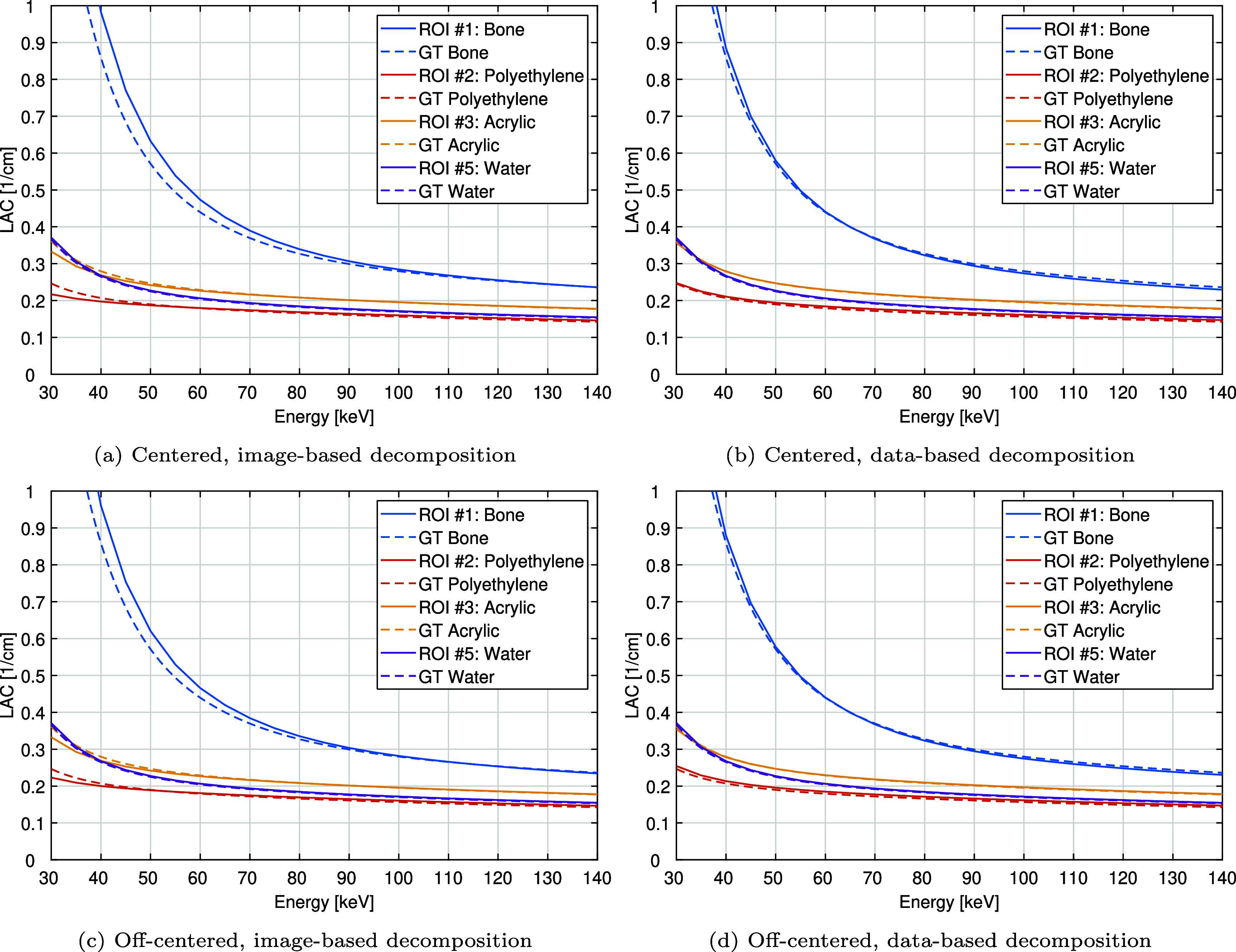 Figure 8
 Figure 9
 Figure 10
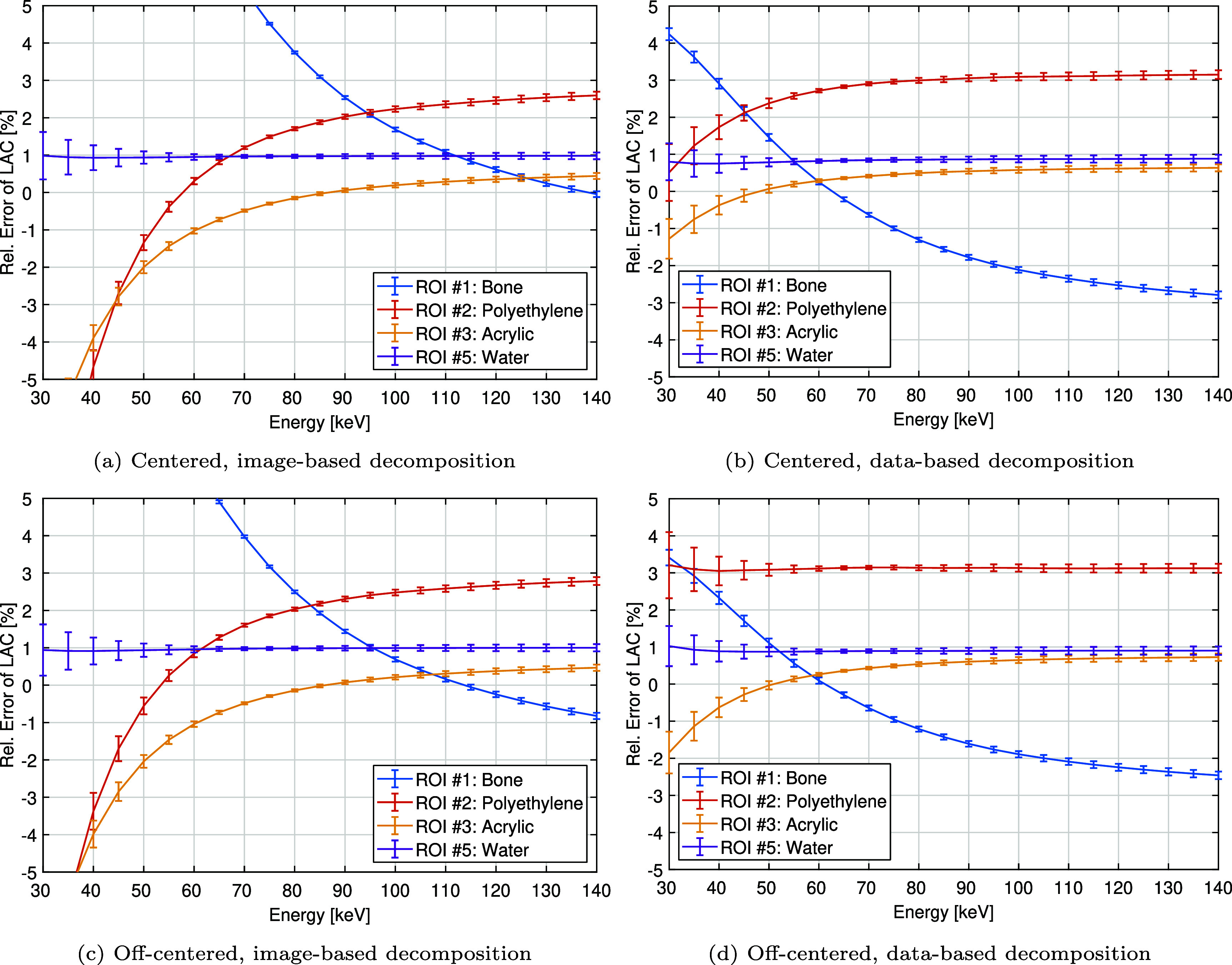 Figure 11
 Figure 12
 Figure 13
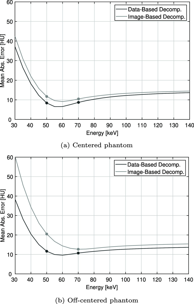 Figure 14
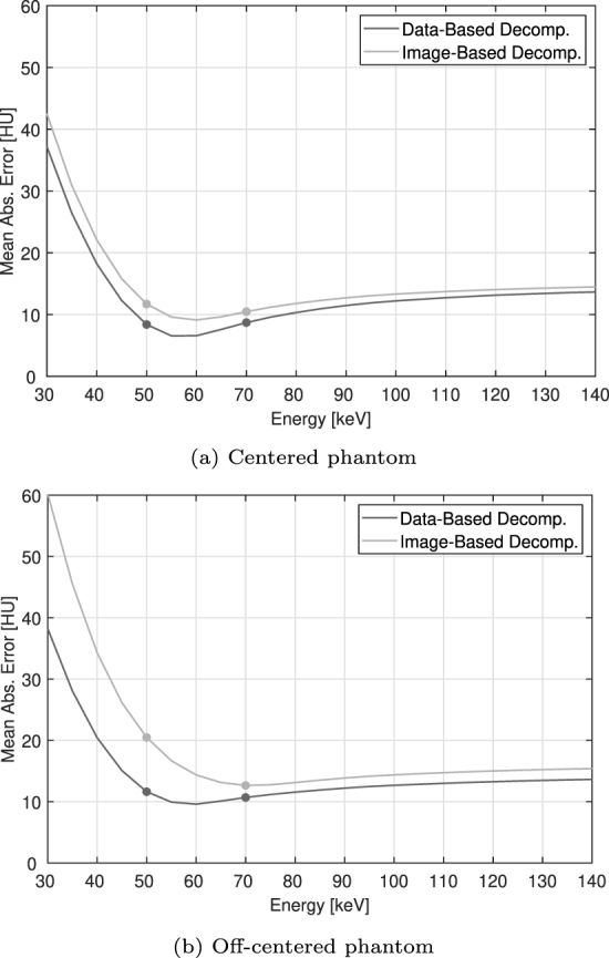 Figure 15
 Figure 16
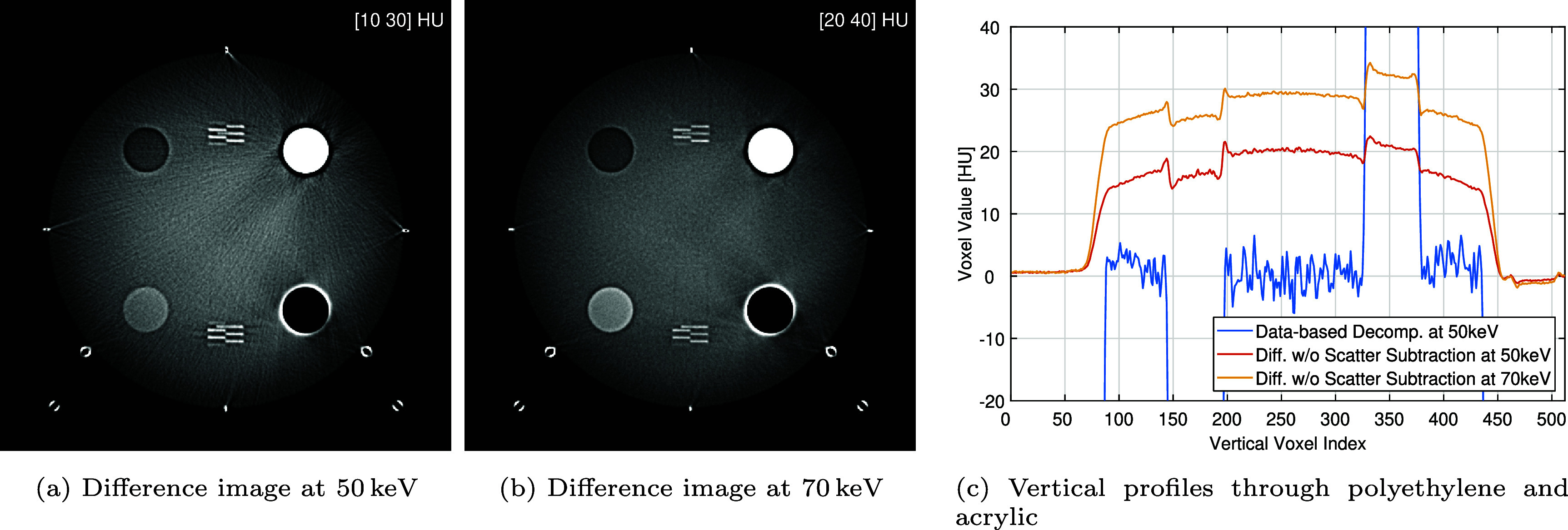 Figure 17
 Figure 18
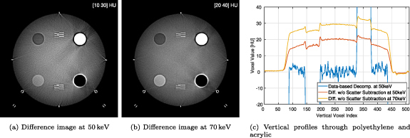 Figure 19
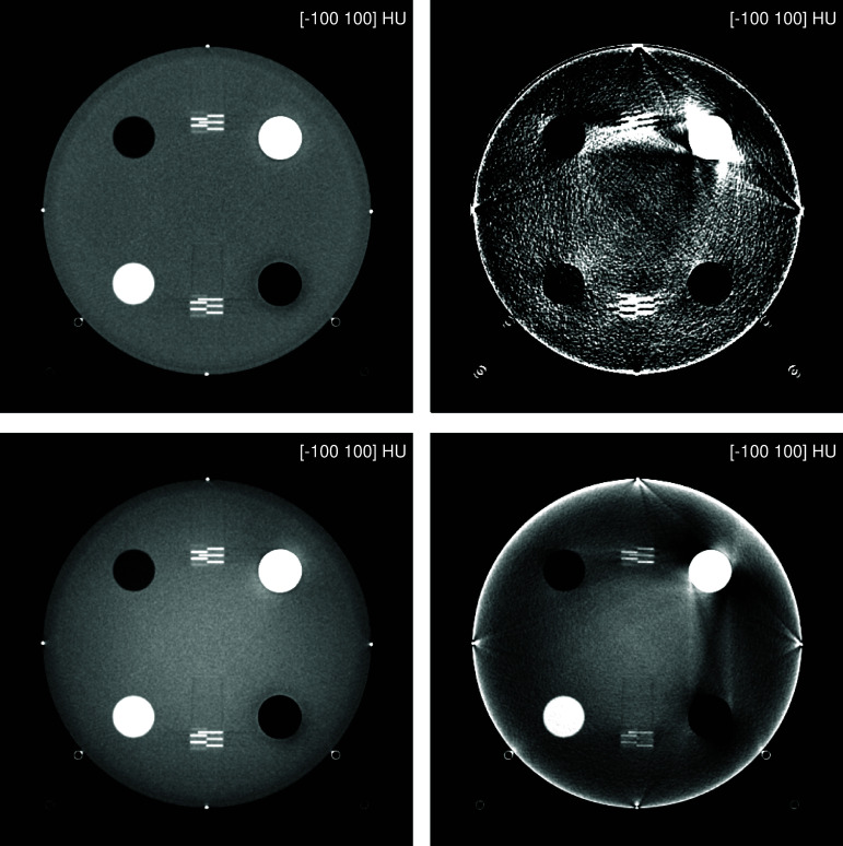 Figure 20
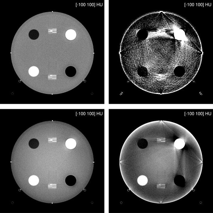 Figure 21
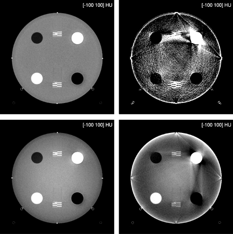 Figure 22
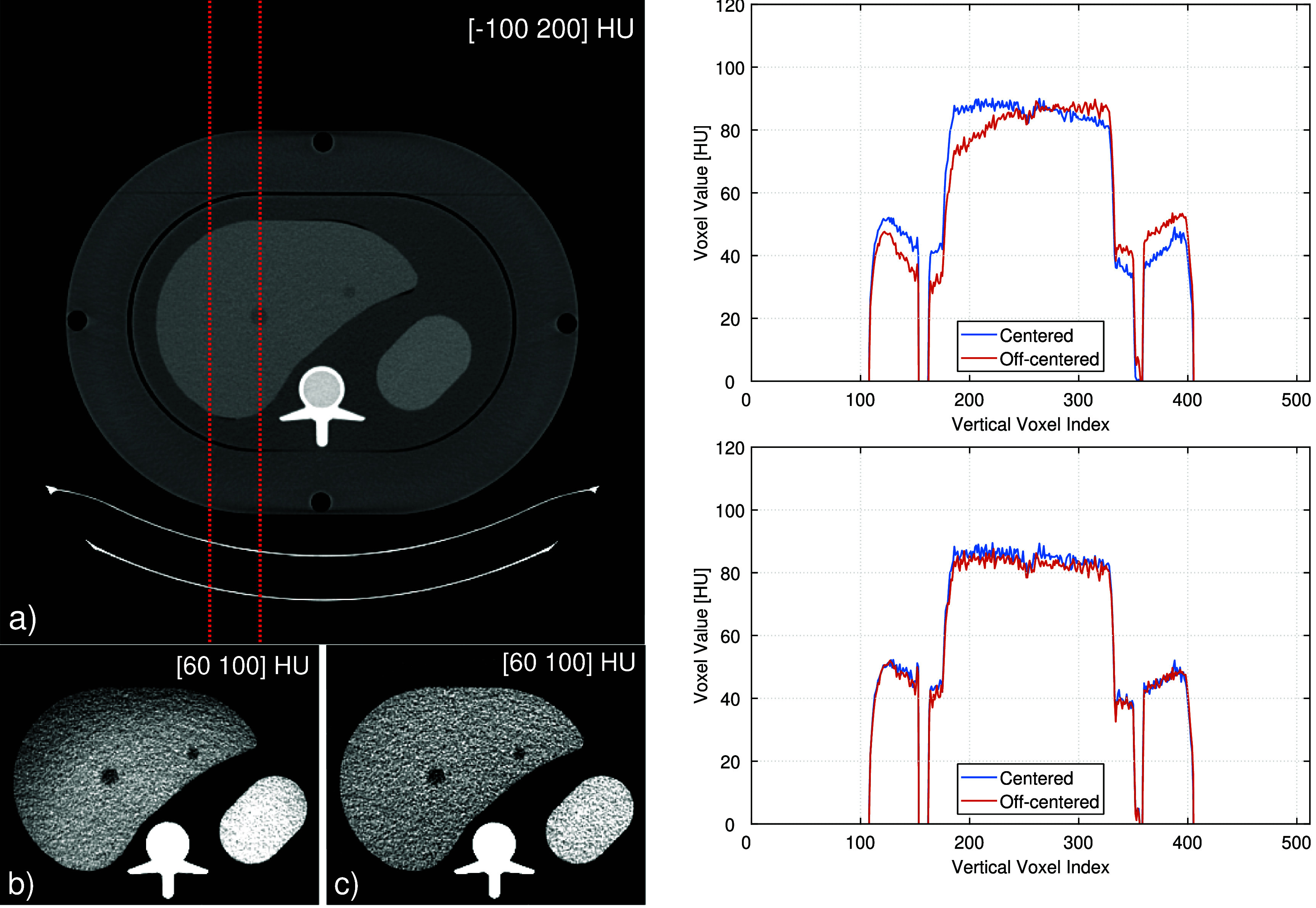 Figure 23
 Figure 24
 Figure 25
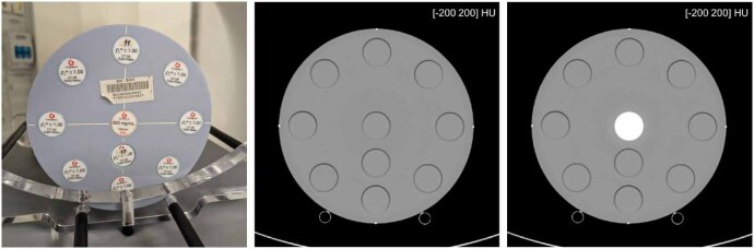 Figure 26
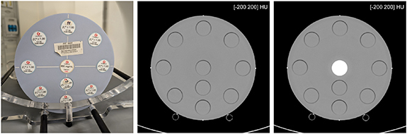 Figure 27
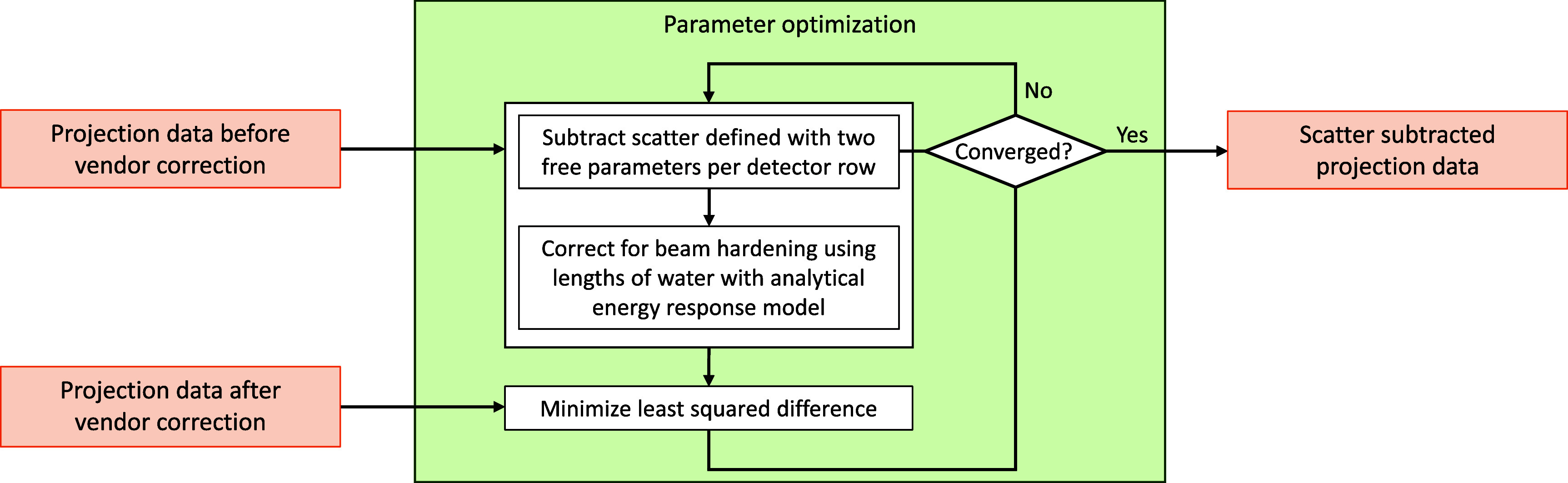 Figure 28
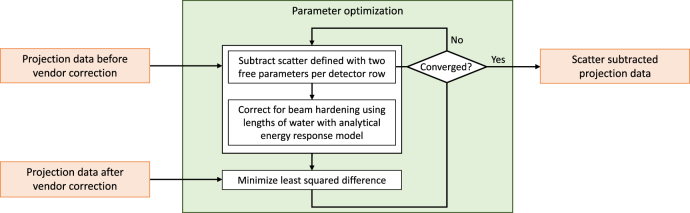 Figure 29
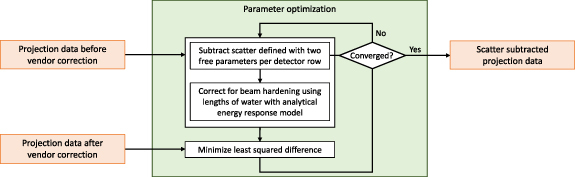 Figure 30
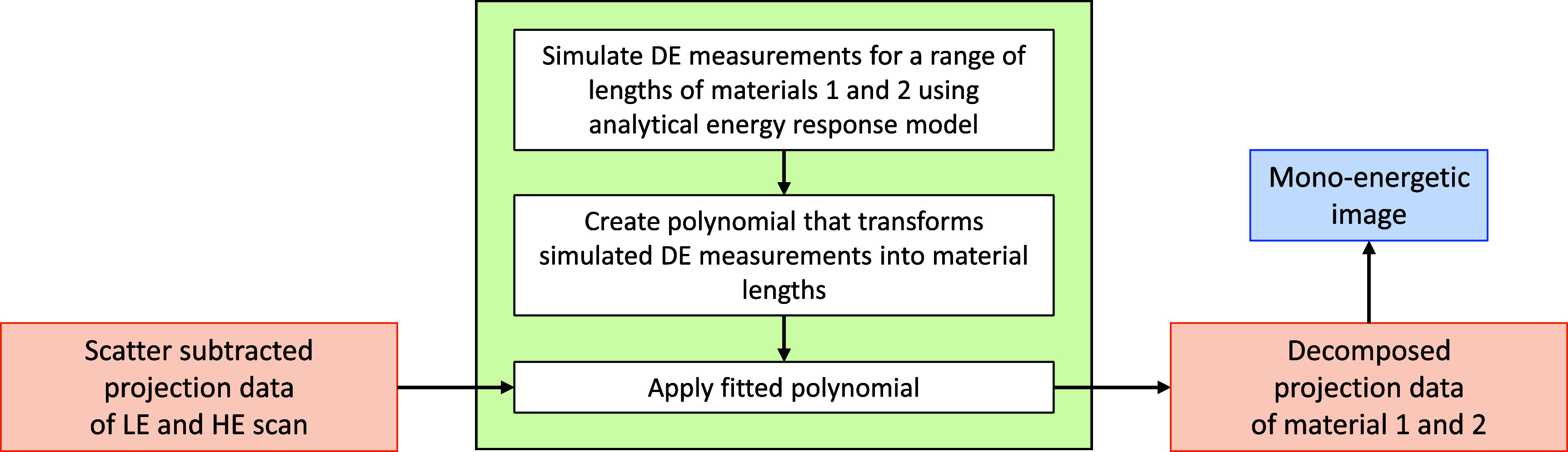 Figure 31
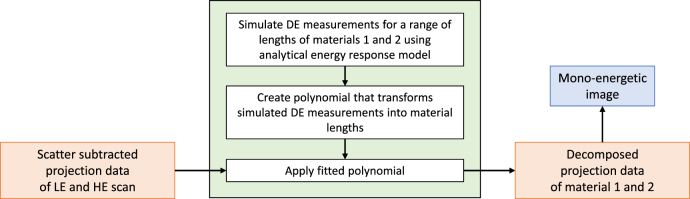 Figure 32
 Figure 33
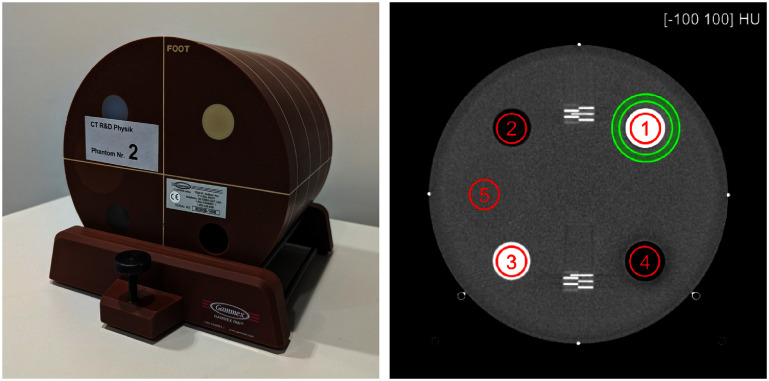 Figure 34
 Figure 35
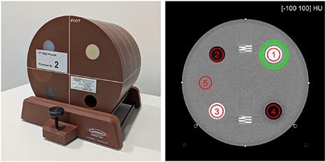 Figure 36
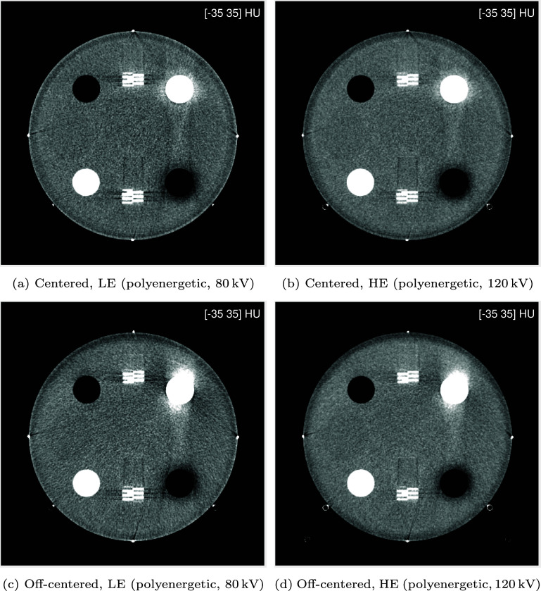 Figure 37
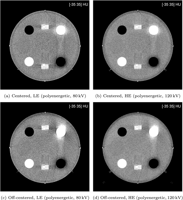 Figure 38
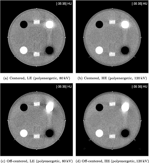 Figure 39
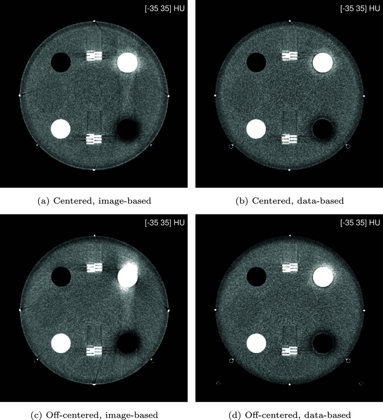 Figure 40
 Figure 41
 Figure 42
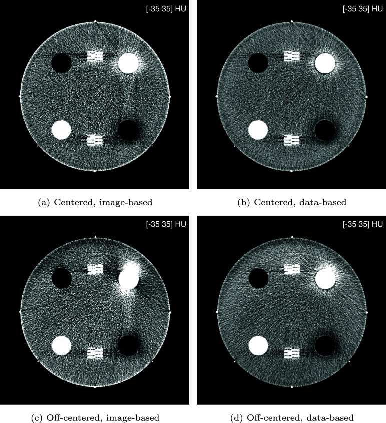 Figure 43
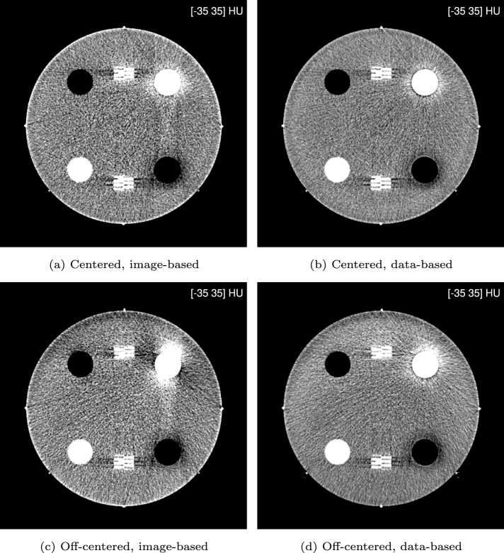 Figure 44
 Figure 45
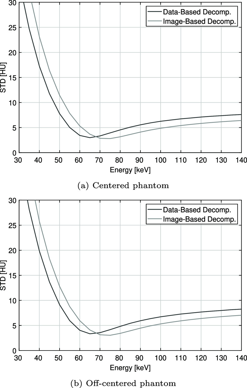 Figure 46
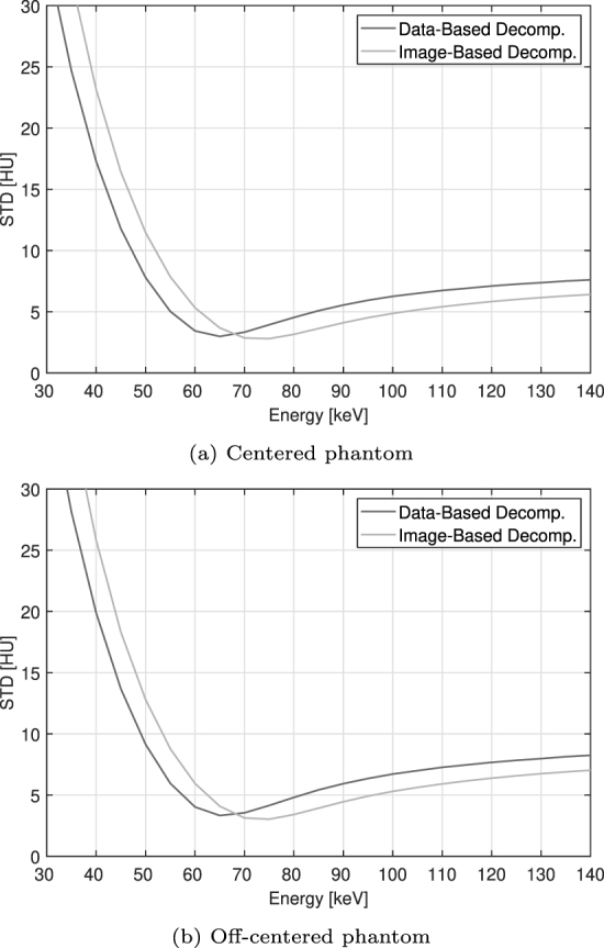 Figure 47
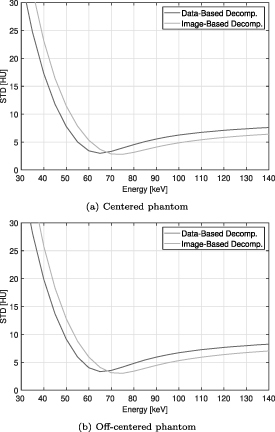 Figure 48
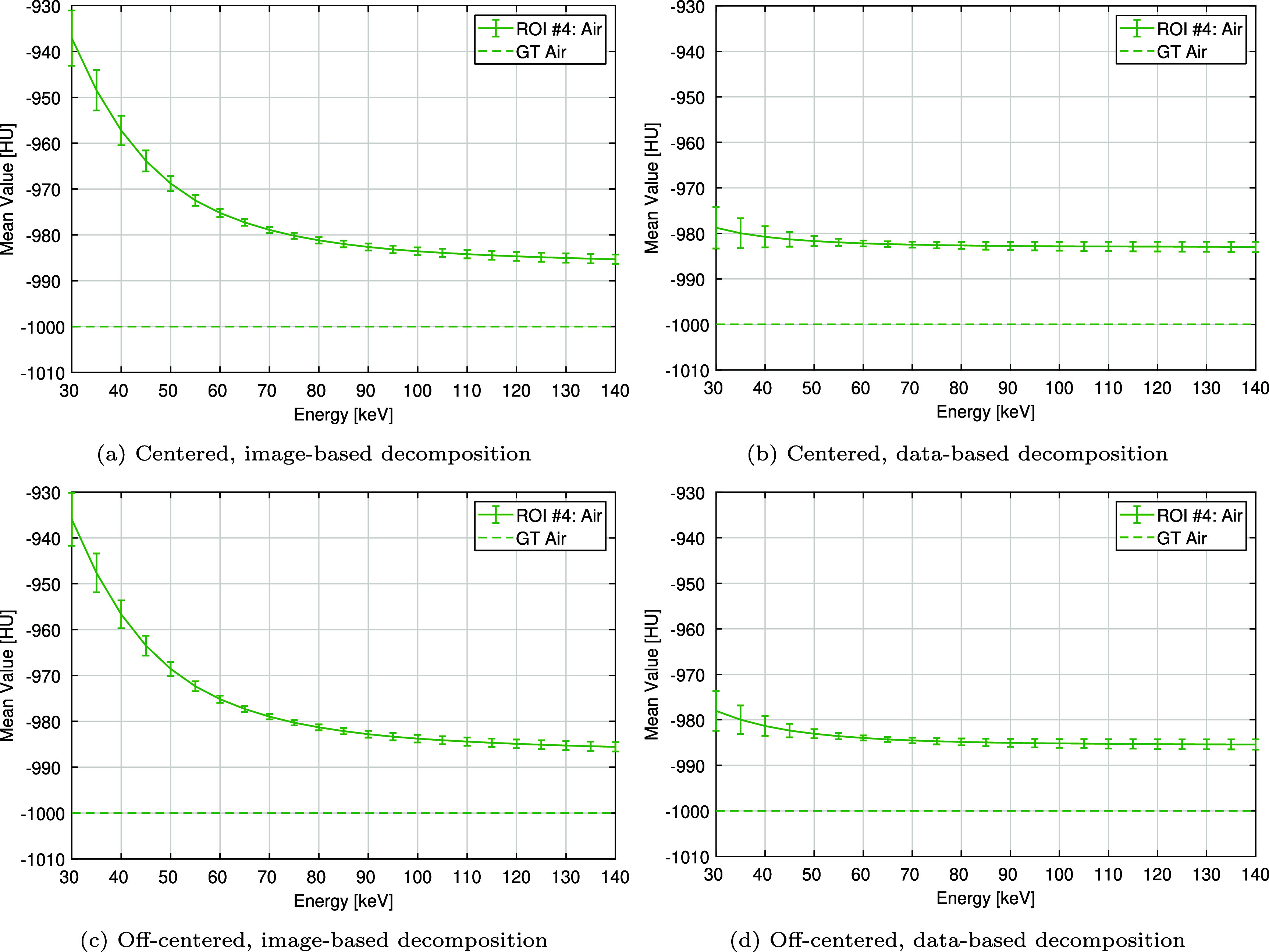 Figure 49
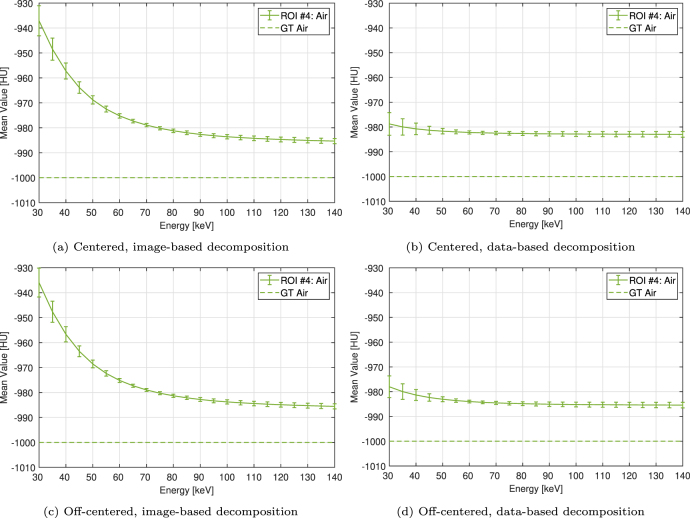 Figure 50
Peer Reviews
No public reviews on file for this paper yet. If you reviewed it on a platform where reviews are public (OpenReview, ICLR, NeurIPS, ICML), you can paste yours below so the community can read it here.
Videos
No videos yet. Explain this paper in a talk, walkthrough, or lecture? Add one.
Taxonomy
TopicsAdvanced X-ray and CT Imaging · Advanced X-ray Imaging Techniques · Radiation Dose and Imaging