Fully automated volumetry of ventricular subregions on computed tomography using object detection and semantic segmentation
Raffaele Da Mutten, Olivier Zanier, Alessandro Carretta, Giorgio Palandri, Massimo Bottini, Daniel de Wilde, Ulf C. Schneider, Luca Regli, Carlo Serra, Victor E. Staartjes

TL;DR
This paper presents a machine learning pipeline for automatically measuring brain ventricle subregions in CT scans, which could help detect subtle changes in ventricular volume for clinical evaluation.
Contribution
A novel deep learning pipeline combining object detection and semantic segmentation for automated ventricular subregion volumetry on CT.
Findings
The pipeline achieved high precision in segmenting ventricular subregions on CT scans.
Best mean Dice scores were 0.92 ± 0.1 for the body of the left lateral ventricle.
A user-friendly graphical interface was developed for clinical integration of the method.
Abstract
Our goal was to develop and validate machine learning models that are capable of fully automatic identification and segmentation of frontal, temporal, and posterior horns, the body of the lateral ventricle, the third and fourth ventricle, as well as the atrium on either side. Patients shunted for hydrocephalus were included. Data from two centers was used for development/external validation, respectively. Manual labelling of ventricular subregions on computed tomography (CT) was performed. First, an object detection algorithm (YOLOv5) was trained. This allowed for precise cropping of the subregions that could then be used as input for a 2D U-Net. For comparison, a nnU-Net was also trained. Precision, recall, mean average precision 50 and 50–95 (mAP50; mAP50-95) were used as performance metrics for the YOLO algorithm. Dice score, Jaccard score, and 95th percentile Hausdorff distance…
Genes, proteins, chemicals, diseases, species, mutations and cell lines named across the full text — each resolved to its canonical identifier and authoritative record.
Click any figure to enlarge with its caption.
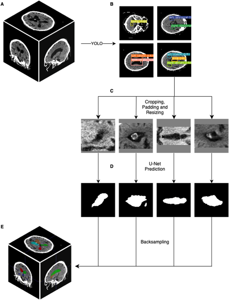 Figure 1
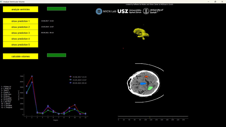 Figure 2
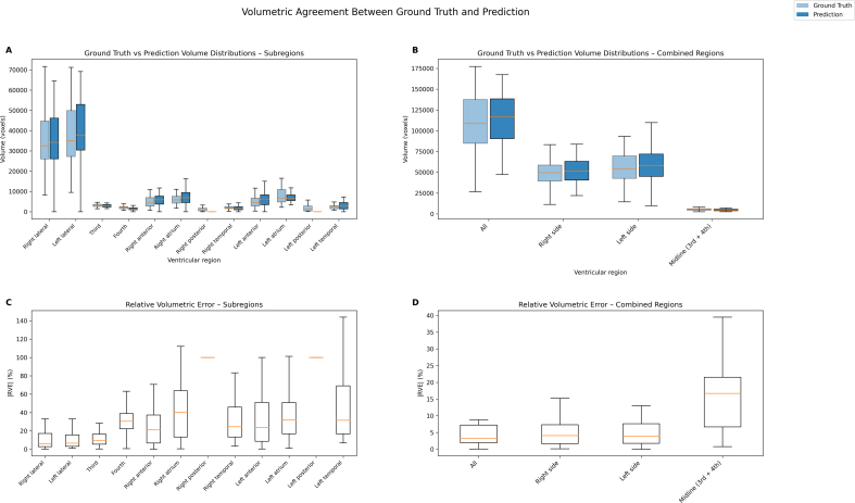 Figure 3
Peer Reviews
No public reviews on file for this paper yet. If you reviewed it on a platform where reviews are public (OpenReview, ICLR, NeurIPS, ICML), you can paste yours below so the community can read it here.
Videos
No videos yet. Explain this paper in a talk, walkthrough, or lecture? Add one.
Taxonomy
TopicsMedical Image Segmentation Techniques · Advanced Neural Network Applications · Medical Imaging and Analysis