Automated segmentation of trunk musculature with a deep CNN trained from sparse annotations in radiation therapy patients with metastatic spine disease: an observational study
Vy Hong, Steve Pieper, Joanna James, Dennis E. Anderson, Csaba Pinter, Yi Shuen Chang, Aslan Bulent, David Kozono, Patrick Doyle, Sarah Caplan, Heejoo Kang, Tracy Balboni, Alexander Spektor, Mario Keko, Ron Kikinis, David B. Hackney, Ron Noah Alkalay

TL;DR
A deep learning model was developed to automatically segment trunk muscles in cancer patients with spine disease using sparse CT data, achieving results comparable to manual methods.
Contribution
A deep CNN trained from sparse annotations enables accurate 3D segmentation of trunk musculature in metastatic spine disease patients.
Findings
The DL model achieved a mean Dice score above 0.769, comparable to manual segmentations.
Radiologist ratings showed noninferiority of DL-generated segmentations to manual ones.
The model can rapidly generate high-fidelity 3D muscle volumes for individualized fracture risk evaluation.
Abstract
Given the high prevalence of vertebral fractures following radiotherapy in patients with metastatic spine disease, torso muscle segmentation is necessary for biomechanical modeling of vertebral loading, permitting individualized evaluation of fracture risk. In this study, we developed and validated a deep-learning model for full volumetric segmentation of the thoracic and abdominal spinal musculature in cancer patients with metastatic spine disease from sparsely annotated clinical CT image data. We obtained CT data for 148 metastatic spine disease patients undergoing radiotherapy treatment, and an external set of randomly selected 30 subjects from the National Lung Screening Trial. We extracted 1924 axial CT images at the midpoint of each vertebral level (T4 to L4) and manually labeled the key extensor and flexor muscles (up to 8 muscles per side) at each level. We trained a 2D nnU-Net…
Genes, proteins, chemicals, diseases, species, mutations and cell lines named across the full text — each resolved to its canonical identifier and authoritative record.
Click any figure to enlarge with its caption.
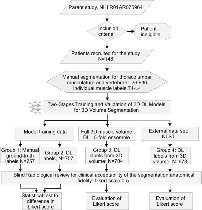 Figure 1
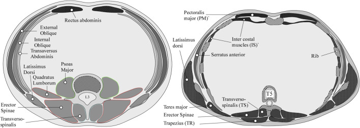 Figure 2
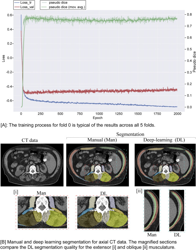 Figure 3
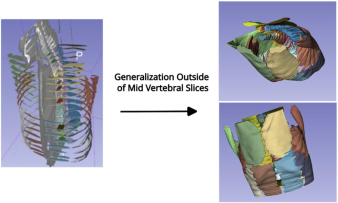 Figure 4
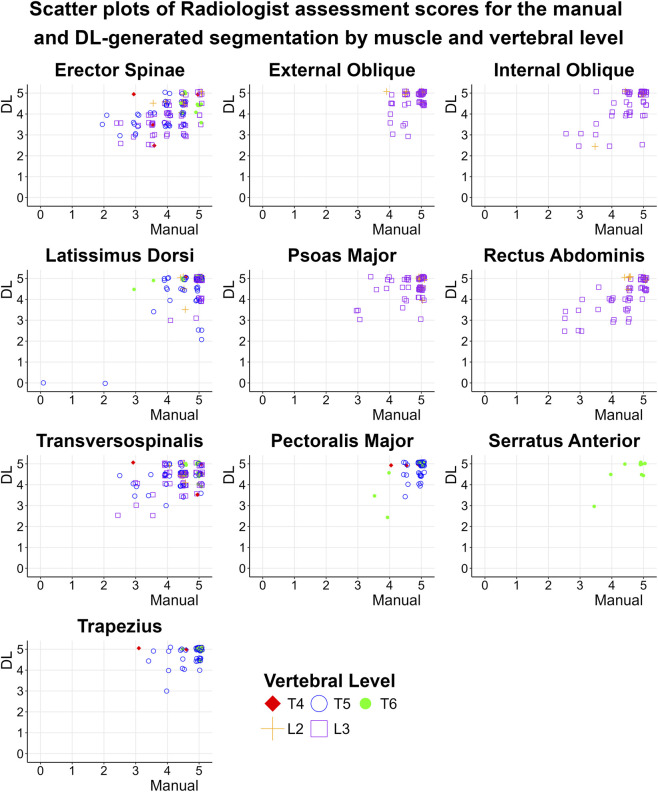 Figure 5
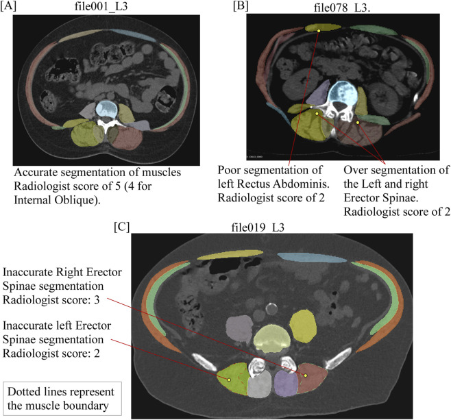 Figure 6
Peer Reviews
No public reviews on file for this paper yet. If you reviewed it on a platform where reviews are public (OpenReview, ICLR, NeurIPS, ICML), you can paste yours below so the community can read it here.
Videos
No videos yet. Explain this paper in a talk, walkthrough, or lecture? Add one.
Taxonomy
TopicsMedical Imaging and Analysis · Management of metastatic bone disease · Advanced Radiotherapy Techniques