Anatomical Study and CT Scan of the Scleral Ring in the Little Owl (Athene noctua)
Omid Zehtabvar, Majid Masoudifard, Hesameddin Akbarein, Arman Shahbazi, Setayesh Karimi, Sara Binesh, Elnaz Khalilzadeh

TL;DR
This study examines the eye structure of little owls, revealing unique features of their scleral ring and differences between male and female owls.
Contribution
The study provides new anatomical insights into the scleral ring of Athene noctua using CT scans and morphometric analysis.
Findings
The scleral ring in little owls typically consists of 15 quadrilateral ossicles, with one specimen showing 16.
The ring has a bipartite structure with a tubular anterior and funnel-shaped posterior segment.
Female owls have significantly larger ocular dimensions than males.
Abstract
Athene noctua, commonly known as the little owl, thrives across the warmer climates of Europe, Asia and North Africa. One of the anatomical features of birds is the presence of a bony scleral ring in the eye. In avians, this configuration comprises ossicles that are affixed together in diminutive plates and are not articulated to other components of the skeleton. The morphology, number, development and location of the scleral ring vary among different vertebrate groups. The objective of this research is to furnish a comprehensive elucidation of the morphology of the scleral ring in the A. noctua predicated on CT scan results and anatomical examination. The overall shape of the scleral ring, the number and shape of the ossicles and their positioning and extensions are notable features that can be used for classification purposes. The study population comprised 10 adult owls (five male…
Genes, proteins, chemicals, diseases, species, mutations and cell lines named across the full text — each resolved to its canonical identifier and authoritative record.
Click any figure to enlarge with its caption.
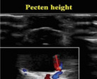 Figure 1
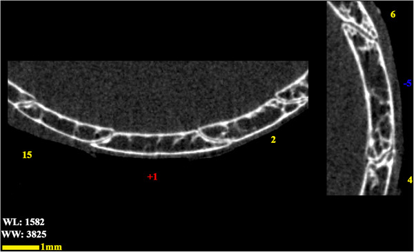 Figure 2
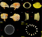 Figure 3
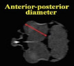 Figure 4
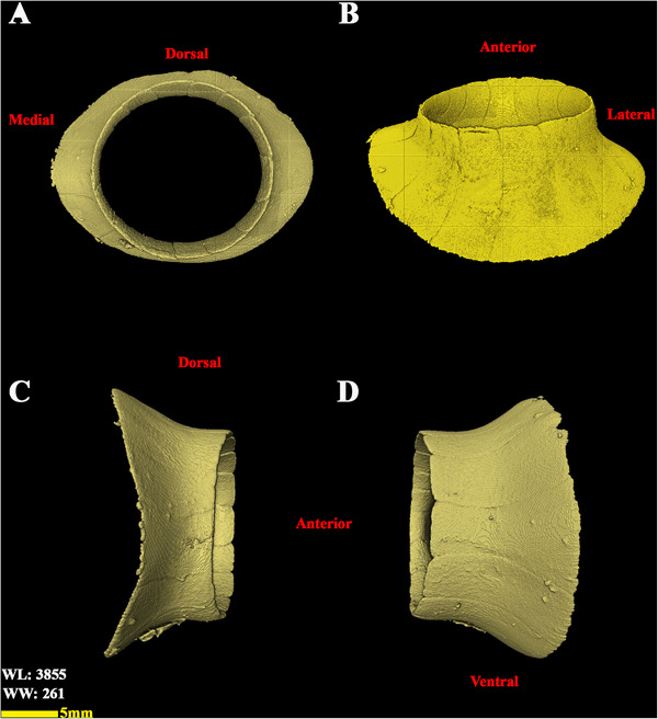 Figure 5
 Figure 6
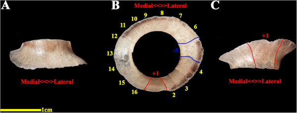 Figure 7
 Figure 8
 Figure 9
 Figure 10
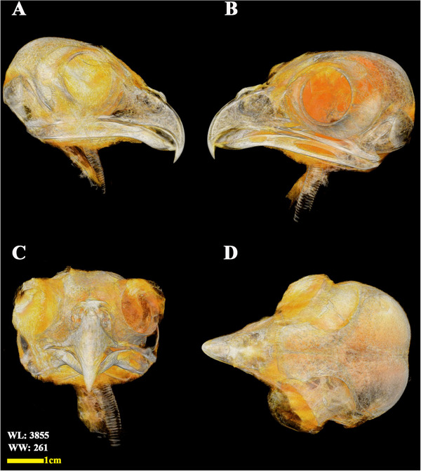 Figure 11
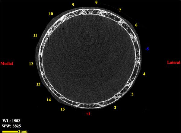 Figure 12
 Figure 13
 Figure 14
 Figure 15
 Figure 16
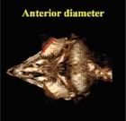 Figure 17
 Figure 18
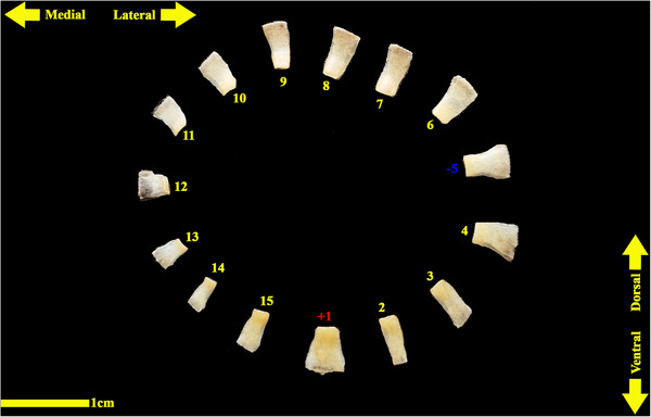 Figure 19
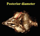 Figure 20
Peer Reviews
No public reviews on file for this paper yet. If you reviewed it on a platform where reviews are public (OpenReview, ICLR, NeurIPS, ICML), you can paste yours below so the community can read it here.
Videos
No videos yet. Explain this paper in a talk, walkthrough, or lecture? Add one.
Taxonomy
TopicsComparative Animal Anatomy Studies · Ocular Disorders and Treatments · Paleontology and Evolutionary Biology