Evaluation of choriocapillaris perfusion changes in chronic central serous chorioretinopathy following Half-Dose photodynamic therapy
Giacomo Boscia, Pasquale Viggiano, Giulia Corradetti, Alba Chiara Termite, Alfonso Savastano, Alberto Quarta, Alessandro Feo, Ceren Soylu, Rodolfo Mastropasqua, Lisa Toto, Francesco Boscia, SriniVas R. Sadda

TL;DR
This study shows that half-dose photodynamic therapy improves blood flow and other eye health metrics in patients with chronic central serous chorioretinopathy.
Contribution
The study introduces half-dose PDT as an effective treatment for chronic central serous chorioretinopathy, showing improvements in choriocapillaris perfusion.
Findings
Choriocapillaris flow deficits decreased significantly after 6 months of half-dose PDT.
Best-corrected visual acuity and macular thickness improved significantly following treatment.
Subfoveal choroidal thickness and serous retinal fluid resolved or reduced in most patients.
Abstract
This study aimed to evaluate changes in choriocapillaris (CC) perfusion in patients with chronic central serous chorioretinopathy (cCSC) following treatment with half-dose photodynamic therapy (PDT), using swept-source optical coherence tomography angiography (SS-OCTA). This pilot study included patients diagnosed with cCSC who underwent half-dose PDT using the Visulas 690s PDT Laser System (Carl Zeiss Meditec, Inc., Jena, Germany; 689-nm wavelength, 50J/cm², 83s). Fovea-centered OCTA scans (6 × 6mm) were obtained using the Zeiss Plex Elite 9000 SS-OCT system. OCTA assessments were conducted at baseline prior to treatment and 6 months post-treatment. The primary outcome was the percentage of choriocapillaris flow deficits (FD%) on OCTA. Secondary outcomes included best-corrected visual acuity (BCVA), central macular thickness (CMT), and subfoveal choroidal thickness (SFCT). The study…
Genes, proteins, chemicals, diseases, species, mutations and cell lines named across the full text — each resolved to its canonical identifier and authoritative record.
Click any figure to enlarge with its caption.
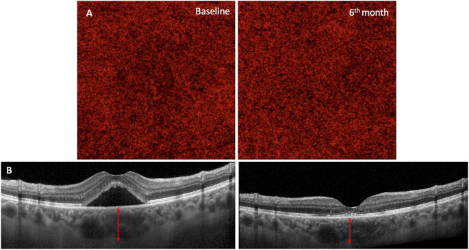 Figure 1
Peer Reviews
No public reviews on file for this paper yet. If you reviewed it on a platform where reviews are public (OpenReview, ICLR, NeurIPS, ICML), you can paste yours below so the community can read it here.
Videos
No videos yet. Explain this paper in a talk, walkthrough, or lecture? Add one.
Taxonomy
TopicsRetinal Diseases and Treatments · Glaucoma and retinal disorders · Retinal Imaging and Analysis